
Classification of Enterobacteriaceae according to Bergeys manual Genus Tripe 1Escherichia 2Edwardsiella 3Citrobacter 4Salmonella 5Shigella 6Klebsiella 7Enterobacter ID: 915480
Download Presentation The PPT/PDF document "Family Enterobacteriaceae" is the property of its rightful owner. Permission is granted to download and print the materials on this web site for personal, non-commercial use only, and to display it on your personal computer provided you do not modify the materials and that you retain all copyright notices contained in the materials. By downloading content from our website, you accept the terms of this agreement.


Slide1
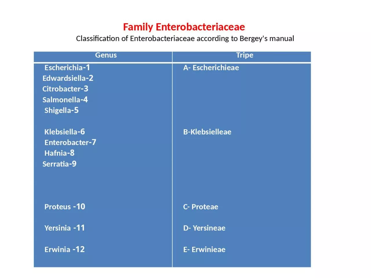
Family Enterobacteriaceae Classification of Enterobacteriaceae according to Bergey's manual
GenusTripe1-Escherichia 2-Edwardsiella3-Citrobacter4-Salmonella5-Shigella 6-Klebsiella 7-Enterobacter 8-Hafnia 9-Serratia 10- Proteus 11- Yersinia 12- Erwinia A- Escherichieae B-Klebsielleae C- Proteae D- Yersineae E- Erwinieae
Slide2Coli form bacilli: Coli form bacillus mean Escherichia, Citrobacter , Klebsiella and Enterobacter (
lactose fermenter ) , and to include the non lactose fermenter Edwardsiella and Serratia . Escherichia The germs is named after Escherich, how was the first to describe this bacillus under the name bacteriun colicommune . (1885) the germs contain only when species name Escherichia coli The bacteria are gram negative motile or non motile, non spore former , grow easily on ordinary culture media, ferment lactose with gas production at 37Ċ and 44Ċ. Indole (+) MR(+) , VP(-) , citrate (-) IMViC = + + - - .
Slide3Slide4Intestinal parasite of man and animals capsules and fimbriae are found in many strains .Cultural characters:Nutrient agar the colonies are large opaque grayish white circular , raised low convex with an entire edge and smooth surface . Blood agar: Some strains produce beta haemolytic
colonies .
Slide5E. coli in MacConkey agar
Slide6MacConkey's medium : The bacteria produce large bright pink colonies due to lactose fermentation. Nutrient broth: There is profuse growth , producing general turbidity and deposit
. Biochemical reaction: The bacteria ferment glucose , maltose , xylose , arabinose and glycerol with gas formation . most strains (80%) ferment lactose , indol (+), MR(+) , VP(-), Citrate (-) : IMViC(+ + - - ) H2S (-) . Eijkman test is (+). Antigentic structure : At least 160 types of O antigen , 90 K antigen and 50 H antigens have been recognized . the antigenic pattern of a strain is recorded as the number of the particular antigen it carries , as for example O 111, K 56 , H 4 . The K antigen : is the envelope antigen located in the capsule or microcapsule covering the O antigen and renders the strain in agglutinable by the O antiserum . Three kinds of K antigens have been described as L , A and B . H- antigen: the flagellar antigens are some times poorly developed, at least on primary isolation. O- antigen: agglutination tests must be curried out on boiled or autoclaved cultures to overcome in agglutinability caused by K-antigens . numerous cross reaction occur between individual E.coli antigens and between O-antigens of Salmonella , Shigella and Citrobacter , organisms . Toxins : In addition to the classical endotoxic activity associated with O-antigen , E. coli produce several soluble toxins . the important being two types of exotoxins , which are : 1- Enterotoxin 2- Haemolysin .
Slide7Enterotoxins : This toxins is produced by few strains of E. coli . the great majority of enteropathogenic strains of E. coli produce an enterotoxin which mediates the movement of water and Ions from the tissues in to the bowel lumen to give rise to diarrhea . The process is similar to that occurring with cholera enterotoxin
. Enteropathic strains produce two such toxins : thermostable(ST) and thermolabile (LT). There is a widely held view that the toxin having bound to sensitive cells acts in a manner analogous to the cholera toxin by activating cellular adenyl cyclase in epithelial cell of the small intestine lining to increase the level of cyclic adenosine -5- monophosphate (cAMP) , which in turn brings about water and ion loss by increasing the permeability of the intestinal lining and thus produce diarrhea . The ST does not stimulate adenyl cyclase activity or antibody production , it stimulates fluid secretion in the gut through the medication of cyclic – guanosine – monophosphate (cGMP) . the test is usually performed in the ligated ileal loop. Haemolysin : Two kinds of haemolysins are produced . ά haemolysin . which is a soluble haemolysin found in cell free culture supernatants . the second , Beta haemolysin is a cell- bound haemolysin . these haemolysin do not appear to be associated with pathogens of infection with haemolytic strains . Colicins : The colicins are antibiotic like principles , broadly known as bacteriocins but they are often given more specific names to indicate the nature of the organism which produces them and those produced by E. coli are known as colicins . Colicins are proteins , but are some times associated with lipopolysaccharide of the O antigen complex . they act against other sensitive strains of E. coli as well as some closely related enterobacteria . Colicins bind to receptors on the outer membrane of sensitive cells, following this they contact the cytoplasmic membrane and disturb its function is some way . there appears to be no signification relation ship between colicins and pathogenicity in E. coli .
Slide8. Human pathogencity: This bacteria is part of the normal intestinal flora (NIF) of man , and except for relatively few enteropathic strains are considered to be commensals at this site . It is potentially pathogenic at other sites in the body , where they may produce pyogenic infection . the following type of infections by
E. coli may be produced : 1- Urinary tract infection (UTI) 2- Diarrhea 3- Pyogenic infections 4- Septicaemia ***************there are five groups of E. coli which can cause diarrhea particularly in infants, but also in children and adults . these groups have been labeled as : 1- Enteropathogenic E. coli (EPEC) 2- Enterotoxigenic E. coli (ETEC)3- Enterovasive E. coli (EIEC) 4- Enteroadherant E. coli (EAEC) 5- Enterohaemorrhagic E. coli ( EHEC) Laboratory diagnosis:The common clinical material referred to the laboratory may be : 1- Urine 2- stool 3- pyogenic material : a) pus b) swab c) C.S.F 4- septicaemia Eijkman test : The test is based on the production of gas in MacCokey's broth at 44Ċ and Indole from peptone at 44Ċ which is a characteristic of E. coli .
Slide9Genus Klebsiella Friedlander , 1883 first described this bacillus as an aetiologic agent of pneumonia and so the formal name of
Klebsiella pneumoniae was given to this organisms . These organisms have also been called Aerobacter aerogenes , but it is generally accepted that the name Klebsiella aerogenes is preferable . The most commonly isolated members of the tribe klebsielleae is Klebsiella aerogenes (K. pneumoniae).originally it was thought to be the cause of the classic lobar pneumonia , the true agent of which is pneumococcus . The remaining two species of Klebsiella K. ozaenae and K. rhinoscleromatic are causative agent of chronic infection of the nasal mucosa and pharynx and are quite rare incidence . Klebsiella aerogenes : ( Klebsiella pneumoniae , Aerobacter aerogenes , Friedlander bacillus ) non motile , gram negative bacilli, capsulated , ferment common sugars with abundant gas production . Indole (-) , MR(-) VP(+), citrate (+) : IMVIC: - - + + , do not liquefy gelatin found in the bowel and respiratory tract of man and animals and in soil and in water . Mucoid strains produced the largest amount of polysaccharide and most of it was present as capsule or slime .
Slide10Slide11Cultural characters : Klebsiella grow well on ordinary culture media . on nutrient agar the colonies are grayish – white , mucoid . the bacteria are aerobic, optimum growth temperature is 37Ċ. On MacConkey's medium the colonies are large ,
mucoid and pink .there is no haemolysis on blood agar , and it is urease positive , H2S negative .
Slide12Klebsiella aerogenes on MacConkey agar
Slide13Proteus
They were named proteus because of there pleomorphism , after the Greek God ( proteus ) who could assume an shape . The bacteria are gram negative bacilli , usually motile by peritrichate flagella , many strains produce swarming growth on agar media , non lactose ferment , decompose urea , convert phenyalanin to phenylpyruvic acid . it is one of the most common bacteria in the soil and water containing decaying organic matter of the animal origin and usually occurs in large numbers in sewage .
Slide14Cultural characters:Growth occurs freely on ordinary nutrient media. One of the most characteristic properties of Pr. vulgaris and Pro. mirabilis is their ability to swarm on solid media like simple agar or blood agar . The growth has fishy or seminal odor . On
Macconkey's agar , the colonies are pale , non lactose fermenter , large , entire or finely radially striated edge . Biochemical reaction :The genus Proteus can be divided in to species on the basis of biochemical reaction . All the strains ferment glucose and do not ferment lactose . MR(+) , VP(-) , Citrate(+) , Indole(+) , Catalase (+). Phenyl pyruvic acid reaction ( PPR ) : This test is specific test for the identification and differentiation of Proteus . the test is based on the ability of Proteus to transformed phenylalanine to phenylpyruvic acid . No other members of the family enterobacteriaceae possess such activity . Antigenic structure :The presence of the flagellar and somatic antigens was demonstrated by Weil and Felix (1918) . non motile strains contain O-antigen. Certain Proteus strains are agglutinated by the serum of patients having typhus fever . These so called X strains contain an antigen common to the typhus rickettsiae and cause agglutinated of these bacilli . Weil and Felix reaction is of diagnostic value in typhus fever . in the Proteus OX-19, OXK, OX2 strains , the antigen is a part of the O-antigen and its specificity is determined by an alkali-stable carbohydrate hapten , which is also found in Rickettsia .
Slide15When 10% ferric chloride is added to phenylpyruvic
acid causes
Slide16. Pathogenicity :Proteus mirabilis accounts for the majority of Proteus infections in man , causing community as well as hospital – acquired urinary tract infections. Wound and burn infections are common occurrence . pneumonia and septicaemia can also occur . There is evidence that
Proteus urease is nephrotoxic favoring the intracellular infection of tubular epithelium and creating alkalinity in the kidney which leads to necrosis of renal tubular epithelium with the precipitation of magnesium and calcium salts and the formation of calculi. Previously genus Proteus were divided in to : Pr. mirabilis H2S (+) Indole (-) , maltose (-) Pr. vulgaris swarming Indole(+) , maltose (+) Pr. morganii H2S (-) Citrate (-) , nicotinic acid requirement (+) Pr. rettgeri no swarming Citrate (+) nicotinic acid (-) Now genus Proteus are classified as:Pr. mirabilis Pr. vulgaris Pr. penneri Pr. morgani changed to ( Morganella morgani ) Pr. rettgeri changed to ( Providencia rettgeri )
Slide17Genus Salmonella
Salmonella on Macconkey agarGram negative bacilli , non spore forming , non capsulated , most strains are motile , grow very easily on ordinary culture media , ferment glucose and mannitol with acid or acid and gas . Do not ferment lactose and sucrose , VP(-) , Indole is (-) exception is in rare strains , H2S (+) , Urea (-) , citrate (+) . intestinal pathogens of man and animals . More than 1800 types are included in this genus . Human pathogens produce typhoid and paratyphoid fevers and food poisoning , with the exception of Sal. gallinarum and Sal. pullorum all are motile by peritrichate flagella .
Slide18Cultural characters:On nutrient agar:The colonies after 24h . incubation are large , grayish white , moist , convex , smooth, circular , entire edge. On Macconkey's medium: Non lactose fermenters and thus produce pale colonies.
On Deoxycholate citrate agar medium (DCA) : Colonies are pale due to absence of lactose fermentation.On Wilson and Blair medium : Brilliant green in the medium inhibits the growth of coliform and dysentery bacilli , but does not affect the growth of Salmonella . Salmonella typhi gives rise to black colonies with a metallic sheen. reduction of sulphite to sulphide occurs in the presence of glucose .Sal .paratyphi – A and other species that do not form H2S produce green colonies . Selenite F and Tetrathionate broth : These media inhibit the growth of coliform and other intestinal opportunists and thus may give pure growth of Salmonella and are thus enrichment media for Salmonella . Biochemical reaction:Maltose, glucose and mannitol are fermented with the production of acid or acid and gas. Salicin , lactose and sucrose are not fermented. Gelatin is not liquefied . Indole (-) , MR(+) , VP(-) , - Citrate (+) except Sal. typhi and Sal. paratyphi-A (-)(- + - -) -H2S (+) except Sal. paratyphi –A (-) - Gas is not formed by Sal. typhi and Sal. gallinarum–pullorum – Indole (-) except Sal. enteritidis and Sal. panama (+) (+ + - +)
Slide19Antigenic structure:Antigenic structure of Salmonella has been studied in great detail by Kauffman and White . At least 12 antigens have been identified in Sal. typhi by haemagglutination test .
The Salmonella are divided in to groups on the bases of somatic (O) antigens and the species with in the groups are often differentiated on the bases of the flagellar (H) antigens . some of these species notable Sal. typhi also possess further somatic envelope antigen original described by Felix and Pitt (1934) as Vi antigen (Vi for virulence ) which inhibits agglutination by the O- antibody . Several strains may have fimbriae . fimbrial antigens are important in identification but cause confusion due to their non specific nature and wide – spread sharing among enterobacteria . The O – antigen is less immunogenic than H – antigen the titre of O- antibody induced often infection or immunization is generally lower than that of the H- antigen, the H- antigen is strongly antigenic and induced antibody formation rapidly and in high titre following infection or immunized . The Vi antigen tends to be lost on serial subculture . It is poorly immunogenic and only low titres of antibody are produced following infection . Detection of Vi antibody is not helpful for diagnosing of cases typhoid fever, and hence Vi antigen is not employed in the widal agglutination reaction .
Slide20Human pathogenicity:Salmonellae cause the following clinical syndromes in man : 1- Enteric fever . 2-Septicacmia .3-Gastro enteritis or food poisoning .Salmonella infection in man is almost always acquired by ingestion of the microorganisms , usually as contamination of food , milk or water . Enteric fever :
The term enteric fever includes typhoid fever caused by Sal. typhi and paratyphoid fevers caused by Sal .paratyphi A,B,C. The frequency of the paratyphoid fevers as compared with typhoid fever varies indifferent localities ,but most hospital records give a ratio of less than 1:10 . in some regions the proportion of paratyphoid my be as high as 1:5 even more. The infection is acquired by ingestion and is thus through faecal oral route, it is possible that the bacilli may enter the body through the lymphoid tissues of the pharynx ,but it is not known how frequent is this mode of entry is on reaching the gut, the bacilli attach themselves on the epithelial cell, of the small intestinal villi. The biochemical mechanism of penetration is not known , but the process appears to be similar of phagocytosis , as the bacteria approach the epithelium, the brush border begins to degenerate and the bacteria enter the cell. They are then surrounded by inverted cytoplasmic membranes similar to phagocytic vacuoles. The Salmonella pass through the epithelial cells in to the lamina propria and submucosa. They are phagocytosed there by polymorphs and macrophages. The ability to resist intracellular killing and to multiply with in these cells is a measure of their virulence. The organism enter the mesenteric lymph nodes, where they multiply and enter the blood stream. A transient bacteraemia follows during which the bacilli are seeded in the liver, gall bladder bone marrow, spleen, lymph nodes, lungs and kidneys where further multiplication takes place. The incubation period is usually 15 days, but may range from 5-20 days and appears to be related to the does of infection. During the first week of the infection, the symptoms consisting of headache, fever, malaise and general pains can be confused with a variety of other illnesses. As bile is a good culture media for the bacillus, it multiplies a abundantly in the gall bladder and is discharged continuously into the intestine. The clinical course of the disease is variable from a mild undifferentiated pyrexia to a rapidly fatal disease. A soft palpable spleen is a constant finding. Hepatomegaly also is a common feature. Rose spots that fade on pressure appearance on the skin during the second or third week.
Slide21Laboratory diagnosis:1- Direct evidence of isolation of the bacilli from the patients urine or bile. 2- Indirect evidence by the demonstration of signification titre of antibodies in the serum ( widal test ). Isolation of organism from blood ( Blood culture ) of the patient constitutes a positive laboratory diagnosis.
Bacteremia occurs early the disease and blood cultures are pos. in approximately 90% of cases in the first week of fever, 75% of cases in the second week, 60% in the third week 25% there after till the surer of pyrexia. This is method of the choice in all suspected case of typhoid fever. Blood cultures rapidly become negative on treatment with chloramphenicol . bone marrow cultures may positive for Sal. typhi after the blood becomes negative about 4-5 ml of blood is collected by vein puncture under strict precaution and inoculated into a culture bottle containing 30-50ml of 0.5% bile broth. This is mix and incubated at 37Ċ. The large volume of blood is collected to trap more organisms the large volume of the culture medium neutralized inhibitor substances in the blood. After 24h. incubation, the bile broth is subcultured on Macconkey's agar or Shigella Salmonella(SS) agar. On these media the non lactose fermenting Salmonella produce colorless, opaque or translucent colonies. The colonies are picked up for motility and biochemical reaction, then diagnosis confirmed by using antisera.
Slide22. Widal agglutination reaction: ( widal test ) The antibodies against the causative organisms appear in the blood of a patient from 7th to 10
th day after the onset of the typhoid and rarely in salmonella gastro enteritis. Tests should be curried out for both H and O antigen of enteric organisms likely to be prevalent and encounted in the area. In India it is common to test against Sal. typhi and Sal. paratyphi –A, whereas great Britain Sal. typhi and Sal. paratyphi –B are common organisms responsible for enteric infection. The antigens used in the test are the H and O antigens of Sal. typhi and the H antigens of Sal. paratyphi –A and Sal. paratyphi-B the paratyphoid O antigens are not employed as they cross reaction with the typhoid O – antigen. In widal test the titre of the antibody, from the first appearance in the serum gradually rises, may reach its maximum about the end of the third weeks after which it declines gradually. A rising titre on repeat testing is there fore of high significance in the sero- diagnosis of infection. H antibodies persist longer than O- antibodies and it is more significant and reliable a total leucocytic count may be helpful, a leucopaenia with a relatively lymphocytosis in seen. Eosinophils are markedly decreased or even absent.