
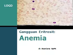
dr Bastiana SpPK wwwthemegallerycom Company Logo Gangguan Eritrosit Anemia Polisitemia ANEMIA Definisi Anemia Sindroma klinis yang disebabkan penurunan massa ID: 907951
Download Presentation The PPT/PDF document "Gangguan Eritrosit : Anemia" is the property of its rightful owner. Permission is granted to download and print the materials on this web site for personal, non-commercial use only, and to display it on your personal computer provided you do not modify the materials and that you retain all copyright notices contained in the materials. By downloading content from our website, you accept the terms of this agreement.


Slide1
Gangguan Eritrosit: Anemia
dr.
Bastiana
SpPK
Slide2www.themegallery.comCompany LogoGangguan
Eritrosit
Anemia
Polisitemia
Slide3ANEMIADefinisi Anemia:
Sindroma
klinis
yang
disebabkan
penurunan massa
eritrosit total dalam
tubuh
.
Keadaan dimana
massa
eritrosit dan
atau
massa hemoglobin tidak dapat memenuhi fungsinya untuk menyediakan oksigen bagi jaringan tubuhPenurunan di bawah normal kadar Hb, hitung eritrosit, dan hematokrit
www.themegallery.com
Company Logo
Slide4www.themegallery.comCompany LogoANEMIA
Penurunan
Hb
dan
Hct
:
<
batas
bawah 95% interval
referens
dari kelompok
usia
,
jenis kelamin dan lokasi geografis (ketinggian)Hb12-14 g/dl ; (Hct 36-41%), Hb7g/dl symptom (+)Akut: hipovolumia (pucat, ggn penglihatan, syncope, tachycardia
) ;
Kronis
: tissue hypoxia (
fatique
,
dyspnea
,
Headache, angina)
Anemia
Slide55
ANEMIA
→ symptoms / syndrome
Hb
↓
PCV
↓
Hypoxia
→ Otak ,
Otot
RBC ↓
Kompensasi :
- heart rate
↑
→ tachycardia → flow rate ↑ → cardiomegaly → heart failure → † - blood flow priority (pallor) - RBC 2,3-DPG content ↑→ O2 dissoc.curve shift to the right → O2 release to the tissues ↑ .
Slide6Klasifikasi Anemia
Berdasarkan
patofisiologi
:
I.
Kegagalan
produksi
sel darah
merah:
A. Gangguan
sel induk
hematopoesis
Anemia
Aplastik B. Gangguan sintesis DNA Anemia Megaloblastik C. Gangguan sintesis Hemoglobin (Hb) Anemia Defisiensi Besi, Thalasemia D. Gangguan sintesis
eritropoetin
Anemia
karena
GGK
www.themegallery.com
Company Logo
Slide7Lanjutan…..anemia berdasarkan
patofisiologi
E.
Gangguan
karena
mekanisme lain:
Anemia
karena
penyakit
kronis,
anemia sideroblastik
Anemia
karena
infiltrasi sumsum tulangII. Peningkatan destruksi sel darah merah: Anemia HemolitikIII. Kehilangan darah (Blood Loss) Anemia karena
perdarahan
akut
www.themegallery.com
Company Logo
Slide8Anemia
Anemia
berdasarkan
morfologi
Anemia sec.
morfologi
eritrosit, dilihat
dari:
- ukuran
dan
warna
di bawah
mikroskop
atau
-
indeks eritrosit (MCV, MCH, dan MCHC)Kriteria Ukuran (size): Normositik, Mikrositik, Makrositik Kriteria Warna (pucat): Normokromik, Hipokromikwww.themegallery.comCompany Logo
Slide99
Cara
Mengetahui
Ukuran
eritrosit:
* membandingkan
dengan inti
sel
limfosit kecil
(di
bawah
mikroskop
) : → ukuran sama = normositik lebih kecil = mikrositik lebih besar = makrositik * Menghitung MCV (Mean Cell Volume) MCV= PCV/Ery X 10 (fL
)
(1
fL
=10
-12
L= 1
μ
m
3)
N :
dewasa
= 80-100
fL
,
di
bawah
1
thn
= 76- 86
fL
MCV :
normositik
,
mikrositik
,
makrositik
*
Eritrosit
dengan
variasi
ukuran
yang abnormal
anisositosis
Slide1010
Bandingkan
ukuran
sel
eritrosit dengan
inti limfosit
Slide1111
Slide1212
Perhatikan
Warna
sel
eritrosit
:
- Bandingkan
diameter central pallor(CP)
dengan diameter
sel eritrosit
tersebut .
-
Normal,
bentuk sel eritrosit adalah seperti cakram bikonkaf (biconcave disk) → pada hapusan darah tepi terlihat bulat, Ø 7-8 μ dengan area central pallor di
bagian
tengah
CP≤ 1/3 Ø
Eri
=
normokromik
CP> ½ Ø
Eri
=
hipokromik
Slide1313 Eritrosit
dengan
central
palor
(CP)
Bandingkan diameter CP
dengan diameter sel
eritrosit
Slide1414
Warna
,
dapat
diketahui
juga
dari MCH
(Mean Cell Hb
)
MCH=
Hb/RBC x 10 (pg)
Dewasa: MCH=27-32 pg, Anak-anak
: MCH=23-31 pg (1pg=10
-12
g=1
μμg) MCH normal → normokromik MCH < normal → hipokromikMCHC (Mean Cell Hb Concentration) : MCHC=Hb/PCV x 100 (g/dL) Normal: MCHC = 32-36 g/dL
Slide15www.themegallery.comCompany LogoKlasifikasi Anemia secara
morfologi
1.
Anemia
Hipokromik-Mikrositik
.
2.
Anemia
Normokromik-Normositik
3.
Anemia
Makrositik
Slide16www.themegallery.comCompany Logo
1
Contoh
:
- Anemia
defisiensi
Fe
Thalasemia
Anemia
akibat
Penyakit
Kronik Anemia sideroblastik
2
Contoh
:
Anemia
pasca
perdarahan
akut
Anemia
aplastik
Anemia
hemolitik
Anemia
akibat
penyakit
kronik
Anemia
pada
GGK
Anemia
pada
mielofibrosis
dll
3
Megaloblastik
,
contoh
:
- Anemia
defisiensi
Folat
,
- Anemia
defisiensi
vitamin B12
B.
Nonmegaloblastik
contoh
:
- Anemia pd
peny
.
Hati
kronis
- Anemia pd
hipotiroid
,
dll
MCV <80 fl; MCH <27 pg
MCV 80 -95 fl MCH 27-34 pg
MCV > 95 fl
Anemia
hipokromik-mikrositik
Anemia
normokromik-normositik
Anemia
makrositik
Slide1717
Hipokromik-Mikrositik
Slide1818
Normokronik-normositik
Slide1919
makrosit
-oval
(Anemia
megaloblastik
ditandai
oleh
makrosit oval ini
)
Makrositik
Slide2020Pendekatan
diagnostik
Anemia
:
Anamnesis
:
onset /bleeding tendency / routine medicinal / occupation / hobby / travel history / family / diet / GI symptoms / menstruation cycle / history of previous pregnancy-delivery / alcohol consumption , etc
Pemeriksaan fisik :
conjunctiva & lips (pallor) / mouth (cheilosis) / tongue (glossitis) / gum / nails (koilonychia) , hair (
signa de bandera
, alopecia) , jaundice , petechiae , liver & spleen , lymphenodes ,rectal / vaginal toucher , feet (
ulcer,arthritis)
Slide2121
Pemeriksaan
Laboratorium
-
CBC (complete blood count )→
to confirm anemia (
Hb, PCV, RBC) & the type of anemia
(MCV; MCH; MCHC), RDW
-
Reticulocyte count
→ reflects marrow’s responses .
- PBS : to look for the RBCs’ shape and any abnormalities of
RBCs besides the other blood cell lines
- Iron status ( Serum Iron ,TIBC, % Transferrin saturation , Iron storage ) - Blood chemistry ( direct/total bilirubin,LDH and stool examination for occult blood test , etc) .PBS: Pheripheral blood smear
Slide2222
-
Radiological examinations
( Chest X-ray,
USG , MRI )
-
Cardiological
examinations (EKG,Treadmill
, Echocardiography)
Notes
! :
- First confirm
Anemia ( Hb , PCV , RBC
)
-
Classify the anemia (MCV, MCH, MCHC) - Causes of anemiaLanjutan…. Pendekatan Doagnostik…
Slide2323
Slide2424
Slide2525Anemia
Hipokromik-Mikrositik
Setiap
kondisi
yang
menimbulkan
gangguan sintesis
Hb
gambaran hipokromik
mikrositik
Anemia Defisiensi
Besi
penyebab tersering dari anemia Hipokromik-MikrositikPerhatikan penyebab lain (DD=diff diagnosis) sebelum mendiagnosis Anemia def. besi, spt: - anemia akibat penyakit kronis - Thalasemia - anemia Sideroblastik, dll
Slide2626
Slide27ANEMIA DEFISIENSI BESI
Definisi
:
Anemia yang
timbul
akibat kosongnya
cadangan
besi tubuh
besi
utk
eritropoeisis
pembentukan
Hb
Anemia def. Fe, ditandai dgn: - anemia hipokromik mikrositik - besi serum - TIBC (Total Iron Binding Capacity) - Saturasi transferin - Feritin serum - Pengecatan Besi sumsum tulang negatif
-
Respon
terhadap
pengobatan
dengan
preparat
Fe
www.themegallery.com
Company Logo
Slide28www.themegallery.com
Company Logo
Slide2929Faktor
Penyebab
(
Etiologi)
I.
Keseimbangan
negatif Fe (
Negative Iron balance)
:
- Asupan Fe ↓
(inadequate diet , impaired absorption)
- Fe loss ↑
(GI bleeding, excessive menstrual flow,
bleeding diathesis) - ↑ demands (infancy, pregnancy, lactation)
Slide3030II. Inadequate presentation to
erythroid
precursors
:
-
atransferrinemia
- Anti
TrfR
Ab
III. Abnormal Fe balance
:
-
Aceruloplasminemia-
Autosomal dominant hemochromatosis
( mutations in
ferroportin
)Lanjutan….Faktor Penyebab
Slide3131Patogenesis
desifisiensi
Fe
3
pathogenetic
factors
:- Impaired Hb
synthesis (consequence of reduced Fe supply)
Transferin saturation< 16%
inadequate Fe-supply to marrow → Hb
contents of RBC ↓ →
hypochromic & microcytosis
- Generalized defect in cellular proliferation
- Fe-deficient → oxidative damage to the red cell’s membrane → RBC deformability ↓ → RBC viability ↓→ RBC destruction ↑ especially in spleen → reduced RBC survival
Slide3232Status
besi
tubuh
:
Serum Iron = SI
Total Iron Binding Capacity (TIBC
)%
Transferrin Saturation = SI/TIBCx100%
Simpanan besi
(Iron storage):
- Hemosiderin
→produk
degradasi feritin
yang
tidak
larut dalam air → mayoritas tdd aggregat kristal ferric oxyhydroxide, FeOOH (di Hepar danSutul→ dideteksi dengan biopsi/aspirasi dan pengecatan besi (prosedur invasif)
-
Ferritin
→
kompleks
garam
Fe3+dan
apoferitin
yang
larut
dalam
air,
dengan
jumlah
yang
sangat
kecil
di
serum.
(
dideteksi
dengan
metode
imunoasai
)
Slide3333Kandungan
besi
tubuh
= 35-50 mg/kgB
B:
±80
% - Fe fungsional
, sebagai
heme
-Iron
(65%
Hb, myoglobin, enz
im
heme : cytochrom-C,A,A3,B, catalase , peroxidase) - Non-heme-Fe (sebagian kecil) 20% - simpanan besi / Iron storage (ferritin, hemosiderin) hanya ± 15%
pada
wanita
0.2%
-
ci
r
c
ula
t
i
ng
(
terikat
pada
Transferrin
)
Slide3434Iron Cycle in the body :
Fe-diet
→ as
heme
-Fe
(
Hb
, myoglobin,
enzyme-Fe), 5-35% adsorbed
from animal/meat sources , adsorbed easily .
→ as
non-heme
-Fe (vegetables , legumes), 90% of diet-Fe but
only 2-20% of it absorbed →
depends on the iron-status and
the ratio of
Enhancer:Inhibitor
Slide3535
Enhancers
(
zat
yang
menstimulasi
penyerapan (absorbsi
) :
Ascorbate, Cytrate
, organic acids / other
amino acids , by reducing Fe3+
to Fe2+.
Inhibitors (
zat
yang
menghambat absorbsi) : Carbonate, Phytate, Tannins, Phosphate, Oxalat chelate Non-heme-Fe → unabsorbable
Slide3636
Bahan
makanan
yang
menghambat
absorbsi
besi non heme
(Non-heme
Iron) :
-
Phytate (
dari legumes
, sayuran
)
-
Tannin & Polyphenol (dari teh, kopi, wine, coklat ) - Phosphate/phosphoprotein dari kuning telur - Minerals (Ca, Zn, Cd) -
Tetracycline
yang
bereaksi
dengan
Fe →
menghambat
absorbsi
Slide3737
Siklus
Fe
dalam
tubuh :
Diet’s Iron → duodenum / proximal jejunum .
Iron from gut → released into circulation , bound to
transferin → distributed to body’s organ / tissues( to bone marrow as a part of heme
/ Hb
) → circulate inside red blood cells with blood flow
Slide3838The development of IDA
Stage-1
(
prelatent
Fe-deficient):
- progressive loss of storage-Fe
- body’s Fe reserve is still sufficient to
maintain both the transport and functional
compartment , so RBC development is
still normal .- peripheral blood picture is normal , no
symptoms of anemia , but
ferritin is ↓ .
*IDA= Iron Deficiency Anemia
Slide3939
* Stage-2 (latent Fe-deficient)
- Exhaustion of storage-Fe , RBC
production is still normal ,
Ferritin
↓↓
- Circulating-Fe (SI) begin ↓ ,
Transf
-
Receptor ↑ .
* Stage-3 (Fe-Deficiency Anemia)
- Stadium of Iron Deficiency Anemia
Slide4040
Stage-1
(
prelatent
)
Stage-2
(latent)
Stage-3
(IDA)
Marrow
Ferritin
Transf-Sat
sTrfR
Retic Hb
content
Hb
MCV
Symptoms
↓
↓
N
N
N
N
N
fatigue
(
-
)
<12ug/L
<16%
↑
↓
N
N
fatigue
( - )
<12ug/L
<16%
↑
↓
<
<
pallor
Slide4141
Symptoms
Morphology
SI - TIBC
Ferritin
I D A
Anemia
Hypo – Micro
SI
↓ - TIBC ↑
↓↓
A.C D
Anemia
Hypo – Micro
SI
↓ - TIBC ↓/N
N/
↑
Slide4242Pendekatan
Diagnostik
Anemia
Defisiensi
Fe
1. Anamnesis
– pola
menstruasi, kehamilan
/
persalinan
, tendensi
perdarahan,
penyakit
kronis, diet, pekerjaan, riwayat bepergian 2. Pemeriksaan fisik – sistematik dari seluruh permukaan tubuh sampai ke organ dalam ( hati, limpa, kelenjar getah
bening (
lymphnodes
)
Slide4343
3.
Laboratorium
-
Hema
(DL, LED,
Hapusan
darah tepi
, Retikulosit
)
- Serum (SI,TIBC,Ferritin,
Bilirubin)
- BMA (Bone Marrow Aspiration) - Pemeriksaan
Urine
dan
tinja4. Penunjang - Radiology (EKG, USG) - Endoscopy
Slide4444
S I
TIBC
Normal
N
(1/3
mol.Trsf
)
N
I D A
↓
↑
An.of
Chronic Disease
↓
N /
↓
Fe Overload
↑↑
N /
↑
Slide4545Pemeriksaan
Lab. Anemia def. Fe
1.
CBC
– confirm Anemia & find
hypochromic
microcytic picture from BSE and Red
Cells Indices ( Hb, PCV ,MCV , MCH ,
MCHC)
2
.
SI – Fe2+ released from
Transferrin
+
ferrozine (chromagen) → measured colored complex TIBC – serum + excess FeCl2 → to fill all Transferrin- binding sites → the excess Fe is fixed by Mg- carbonate → Fe-saturated Transferrin is measured with Ferrozine (= TIBC)
Slide4646
%
Saturasi
Transferrin
= SI/TIBC X 100%
Erythropoeisis impaired when %
Tf.Sat < 15%
3.
Ferritin
Serum : Serum
Ferritin level ~ Fe-storage
Ferritin
<15
ug
/L → Definitive Fe-Deficient N/↑ Ferritin in IDA , if : - impaired liver function ( damaged hepatocyte), hemolysis, inflammation / infection / malignancy ( Ferritin = acute-phase protein )
Slide4747
4.
Transferrin
Serum :
measured by
immunodiffusion
methode
Normal value : 2-4 g/L
5. Bone Marrow’s Aspirate evaluation
: ( using
Perls
or Prussian Blue stain )
Slide4848Anemia of Chronic In
fection
Gejala
klinis
miripdengan anemia def.Fe
Gambaran lab. hematologi = Anemia def. Fe (An.Hypo
-Micro, MCV↓, MCH↓, SI↓) , tapi
TIBC N/↓ and Ferritin
N/↑)
Pathogenesis : Fe →
storage // Transferrin
Tissues / RES
Slide4949
1.
Impairment of Fe release from
macrophage in competing with
lactoferrin, phagocyte’s product , even
storage-Fe is still enough .
2. Inadequate EPO Respons
towards
anemia (effects of cytokine production by
macrophage) .
Penyebab menurunnya ‘circulating Fe’
:
Slide5050Diagnosis Anemia
akibat
penyakit
kronis
:
lab hematologi
: - Anemia
hipokromik mikrositik
- SI ↓ , TIBC ↓/N ,
Ferritin
N/↑ (
jika Ferritin ↓,
An.
Def.Fe ) - Inflamasi / infeksi (+) : CRP and LED ↑Problem: IDA with inflammation → ferritin ↑ (falsely diagnosed as ACD) ; it can be differentiated by sTfR exam (serum transferrin receptor) that ↑ in IDA but normal in ACD .
Slide5151Anemia
Sideroblastik
Defek
pada
sintesis
Heme → akumulasi Fe
di mitochondria →
degenerasi Fe →
granula Fe di
sekitar
inti normoblast
,
membentuk
struktur spt cincin {paling jelas terlihat dengan pengecatan Perl (Perls’ stain) } → Ringed Sideroblast (karakteristik anemia Sideroblastik)Sideroblast bisa dijumpai secara normal di sutul
Slide5252Sideroblast
and Ringed
Sideroblast
( in
Sideroblastic
Anemia )
Slide5353
Slide5454Classification of
Sideroblastic
Anemia
Hereditary
: X-linked, defect in
heme-synthesis enzyme pathway
Fe absorption ↑ → % of Transferrin saturation and Ferritin
level ↑
Slide5555
2
.
Acquired
:
- Primary
:
Stem cell clonal
mutations(MDS =
MyeloDysplastic
Syndromes , RA-RS)
Normochromic-macrocytic anemia .
Marrow : erythroid hyperplasia with
dysplastic or
megaloblastic
appearance - ringed sideroblast in normoblast .
Slide5656 -
-
Secondary;
Abnormal metabolism of Vit.B6 (alcoholism,
malabsorption
) , impairment of
heme
synthesis ( Pb intoxication) ,
Rhematoid Arthritis , or An.megaloblastik
.
Usually related to
myeloproliferative diseases ( AML, Myelofibrosis
, Polycythemia or another types of MDS )
Slide5757Macrocytic
Anemia
- Non-
Megaloblastic
Macrocytic
Anemia :
Reticulocytosis
Liver disease / Alcoholism
Myelodysplastic Syndrome
Erythroleukemia (FAB-M6)
- Megaloblastic
Macrocytic Anemia
Slide5858
macrocyte
= erythrocyte with MCV > normal .
macrocyte
/
microcyte depend on the balance between nuclei & cytoplasmic
maturation .
(nuclear dividing stopped when intracellular Hb
production reach a proper level ) . If nuclear maturation delayed ( in DNA
synthesis’s defect ) or
cytoplasmic maturation ↑ ( increase of EPO’s activities ) → critical level of
Hb
achieved earlier → Macrocyte Megaloblastic Macrocytic Anemia
Slide5959
Megaloblast
= bigger than normal
normoblast
.
Megaloblastic
changes = increased size of
hemopoietic precursor cells in bone marrow ( not only in normoblast
!) Primary defect :
Defect of DNA synthesis ( altered almost all active cells / organs
i.e
: hemopoietic tissue, epithelial cells , mucous cells, etc )
Slide6060
Etiology of DNA synthesis defect
:
deficiency of vit.B12 and
folic acid
→ maturation
dysharmony
between nuclei & cytoplasm (delayed nuclei maturation) → increased
cels (megaloblastic
changes) → marrow’s ineffective erythropoiesis →
intramedullary
hemolysis → total/indirect
Bili and LDH ↑.
Slide6161Deficiency of Folic acid
:
- Inadequate diet
(intake < / demand ↑ in pregnancy -
lactation , child’s growth /
malabsorption
in tropical
sprue
/ bowel resection / small intestine inflammation )
- Drug’s effect (anti-
epilepsi)- FA
loss ↑ (dialysis)
Slide6262Deficiency of Folic acid
:
- Inadequate diet
(intake < / demand ↑ in pregnancy -
lactation , child’s growth /
malabsorption
in tropical
sprue
/ bowel resection / small intestine inflammation )
- Drug’s effect (anti-
epilepsi)- FA
loss ↑ (dialysis)
Slide6363Deficiency of Vit.B12:
- Inadequate diet :
Intake < in vegetarians , demand ↑ ,
impaired absorption caused by
decreased Intrinsic Factor
(
gastrectomy
,
pernicious anemia
)
Malabsorption (bowel infection , worms
/ blind loop syndr )
Slide6464
VITAMIN B12
ASAM FOLAT
-Food from animal products
-Heat stabile
-Storage : enough for 3 yrs
-Relatively low needs (only 1% of
folate
requirements)
-Limited sources (vegetable ,
fruits)
-Heat labile
-Storage enough only for 3
mths
-Higher
folate needsCAUSE OF DEFICIENCYCAUSE OF DEFICIENCY
-Vegetarian (seldom)
-Impaired Intrinsic Factor
(pernicious anemia)
-
Gastrectomy
-
Atropic
Gastritis
-Anticonvulsant, alcoholism
-Nutrition (alcoholism, goat’s
milk diet)
-Prematurity
-
Hemodyalisis
-Bowel resection
-Pregnancy
-Anticonvulsant , MTX
Slide6565Pathogenesis of
Megaloblastic
Anemia
:
Megaloblastic
changes
atrophy of tongue papilla & mucosal GI →
glossitis , gastritis, nausea , constipation.
B12 defic →
demyelinisation of spinal cord & peripheral nerve → loss of foot’s balance / sensory (Neuropatia
)
FA defic
→ hyperhomocysteinemia → thrombosis and vascular occlusion .
Slide6666B12 Metabolism
Vit.B12 →
purine
&
pyrimidin
synthesis → synthesis DNA & RNA → mitosis and
maturation
Vit.B12 made from microbiological source because plants do not produce B12 ( meat ,
liver, eggs and milk are rich of
Vit B12 ).
Vit.B12 content in the daily diet is
5-3ug , daily requirement of B12 is 1-3 ug
, and B12 body’s storage is
2-5 mg (enough for 3 yrs)
Slide6767Vit.B12 absorption
B12 diet → in
gaster
bind by
IF (Intrinsic Factor)
produced by parietal cells → IF-B12 complex → ileum : B12 absorbed , IF freed into the lumen
impaired IF :
gastrectomy
/gastritis/ Auto-Ab
-antiIF or Auto-
Ab
-antiparietal) → no absorption of B12 →
impaired DNA synthesis →
(Pernicious Anemia with
Achlorhydria
)
Pernicious Anemia = autoimmune disease → auto-Ab to parietal cells (Anti-IF or Anti-Parietal)
Slide6868Hematological pictures of
Megaloblastic
Anemia
Bone Marrow :
-
megaloblastosis
- ineffective
erythropoiesis
Peripheral blood :
- Oval macrocytosis
-
Hypersegmented neutrophil
( five 5-lobed
cells or one 6-lobed cell)
or the mean lobes
of 100 neutrophils is > 3.4
Slide6969Megaloblastic
Anemia
find oval-
Macrocyte
cell and
hypersegmenteneutrophil
.
Slide7070Diagnosis of
Megaloblastic
Anemia
Screening
:
- CBC ,
Neutrophil’s
lobe count
- Serum Indirect Bilirubin , LDH (lactate
dehydrogenase)
Spesific
tests :-
Bone Marrow Aspiration: megaloblastosis &
megaloblastic
changes,
erythropoietic activitiy ↑ ( ineffective erythropoiesis)- Folate & Vit.B12 assay- Gastric juice analysis- Schilling Tests- Antibody Assay
Slide71Anemia Hemolitik
Anemia
hemolitik
: anemia yang
disebabkan
oleh
proses
hemolitik. Hemolisis
: pemecahan
eritrosit
sebelum
waktunya (sebelum
masa
hidup
rerata eritrosit, yaitu 120 hari). (Proses pemecahan eri karena sdh waktunya senescence=penuaan) Hemolisis dapat terjadi di dalam pembuluh darah (hemolisis
intravaskular
)
dan
di
luar
pembuluh
darah
(
hemolisis
ekstravaskular
).
www.themegallery.com
Company Logo
Slide7272HEMOLYTIC ANEMIA
Normal red cell’s survival = 110-120 days → destructed by macrophage in marrow and spleen .
When the survival are shortened → EPO production is stimulated (compensated) → no
Hb
changes → anemia (–) .
If the destruction is acute or chronic with very shortened life of red cells , there will no compensation → anemia (+) .
Slide7373
Definition of
Hemolytic Anemia
:
anemia caused by shortened red cell’s survival as a result of excessive uncompensated destruction of red cells .
Hemolytic process = every process of red cells destruction with still / without compensated by bone marrow → anemia is not always present .
Slide7474-
C
ompensation ability of bone marrow
:
Ability to ↑ red cells production ( 6-8 x normal ) :
- survival shorten ½ → production ↑ 2x
- survival shorten ¼ → production ↑ 4x
- survival shorten 1/6 → production ↑ 6x
- survival shorten 1/8 → production ↑ 8x
↑ of production 6-8 x is
maksimum
.If red cells live only 20 days → anemia (+).
Slide7575
D
iagnostic approach in Hemolytic Anemia
:
Confirm anemia (
Hb
/PCV/RBC)
an acute case usually acquired , and chronic case is mostly hereditary .
To find the signs of hemolytic process .
Extra or Intravascular ?Hereditary or acquired ?
The cause of
hemolysis episodes .
Slide7676The signs of Hemolytic process :
1. Increased of red cells destruction
-
Unconjug.bilirubin
serum ↑ → jaundice
-
Urobilinogenuria
- Hb-uria
→ sign of intravascular hemolysis
- Abdom.pain →
splenomegaly, spleen infarction
- Leg’s Ulcer → intrinsic defect of erythrocyte -
Haptoglobin
serum ↓↓/
neg → intravascular hemolisys .
Slide77772.Destruksi
eritrosit
:
Microspherocyte
,
Fragmentocyte
, Poikilocyte
Erythrocyte Osmotic Fragility ↑Positive
Autohemolysis test
Shortened of red cells’ survival
3.
Tanda Peningkatan
Eritropoisis:
Reticulocytosis
Normoblastosis
Erythropoietic Hyperplasia in bone marrow
Slide7878
Slide7979
Slide8080
Slide8181
Slide82Hemolisis ekstravaskular
lebih
sering
dijumpai
dibandingkan hemolisis
intravaskular
Hemolisis terjadi
di sel
makrofag dari
sistem retikuloendothelial (RES)
terutama pada Lien,
hepar
dan sutul karena sel ini mengandung enzim heme oksigenaseLisis terjadi karena kerusakan membran eritrosit (misal Akibat reaksi Ag-Ab; presipitasi hb di sitoplasma, menurunnya fleksibilitas eri,dll
)
Hemolisis
Ekstra
vaskular
Slide8383
Slide8484
Slide8585
Slide86Klasifikasi Anemia Hemolitik
Dibagi
atas
2
golongan
besar, yaitu
:1. Anemia hemolitik
karena
faktor
di
dalam eritrosit
sendiri (gangguan
intra
korpuskuler
)2. Anemia hemolitik karena faktor di luar eritrosit (gangguan ekstra korpuskular)www.themegallery.comCompany Logo
Slide8787
lanjutan
….
Klasifikasi
anemia
hemolitik
:
Gangguan
intra korpuskular
(Hereditary Hemolytic Anemia )
- Membrane abnormality
(hereditary
spherocytosis , hereditary ovalocytosis
)
- defect of
globin chain (Thalassemia, Hb- pathia)- enzyme defect ( G-6PD deficiency , PK- deficiency)
Slide8888Hereditary Spherocytosis :
Slide8989Hereditary Ovalocytosis :
Slide9090
2.
Gangguan
ekstrakorpuskular
(Acquired Hemolytic Anemia)
:
- physical / chemical substances
- infections (bacteria, parasites, viruses, fungi)
- mechanical trauma (
prostetic heart valves)
- Immune mechanism (Alloimmune
/ Autoimmune / Drug-Induced HA)
Lanjutan
……
klasifikasi anemia hemolitik
Slide9191-
Hereditary
Spherocytosis
:
autosomal
dominant
Spherocytosis
, decreased membrane surface area relative to cell volume → osmotic fragility test (OFT)↑ among the family member .The primary lesion is caused by membrane protein defects
(↓of spectrin)
→ cytoskeleton instability .60% - chronic anemia , jaundice,
splenomegaly
, 20% without hemolysis /
splenomegaly .
Bilirubin excretion ↑ ,causing bilestone
in USG.
Slide9292Thalassemia
:
Defect of 1 or more
globin
-chain synthesis
(the amount = quantitatively)
:
- deficiency of
α
globin-chain → α
-
thalassemia
- deficiency of β
globin-chain → β-
thalassemia
- deficiency of
δβ globin-chain → δβ-thalassemia the primary defects in Hb-pathia is in the globin amino acids structure (qualitatively)
Slide9393
Slide9494
Slide9595α
-
Thalassemia
α
-
Thalassemia
= is caused by the impairment of
α-
globin chain production/synthesis .
α-
globin
chain synthesis is directed by 2 pairs of α-gene (
4 locus α-gen
) → depending of the number of defected locus → 3 types of α-
Thalassemia
(α-thal trait , HbH Disease, and HbBart’s Hydrops Fetalis)
Slide9696
Clinical consequences in
α
-
Thalassemia
Deficiency of
α
-globin
chain → excess of β,
γ chain since fetal life to form β
4-tetramers (
HbH) or γ4-tetramers (
HbBart) .
Defect of 1-2 α
-Gen =
α
-trait (clinically good)Defect of 3 α-Gen = HbH disease ( Hb 10-11 g/dl) → excess of β-chain → to form β4-tetramers (HbH) as intracellular inclusion → detected by BCB-stain .
Slide9797HbH
-inclusion (
β
4)
in
HbH Disease as shown in BCB staining (compare with
reticulocyte)
Slide9898Defect of 4
α
-gene (
HbBarts’hydrops
fetalis
) → clinically severe , stillborn baby with hydrops
fetalis ( severe hypoxia ) .
HbBarts =
γ4-tetramers (excess of
γ
-chains that unable to form HbF ) .
HbBarts and
HbH inclusions precipitated in red cell’s membrane → mechanical trapping in spleen → macrophagic
phagocytosis
→ hemolysis .
Slide9999
Slide100100
Slide101101-
β
-
Thalassemia
Clinically consequences in
β
-Thalassemia :
- No problems during fetal life because
HbF
synthesis is normally produced
(normal
α and γ
chains)
- When
HbA
is dominantly needed , the clinically problems exist as incapability to synthesize HbA (α2β2) → excess of α-chain → compensated ↑ of δ and γ production → HbA2 ↑ (in β-Thalassemia minor) and HbF ↑ (in β-Thalassemia mayor)
Slide102102
- severe anemia → repeated transfusion is
oftenly
needed → Fe↑↑ →
hemochromatosis
- chronic ineffective
erythropoiesis
→
medullary hypertrophy in childhood → facial
malformation: * Frontal bossing
* Maxillary hypertrophy
*
Hypertelorism (mongoloid’s eye)
Β-
Thalassemia
mayor :
Slide103103-
β
-chain deletion forms :
β
0
-
Thalassemia
: no β-chain production.β
+Thalassemia :
β-chain production <<
in heterozygous case : medium severe
in homozygous : severe (Cooley’s anemia
)
Slide104104
Slide105105
Laboratory Diagnosis in
Thalassemia
CBC
, Peripheral Blood Smear
Hb
-Electrophoresis :
in Celulose-Acetat (pH 8.4) for
thalassemia and Hb-pathia
screening
Using hemolysate
→ formed bands of different types of Hb ( normal : bands A, F, and A2 , measured
densitometrically)
Slide106106
Slide107107
3
.
HbA2
mesurement
to diagnose
β-
Thalassemia trait using anion-exchange resin
column chromatography
in both
HbELP and chromatography , HbC
, HbE and
HbO can interrupt the conclusion because of the same band location with HbA2 .
4.
HbF
determination : - Alkali Denaturation Test - Acid-elution (Kleihauer) test - RID or ELISA methods Lanjutan…..Lab diagnosis in thalasemia
Slide1081085.
HbH
Inclusion detection
:
-
Supravital
staining using Brilliant
Cresyl Blue (BCB) or NewMethylene
Blue (NMB)
-
HbH inclusion seen as dispersed blue-
green granules in red cells (compare with
reticulocyte as a filament)
-
in
HbH disease : HbH inclusion +++- in Thalassemia-α-trait : HbH inclusion + in 1: 10000 eritrosit .
Slide109109
-
Oxidant → produce H2O2 → oxidizing
Hb’s
free
sulfhydryl → to form
Sulf-Hb →
aggregates that precipitated as Heinz
Bodies → destructed in spleen .
- Oxidant /
Sulf-Hb are controlled by
Reduced Glutathione (GSH)
Defisiensi
G-6PD
Slide110110
Slide111111- X-linked, ± 300 variants .
normal G-6PD genes : - type B (
GdB
)
- type A (
GdA
)
- Abnormal enzyme types :
1. GdA– (type A–)
2.
Gd-Mediterranean (
GdMed)
3. Gd-Canton : many in Asia
- G-6PD deficient red cells are
resistent
to Plasmodium Falciparum .
Slide112112-
Substances causing
lysis
in G-6PD deficiency :
1.
Antimalaria
6. Fava
beans 2. Sulfonamides 7.
Naphtalene
3.
Vit.K, Vit.C
8. Uremia 4. Lung Infection 9. Antibiotics
(virus,bacteria
) (
Penicilline
, 5. Antipyreticum streptomycine
Slide113113The highest G-6PD activity is in
reticulocyte
.
G-6PD screening test
:
Test’s principle
:
G-6PD
G-6P + NADP 6-PG + NADPH
UV
(fluorescence)
Slide114114Acquired Hemolytic Anemia
:
-
Secondary Hemolytic Anemia
caused by
infection / systemic disorders
:
Malignancy – Autoimmune-reacted
hemolysis ,
microangiopathy or hypersplenisme
, appearing Anemia of chronic disease, bleeding tendencies, and marrow’s suppression
Slide115115Disseminated Intravascular Coagulation (DIC):
Systemic intravascular coagulation → fibrin deposit
intravascularly
/ endothelial damage (
microangiopathyi
) caused by sepsis → red cells destruction .
Chronic Liver Disease
:
hemolysis caused by
hypersplenism .
Chronic Renal Disease
: hemolysis
caused by microangiopathy
Slide116116Acquired Hemolytic Anemia (
extracorpusc
.)
Red cell membrane-bound
Ab
hemolysis
.
The speed & hemolysis location depend on
IgG or
IgM, and the ability to activate complement .
Optimal temperature to bind Ab
: 370
C – Warm-IgG-Type
<30
0
C – Cold-IgG-TypeImmune Hemolytic Anemia
Slide117117Cell+IgG
→ destructed by spleen
Cell+IgM
→ enhance the activation of complement’s cascade → intravascular
hemolysis
Immune destruction often cause minimally membrane damage → shape change into
spherocyte
.Lanjutan
….acquired hemolytic anemia
Slide118118Immune Hemolytic Anemia classification
:
1.
Alloimmune
: Transfusion Rx ,
Hemolytic
Disease of the Newborn (HDN)
2. Autoimmune
: Warm/Cold AIHA,
Paroxysmal Cold
Hb-uria (PCH)
3.
Drug-induced HA : penicilline
type,
aldomet, and stibophen type .
Slide119119Hemolytic Disease of the Newborn (HDN) –
Rh-neg
mother , with
Rh
-Pos fetus , during I and second pregnancy
Slide120120Antiglobulin
Tests (Coombs) :
Direct Coombs Test
(Direct
Antiglobulin
Test/DAT) =
Ab
detection test (IgG and or C3d /complement-bound red cells) .
Indirect Coombs Test
= test for serum free Ab
.
DAT usually positive in AIHA (.
Slide121121Drug-Induced hemolytic anemia
:
Penicilline
type
: drug as
hapten
binds red cell membrane → antigenic → stimulate Ab
production against Drug in drug-red cell complex
Phenacetin/Quinidin
type
: Drug (hapten) adsorbed protein → stimulated-
Ab binds drug-protein complex → activate complement → red cell lysis
.
Aldomet
type
: drug change red cell membrane’s structure → detected as foreign cell → Autoantibody production .
Slide122122
Slide123123Aplastic
(
Hypoplastic
?) Anemia
Severe & fatal Anemia because of ↓ red cells/leucocytes/platelet production (
pancytopenia
) caused by Stem Cells impairment (radiation, chemicals, drugs, or genetic matters)
Marrow
aplasia /
hypoplasia-causing substances - radiation , benzene,
cytostatics
(6-MP,
busulfan), arsen
, chloramphenicol,
anticonvulsant (
phenytoin
), analgetic (phenylbutazone) , DDT, etc
Slide124124Symptoms &
Lab.appearance
of
Aplastic
Anemia
fatigue, palpitation, infections, bleeding tendency
Lab : - pancytopenia
- normochromic
normocytic
- ‘dry-tap’ marrow ,
hypocellularity
Prognosis :
- bad especially for < 40 yrs old patients →
marrow transplantation .
Slide125125-
Treatment for
Aplastic
Anemia
:
Avoid every toxic material
Avoid infections / bleeding tendency
Use Washed-Erythrocyte if transfusion is needed or
Plat.Concentrate (PC) for any profuse bleeding ( give corticosteroid if bleeding is minimal)Marrow stimulants (androgenic
hormon )
Marrow Transplantation
Slide126POLISITEMIA(ERITROSITOSIS)
Peningkatan
patologis
massa eritrosit
massa
eritrosit normal : (sea level)
- o : 26 - 32 ml / kg BB - o : 23 - 29 ml / kg BB
eritrositosis
: massa eritrosit
> normal ( PCV : o >51% ; o >48% )
Slide127Klasifikasi :
Primer (
Otonomik
)
Polisitemia
Vera Eritrositosis
Murni (Eritremia)
Sekunder
Fisiologis (Oksigenasi Jaringan
)
Non-fisiologis (Oksigenasi
Jaringan N)
Eritrositosis Relatif
Slide128ERYTHROCYTOSIS - DIAGNOSTIC TESTS
Complete Blood Count
Bone Marrow examination
Arterial Blood Gas analysis
Leukocyte Alkaline
Phosphatase P5O
IVP or renal ultrasound Liver ultrasound or CT scan Erythropoietin level
Erythroid progenitor assay Sleep apnea evaluation
Slide129POLISITEMIA VERA
Proliferasi
klonal
neoplastik sel progenitor
hematopoitik pluripotenKriteria diagnosis P.V. :
Kategori A 1.Massa eritrosit:
Lk > 36 ml / kgBB (PCV >
54%) Pr > 32 ml / kg BB (PCV >
51%) 2.
Saturasi oksigen > 92% 3. Splenomegali
Slide130Kategori B 1.
Trombositosis
(> 400.000 /
m
l) 2. Lekositosis (> 12.000 /
ml) 3.
Skor LAP 4. B12 serum > 900 pg/ml
Diagnosis PV bila : A1 + A2 + A3 atau A1 + A2 +
dan 2 dari kategori B
+
+
+
+
+
+
+
Slide131PRIMARY “PURE” ERYTHROCYTOSIS( ERYTHREMIA )
peningkatan
massa eritrosit
murni
tidak ada penyebab eritrositosis
sekunder kadar eritropoitin normal atau
rendah mungkin akibat mutasi gene
reseptor eritropoitin ® progenitor
eritroid jadi
lebih sensitif terhadap eritropoitin
.
Slide132II. ERITROSITOSIS SEKUNDER
Merupakan
respons
terhadap keadaan lain yang
bersifat :
- fisiologis : akibat oksigenasi jaringan yang
¯ - non fisiologis : tanpa penurunan
oksigenasi jaringan
Slide133III. ERITROSITOSIS RELATIF
Sindroma
Gaisbock
Stress erythrocytosis
Pseudo erythrocytosis
Massa eritrosit tinggi normal
Volume plasma rendah
Slide134Click to edit company slogan .
www.themegallery.com
Thank You !
Slide135www.themegallery.comCompany Logo
1.
Nyonya
Ana,
usia
40
tahun, MRS (
Masuk
Rumah
Sakit
) dengan
keluhan
pusing,
dan
badan terasa lemah. Pemeriksaan fisik: KU lemah, Tensi: 100/60 mmHg, Nadi:90 x/menit, RR: 20 x/menit, suhu:37˚C. Kepala/Leher: anemia (+), tidak dijumpai ikterus, dyspnea dan
sianosis
,
Thorak
/
Cor
dan
Abdomen :
dalam
batas
normal (
dbn
).
Extremitas
:
dbn
.
Hasil
laboratorium
:
Hb
8 g/dl, RBC 3,20 x 10
12
/L,
Hematokrit
24 %, MCV 75 fl, MCH 25 pg, MCHC 33 g/dl.
Jika
anda
adalah
dokter
jaga
di
RS
tersebut
,
dari
data yang
ada
,
kemungkinan
diagnosis
pasien
tersebut
adalah
:
A. Anemia
normokromik-normositik
B. Anemia
hipokromik-mikrositik
C. Anemia
makrositik
D. Anemia
makrositik-megaloblastik
E. Anemia
makrositik
-non
megaloblastik
SOAL LATIHAN :
Slide136www.themegallery.comCompany Logo
2. Dari
kasus
ny
. Ana, 40
tahun
tersebut
, diagnosis
diferensial
untuk
penyebab
anemianya adalah: A. Anemia defisiensi folat, anemia defisiensi Vitamin B12, B. Anemia karena perdarahan akut, anemia
aplastik
C. Anemia
defisiensi
besi
,
thalasemia
, anemia
sideroblastik
D. Anemia
hemolitik
, anemia
pada
penyakit
mielofibrosis
E. Anemia
pada
penyakit
liver, anemia
pada
penyakit
hipotiroid
Lanjutan
…...
soal
latihan
Slide137www.themegallery.comCompany Logo
3. Dari
soal
kasus
Ny
. Ana, 40
tahun
tersebut
,
langkah
pemeriksaan
laboratorium selanjutnya yang perlu dilakukan untuk konfirmasi diagnosis adalah: A. pemeriksaan bilirubin,
haptoglobin
,
hitung
retikulosit
B. Serum Iron, TIBC
dan
Feritin
C.
Pemeriksaan
B12
dan
asam
folat
dalam
darah
D.
Pemeriksaan
T3, T4
dan
TSH
E.
Pemeriksaan
Aspirasi
sumsum
tulang
Lanjutan
…...
soal
latihan
Slide138A 35-year-old man complains of chronic physical fatigue, which began 3-4 weeks ago. He said he felt tired all of the time even through his occupation as a software developer was mentally but not physically demanding. He breathed comfortably at rest but, when he exerted himself, he experienced difficulty in breathing and had hard time catching his breath. He also complained of „more than usual” mental fatigue, confessing an increasing inability to concentrate and focus his attention on tasks at hands. Colleagues noticed his pallor and his inattentiveness at brainstorming sessions and suggested he reschedule his annual physical examination for an earlier date. He complained of vague abdominal pain and sense of abdominal fullness. His appetite was depressed, and he thought perhaps his physical and mental symptoms were caused by poor diet. However, attempts to increase eating resulted in nausea. His stools, he said, were sometimes loose and tarry. Eventually, increased heart palpitations and chest pain made him seek medical advice
CLINICAL CASE
Slide139Laboratory findings revealed the following:
Laboratory test
Patient
Normal
RBC (red blood cell count)
3.5 T
/
L
4.5-6.0 T
/
L
HCT (hematocrit ratio)
28%
40-52%
Hb (hemoglobin)
8.0g
/
dL
13-17g
/
dL
MCV (mean corpuscular volume)
70fL
78-95fL
MCH (mean corpuscular hemoglobin)
22.8pg
29pg
MCHC (mean corpuscular hemoglobin concentration)
28%
34%
Slide140Case history questions:
What general medical condition is suggested by the person’s symptoms?
What fundamental change in function of blood related to the red blood cells could simultaneously affect the function of several systems (cardiovascular, respiratory, gastrointestinal, and others)?
What specific diagnosis is supported by the laboratory findings?
How could the stool be related to the laboratory findings
?
QUESTIONS
Slide141Answers:
Anemia
A reduction in oxygen-carrying capacity of the blood and thus a reduction in the delivery of oxygen to various body tissues
An iron defficiency anemia
Most cases of iron-defficiency anemia result from internal blood loss. Dark, tarry loose stools suggest bleeding from the gastrointestinal tract and warrant further tests to determine the exact cause
ANSWER
Slide142Click to edit company slogan .
www.themegallery.com
Thank You !