
Gray matter located centrally cellular column that extends through out the length of spinal cord Gray matter is composed of Neuronal cell bodies Supportive tissue Nerve fibers Neurons are arranged into nuclei ID: 1033490
Download Presentation The PPT/PDF document "NERVOUS SYSTEM SPINAL CORD" is the property of its rightful owner. Permission is granted to download and print the materials on this web site for personal, non-commercial use only, and to display it on your personal computer provided you do not modify the materials and that you retain all copyright notices contained in the materials. By downloading content from our website, you accept the terms of this agreement.


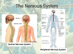
1. NERVOUS SYSTEMSPINAL CORD
2. Gray matter located centrally cellular column that extends through out the length of spinal cordGray matter is composed ofNeuronal cell bodiesSupportive tissueNerve fibersNeurons are arranged into nucleiSeveral multipolar cells are also presentDivided into root cells & Tract cellsRoot cells are motor in activityConnect to effectorsTract cells from ascending & descending fiber bundlesWhite matterSurrounds the gray matterComposed of nerve fibers and supportive tissuePresents large fiber tracts that project rostrally and caudallyFUNCTIONAL ANATOMY OF SPINAL CORD
3. SENSORY SYSTEMS IN THE SPINAL CORDCell that contribute to sensory systemsLocated in the dorsal gray columnsCells and their process confined entirely to CNSReceive collaterals and direct terminations of dorsal root fibersSend axons to ventral horns for reflex connectionsOr into white matter where they bifurcate and form inter segmental pathwaycomposed of myelinated and unmyleinated fibersMyelinated fibers carry signals from stretch receptorsUn-myelinated fibers carry pain, temp. & tactile senses
4. MOTOR SYSTEMS IN THE SPINAL CORDNuclei serving motor systems are limited toVentral hornsIntermedio-lateral column (ILC)Motor systems are divided intoSomatic and visceral systemsVisceral motor system limited to ILCSomatic motor system occupies the remaining ventral columnMedial motor column supplies muscles of axial skeletonLateral motor column supplies remaining somatic muscles
5. FIBER TRACTS OF SPINAL CORDAscending and descending tracts are arranged into distinct bundles that occupy specific areas in the white matterFiber bundles having same origin, course and termination are called tracts/fasciculiSeveral fasciculi make up a funiculusWhite matter is customarily divided intoDorsal, lateral and ventral funiculi
6.
7.
8. FUNCTIONS OF SPINAL CORDPassage way for activity moving to and from brainOperates several reflexesHigher centers alter the activity of these cord reflexes to control bodily activitiesIn domestic animals spinal cord activity is 10X more than that of higher centerThus spinal cord is more important in animals
9. SPINAL ANIMALSpinal cord is cut at the spinomedullary junction OR Brain is destroyed by ischemiaThus in a spinal animal only spinal cord is functionalSuch animal must be maintained on artificial respiration
10. SPINAL SHOCKIn any fresh spinal animalSomatic and visceral reflex activity caudal to leison is abolished for various periods of timeSpinal shock is not due to the injuryBut due to sudden loss of descending influences from higher centersSlowly most of reflex activity inherent to SC shall returnShort spinal reflexes such as myotatic reflex & withdrawal reflex return within a few hoursLong spinal reflexes such as scratch reflex take several daysInitially there is complete atony of urinary bladder and urine accumulates and overflowsMicturition reflex returns in ~ 7 daysBut the bladder is not completely emptied Initially there is diarrhea but by the time micturition reflex returns, defecation becomes depressedInitially there is extreme vasodilatation, but within a week normal vasomotor tone is re-establishedSome disturbances in CVS function and acid –base regulation persist in a spinal animal
11. REFLEXA reflex is an involuntary activity in an effector elicited by stimulation of a receptor without the involvement of brainReflex arcDiagramatic representation connecting receptor to effector through the CNSFive components in any reflex arcReceptor located in soma or visceraAfferent neuron located in the dorsal root gangliaInternuncial neuronlocated in the spinal cord / medullaEfferent neuron located in various portions of CNS Effector located in the soma or viscera
12.
13.
14. Reflex arcsSometimes internuncial neuron may be missingMonosynaptic reflexes Eg: myotatic reflexPoly synaptic reflex – internuncial neuron present Eg: Scratch reflex, micturition reflex etcReflex arcsDrawn only for convenienceNot isolated from general net work of nervous systemNot dedicated to operation of a single reflexStrong stimulation of one reflex path is likely to stimulate adjoining reflex paths
15. SPINAL REFLEXESMyotatic reflexInverse myotatic reflexWithdrawal reflexCrossed extensor reflexScratch reflexExtensor thrust reflex
16. MYOTATIC REFLEXAlso called stretch reflex / Muscle spindle reflexWhen a muscle is suddenly stretched the muscle shall contract immediatelyOrganized to initiate muscle contraction that resists stretchingWhen a joint is forceibily flexed muscular activity is elicited that resists displacement This reflex maintains muscle tone and postureMyotatic reflex induced muscle contraction in a group of muscles is accompanied by relaxation of the antagonist musclesReciprocal innervationCollaterals from afferent neurons terminate on inhibitory motor neurons of the antagonists in the ventral gray horns
17. MYOTATIC REFLEX
18. INVERSE MYOTATIC REFLEXAlso called clasp knife reflexIf a limb is forcibly flexed, muscle tone is increased in the extensors and decreased in the flexors myotatic reflex occurs in the extensorsBut if flexion is continued, suddenly the extensor tone is decreased and flexor tone is increased Limb undergoes active flexion / collapsesReceptor organ is the GTA located in muscle tendonsGTA is stretch sensitive and excited whenever a tendon is stretched due to contraction of the muscleGTA thus serve a reflex that is antagonistic to myotatic reflexInverse myotatic reflex is important for movement
19. WITHDRAWL REFLEXIf a noxious stimulus is applied to distal portion, the limb is quickly withdrawn away from the stimulus.But the activity involved is not always flexionIf the noxious stimulus is applied to elbow joint, the limb is extended to move it away from the stimulusThus the activity elicited depends on the location of the stimulus.Called local sign of a reflexSome times called (wrongly) flexion reflex
20.
21. CROSSED EXTENSOR REFLEXWhen the withdrawal reflex is elicited in a limb by application of noxious stimulus at its distal end, the contra lateral limb is extendedResults from simultaneous excitation of extensors and inhibition of flexors in the contra lateral limbOccurrence of this reflex indicates removal of descending inhibitory influences from higher centers.
22.
23. SCRATCH REFLEXIf tactile stimuli are applied over dorsal and lateral surfaces, the animal extends ipsi-lateral limb and scratches near the stimulated area If the stimulus is moved to another area, scratch is also moved to the new area appropriately.Thus this reflex also exhibits local sign
24. Extensor thrust reflexIf pressure is applied to palamar / plantar surfaces, the limb is extended into a rigid column by simultaneous contraction of both extensors and flexorsCutaneous pressure receptors and muscle stretch receptors provide the inputImportant in providing support when a limb is placed on the ground
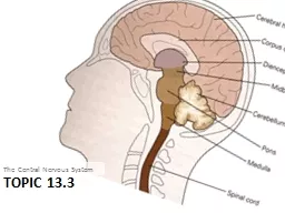
25. NERVOUS SYSTEMBRAIN STEM
26. BRAIN STEM – FUNCTIONAL ANATOMYIncludesMedulla OblongataPonsMid brainHypothalamusThalamusEpithalamus
27. MEDULLA OBLONGATANuclei in medulla are in divided intoMotor columsSomaticVisceralBranchialSensory columnsSomatic VisceralIn these columns there are 14 nuclei on each side
28.
29. MEDULLA OBLONGATASOMATIC MOTOR COLUMNSupplies to all skleletal muscles of head extrinsic muscles of eye and muscles of tongueIncludes 4 nucleiocculomotor TrochlearAbducentHypoglossal
30. MEDUALL OBLONGATA BRANCHIAL MOTOR COLUMNSupplies skeletal muscles derived from branchial arches in the embryoIncludes 4 nucleiTrigeminalFacialNucleus ambigusSp. acc. nerve nucleus
31. MEDUALL OBLONGATA –VISCERAL MOTOR COLUMNSupplies PS innervation toSm. muscles & glands in head, cerv, thor & abd visceraIncludes 4 nucleiRostral Salivary nucleus all major glands except parotid Caudal salivary nucleus parotid salivary glandEdinger Westphal Nucleus intrinsic smooth muscles of eyeDorsal motor nucleus of vaguscervical, thorasic & abd. viscera
32. MEDUALL OBLONGATA VISCERAL SENSORY COLUMNHas only one nucleusNucleus fasciculus solitariusReceives taste afferents and general visceral afferents fromMouthPharynxLarynxThorasic and abdominal viscera
33. MEDUALL OBLONGATA SOMATIC SENSORY COLUMNReceives general somatic afferents fromFace and meningesIncludes one nucleusSensory Nucleus of trigeminal
34. OTHER NUCLEI IN THE BRAIN STEMNucleus gracilisNucleus cuneatusCaudal Olivary complexPontine gray nucleusReticular formation
35. FIBER TRACTS INBRAIN STEMMedial Lemniscal systemExternal aurcate systemSpinotectal tractSpinothalamic tractLateral lemniscal systemSpinoreticular tractSpinocerebellar tractSpinoolivary tractCortiocpsinal tractCorticomedullary tractVestibulospinal tractMedial longitudinal fasciculusCentral tegmental fasciculusFasciculus solitariusCentral pathways of trigeminal nerveASCENDING TRACTSDESCENDING TRACTS
36. FUNCTIONS OF MEDULLA OBLONGATAMedulla and pons are similar to SCReflex centersReflexes are controlled fromhigher centers of brain and Ascending influences from spinal cord
37. MEDUALLRY REFLEXESSalivary ReflexDeglutition reflexMastication reflexSuckling reflexVomition ReflexCough reflexSneeze ReflexOcculo-cardiac reflexBlink ReflexLacrimation reflex
38. Material in mouthStimulation of taste & Gen.Sen.ReceptorsInterneurons in the medullaRostral Salivary NucleusCaudal Salivary NucleusParotid Salivary GlandMandibular & SublingualSalivary glandsCopius salivationGlossopharyngelFacialTrigeminalFacialGlossopharyngelSALIVARY REFLEX
39. SALIVARY REFLEXReflex activity elicited depends on the nature of substance placed in the mouthEdible substance – saliva rich in enzymes & mucusInedible substance-Saliva rich in water & organic contentEasily becomes a conditioned reflex
40. DEGLUTITION REFLEXSwallowing ReflexSomatic stimulation of soft palate, dorsal epiglottis, post. Wall of PharynxGlossopharyngealTrigeminalVagusInterneurons in the deglutition ctr.Nucleus tractus solitariusMotor nuclei ofHypoglosusVagusGlossopharyngealSequential Muscular Activity in Pharynx, Larynx, Hyoid apparatus, tongue & EsophagusPropulsion of material caudally into esophagus,which inturn moves into stomach due to peristalsisInhibition of respiration
41.
42. MASTICATION REFLEXSensory Receptors of Oral and Lingual mucosaGlossopharyngelTrigeminalVagusInterneurons in the Reticular formation in medulla & PonsMotor nucleus of TrigeminalMastigatory musclesNormally under voluntary controlModified by reflexes to protect oral mucosa
43. SUCKLING REFLEXLips, tongue, Oral mucosaFacialTrigeminalInterneurons in Reticular formation of Medulla and PonsMotor nuclei ofHypoglosssusFacialTrigeminalSuckling muscles
44. VOMITION REFLEXEmesis / vomition is designed to remove irritating material from stomach and upper intestinePresence of irritating material in these organs is the normal stimulus for vomitionPain signals from abdominal organs (ureter, kidney)Excessive stimulation of vestibular system (motion sickness)General afferents from all over the body under special conditionsPlant alkaloidsPsychic stimuliIrritating leisons in the external auditory canal in dogsMotor activity in vomitionConstricition of pylorus of stomachRelaxation of cardiaContraction of abdominal skeletal muslcelsConstriction of nasopharynxCessation of respiration
45. VOMITION REFLEXNucleus Tractus SolitariusVagusFacialGlossopharyngealVest.NucVestibular nerveCRZVagusRESP.CTRCessation of RespitrationCVS.CTRFALL IN BPFacialSALIV.CTRCOPIUS SALIVATIONN.AmbigusGlossophryngMOTOR ACTIVITY IN PHARYNX, LARYNX & HYOIDGASTROINTESTINAL TRACTSS & PSS
46. COUGH REFLEXAny mechanical stimuls to tracheal/laryngeal mucosaForces air out of lungs through tracheal and laryngeal passageBrought about by a well coordinated muscular activityDesigned to remove irritating material from trachea and lungs
47. COUGH REFLEXTracheal & Laryngeal MucosaCough centreRegulation of RespirationMotor activity in LarynxReticulo-spinalN.ambigus
48. SNEEZE REFLEXNASAL PASSAGESSen. Nuc.TrigemAppropriate Resp. ActivityN.ambigusReticular formationSimilar to cough ReflexDesigned to remove irritating material from nasal passages
49. OCCULO-CARDIAC REFLEXStimulation of the eye/orbitSen. Nuc.TrigemBrody CardiaVagusCardiac depressor Ctr.Opthalmic Div. of Trigeminal
50. BLINK REFLEXLACRIMATION REFLEXFOREIGN / OFFENDING MATERIAL IN THE EYEOpthalmic Div. of TrigeminalRostral Salivary NucleusLacrimal GlandLacrimationMotor Nucleus of FACIALClosure of eyelidsBlink Reflex is used thousands of times every dayLacrimation occurs usually if blink is insufficient