

Hussein Alhawari MD FASN Kidney Structure The kidneys are two beanshaped organs that lie in the retroperitoneal space each weighing about 150 g The functional unit of the kidney is the nephron ID: 1032972
Download Presentation The PPT/PDF document "Structure and function of the kidneys an..." is the property of its rightful owner. Permission is granted to download and print the materials on this web site for personal, non-commercial use only, and to display it on your personal computer provided you do not modify the materials and that you retain all copyright notices contained in the materials. By downloading content from our website, you accept the terms of this agreement.


1. Structure and function of the kidneys and their clinical assessmentHussein Alhawari, MD, FASN
2. Kidney StructureThe kidneys are two bean-shaped organs that lie in the retroperitoneal space, each weighing about 150 g.The functional unit of the kidney is the nephron; each nephron consists of a glomerulus and a long tubule, which is composed of a single layer of epithelial cells. There are approximately one million nephrons in one human kidney. The nephron is segmented into distinct parts—proximal tubule, loop of Henle, distal tubule, and collecting duct.
3.
4. Renal circulationThe renal artery carries about one fifth of the cardiac output; this represents the highest tissue-specific blood flow of all larger organs in the body (about 350 mL/min per 100 g tissue).
5. The renal circulation is unusual in that it breaks into two separate capillary beds: the glomerular bed and the peritubular bed. As blood leaves the glomerulus, the capillaries coalesce into the efferent arteriole. Pressure in the first capillary bed, that of the glomerulus, is rather high (40 to 50 mm Hg), whereas pressure in the peritubular capillaries is similar to that in capillary beds elsewhere in the body (5 to 10 mm Hg).
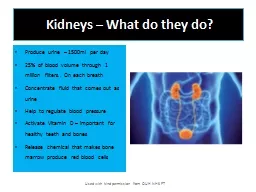
6. Functions of the kidney1. Maintenance of body composition… The volume of fluid in the body; its osmolarity, electrolyte content, and concentration; and its acidity. 2. Excretion of metabolic end products and foreign substances: The kidney excretes a number of products of metabolism, most notably urea, and a number of toxins and drugs
7. 3. Production and secretion of enzymes and hormones: a. Renin is an enzyme produced by the granular cells of the juxtaglomerular apparatus that catalyzes the formation of angiotensin from angiotensinogen. Angiotensin is a potent vasoconstrictor. b. Erythropoietin, is produced by renal cortical interstitial cells, stimulates the maturation of erythrocytes in the bone marrow. c. 1,25-Dihydroxyvitamin D3, is formed by proximal tubule cells.
8.
9. Assessment of renal functionH&PERFP… Including BUN and Cr… Cr-GFRUA with microscopyImagingOthers, if indicated based on the above lab findings.Kidney biopsy.
10. Assessment of Glomerular Filtration RateNormal average GFR values are approximately 130 and 120 mL/min/1.73 m2 for young men and women, respectivelyGFR is often estimated from the serum concentration of endogenous filtration markers.Creatinine is the most commonly used endogenous filtration marker in clinical practice.
11. The most common methods utilized to estimate the GFR are: 1. Measurement of the 24 hour urine creatinine clearance. 2. Estimation equations based upon serum creatinine such as the Cockcroft-Gault equation, the Modification of Diet in Renal Disease (MDRD) study equations, and the Chronic Kidney Disease Epidemiology Collaboration (CKD-EPI) equation.
12. UrinalysisIn conjunction with the history, physical examination, and serum chemistries, the urinalysis plays a central role in evaluating acute and chronic kidney disease. In addition, abnormal findings on a routine urinalysis, often in an otherwise asymptomatic patient, may be the first evidence of underlying kidney disease.A complete urinalysis consists of three components: gross evaluation, dipstick analysis, and microscopic examination of the urine sediment.
13. Normal urine is clear and light yellow in color.The urine dipstick provides a rapid semiquantitative assessment of urinary characteristics on a series of test pads embedded on a reagent strip. Most dipsticks permit the analysis of the following core urine parameters: heme, leukocyte esterase, nitrite, albumin, hydrogen ions, specific gravity, and glucose.
14. Microscopic examination of the urine sediment is an essential part of the urinalysis, as it enables confirmation and clarification of urine dipstick findings and also the identification of structures that are not evaluated by the urine dipstick (eg, epithelial cells, casts, crystals).
15. CrystalsCrystals, such as uric acid crystals, calcium phosphate or calcium oxalate crystals, cystine crystals, and magnesium ammonium phosphate crystals.
16.
17.
18.
19.
20. Bacteria or fungi
21. Red blood cells
22. Dysmorphic RBCs
23. White blood cells
24. Renal tubular epithelial cells
25. RBC casts, which are usually diagnostic of glomerular hematuria
26. WBC casts, which are indicative of kidney inflammation,(eg, pyelonephritis, interstitial nephritis)
27. Muddy brown casts
28. HematuriaGenerally, hematuria is defined as the presence of 5 or more red blood cells (RBCs) per high-power field in 3 of 3 consecutive centrifuged specimens obtained at least 1 week apart.Hematuria can be either gross (ie, overtly bloody, smoky, or tea-colored urine) or microscopic.
29.
30. ProteinuriaTotal urinary protein excretion in the normal adult should be less than 150 mg/day.The normal rate of albumin excretion is less than 30 mg/day.
31. Persistent albumin excretion between 30 and 300 mg/day is called moderately increased albuminuria (formerly called "microalbuminuria"). Persistent albumin excretion above 300 mg/day is considered overt proteinuria or severely increased albuminuria (formerly called "macroalbuminuria"), the level at which the standard dipstick becomes positive.
32. Two semiquantitative methods are available to screen patients for proteinuria. These are the standard urine dipstick and the precipitation of urine proteins with sulfosalicylic acid (SSA). In contrast to the urine dipstick, which primarily detects albumin, SSA detects all proteins in the urine.
33. Patients with persistent proteinuria should undergo a quantitative measurement of total protein excretion.
34. Most commonly, the urine protein-to-creatinine ratio (UPCR) in a spot first- or second-morning urine sample after avoiding exercise is used to estimate 24-hour proteinuria. Usually, the urine protein concentration in a spot sample is measured in mg/dL and is divided by the urine creatinine concentration, also measured in mg/dL (at JUH lab it is measured in gm/L (so x100 to make it mg/dl), yielding a dimensionless number that estimates the 24-hour in grams.
35.
36. Kidney ImagingPlain KUB… Non yielding… I rarely order these days.Renal US. Kidney size, cortical thickness and echogenicity. Renal cysts. Rule out hydronephrosis, Renal calculi greater than 3-5 mm within the renal pelvis with relative insensitivity to ureteric calculi.Renal Doppler to assess patency and flow in renal vasculature
37. CT scan provides the highest sensitivity for detecting fine calcifications within the kidney parenchyma and through out the collecting system, making unenhanced CT the optimal test for detecting stone disease.Contrast enhanced multiphase CT to evaluate kidney masses.
38. 35 yo lady presented to the ER with bilateral LE edema and joint pain. BP was 160/100. Cr was 2.5 with eGFR around 25 ml/min. UA showed 2+ proteinuria, 2+ blood, 5-10 WBCs, +dysmorphic RBCs.What is your provisional diagnosis?Nephrotic syndrome.Acute glomerulonephritisPyelonephritisNephrolithiasisRenal cell carcinoma
39. 75 yo man presented to the ER c/o hematuria. UA showed 3+ blood, 5-10 WBCs, 1+ protein. Monomorphic RBCs on microscopy. Cr was 1.2.What is the best next step?Renal imaging + cystoscopy.Consult nephrology urgently.D/C home and refer to nephrology clinic as outpatient.D/C home and advise to drink more water.No further testing.
40. 45 yo man presented to the ER c/o bilateral LE edema. Cr was 1.4 with eGFR around 45 ml/min. UA showed 3+ protein, 0-5 RBCs, 0-5 WBCs. Urine protein/Creatinine ration UPCR was around 6.5 gm/day.What is your provisional diagnosis?Acute nephritic syndrome.Acute Kidney Injury.NephrolithiasisAcute pyelonephritisNephrotic syndrome.
41. 60 yo lady presented c/o left loin pain and dysuria. UA showed trace protein, 5-10 RBCs, 40-50 WBCs. What is your provisional diagnosis?Acute GN.Acute nephrotic syndromeUTIMusculoskeletal back painAcute appendicitis
42. 35 yo lady prsented c/o LE edema, malar rash and dysuria. UA showed 3+ protein, numerous dysmorphic RBCs and numerous WBCs.What is your provisional diagnosis?Acute GNAcute UTINephrotic SyndromeAcute GN + UTINephrolithiasis.
43. Questions??