
BY DR NILANJANA GHOSH Outlines Definitions and basic physiology Differences between central and peripheral causes of hypotonia Evaluation of hypotonia in infants Management options Relevant terminologies and definitions ID: 1009433
Download Presentation The PPT/PDF document "Approach to floppy infant" is the property of its rightful owner. Permission is granted to download and print the materials on this web site for personal, non-commercial use only, and to display it on your personal computer provided you do not modify the materials and that you retain all copyright notices contained in the materials. By downloading content from our website, you accept the terms of this agreement.


1. Approach to floppy infantBY – DR NILANJANA GHOSH
2. OutlinesDefinitions and basic physiologyDifferences between central and peripheral causes of hypotonia.Evaluation of hypotonia in infants.Management options
3. Relevant terminologies and definitionsTone: is the baseline resistance of muscle to stretch.Phasic tone: The rapid contraction in response to a high –intensity stretch, as in tendon reflex response.Postural tone: It is the prolonged contraction of antigravity muscles in response to the low- intensity stretch of gravity.
4. Hypotonia- reduced resistance to ta passive movements around a jointWeakness is reduction in the maximum power that can be generatedFloppy infants exhibit poor control of movements, delayed motor skills, alteration in postural control, increased range of motion.Weak infants always have hypotonia but hypotonic infants may not have weakness.
5. How muscle tone is maintained?Cerebral cortexCerebellumBasal gangliaVestibular nucleiCerebral cortexCerebellumγ motor discharge of spinal cordBulbo reticular facilitatory area in pons Bulbo reticular area in medulla
6. How muscle tone is maintained?
7. The maintenance of normal tone requires intact central and peripheral nervous system. Hence hypotonia is a common symptoms of neurological dysfunction and occurs in diseases of the brain, spinal cord, nerves and muscles.
8. The term- Floppy InfantIndicates multiple disorders with various degree of hypotonia (axial, appendicular or both).Supraspinal conditions(brain, brainstem, cervical spinal junction)-Central hypotoniaSegmental conditions(anterior horn cell, peripheral nerve, neuromuscular junction, muscle)-Motor unit hypotonia/peripheral hypotoniaVast differential diagnosis-ranging from benign condition with good prognosis to life threatening condition incompatible with life.
9. A systematic approach to a child who has hypotonia,paying attention to the history and clinical examination, is the paramount to localizing the problem to a specific region of the nervous system.
10. Mainly two approaches(1) Based on neuro-anatomical localization i.e Central or Peripheral nervous system involvement.(2) To determine whether or not the hypotonia is associated with weakness.
11. CENTRAL HYPOTONIAPERIPHERAL HYPOTONIACentral causes account for 60% to 80% of hypotonia casesAbnormalities of other brain functions.Global developmental delay.Axial hypotonia> appendicular hypotonia.Dysmorphic features: microcephaly.Fisting of hands.Normal or brisk tendon reflex.Scissoring on vertical suspension.peripheral causes occur in 15% to 30%.Alert, normal cognitionMotor delay.Hypotonia with profound limbs weakness(axial hypotonia=appendicular hypotonia)Hyporeflexia or areflexia.Reduced or absent antigravity movement.Fasciculation.Muscle atrophy or pseudohypertrophy.Respiratory and feeding impairment.
12. Causes of Central hypotoniaBrain malformations :Chromosomal disorders- Down syndrome, Praderwilli syndrome.Genetic disorders- Familial dysautonomia, Lowe syndrome.Endocrine disorders- hypothyroidismMetabolic- rickets, renal tubular acidosis
13. IEM- aminoaciduria, organic aciduria, mucopolysaccharidoses,glycogen storage disorder, fatty acid oxidation defect etcCerebral insult- Hypoxic ischemic encephalopathy, intracranial hemorrhage, Hypoglycemia.Cerebellar disorder- Joubert syndrome, Ponto-cerebellar hypoplasia.Paroxysmal disorder- neonatal adrenoleukodystrophy, Zellweger syndrome.
14. Causes of peripheral hypotoniaAnterior horn cell disorders- Spinal muscular atrophy.Congenital neuropathy- Hereditary motor-sensory neuropathy, congenital hypo myelinating neuropathy, Charcot Marie tooth disease.Neuromuscular junction disorders- Transient neonatal myasthenia, congenital myasthenic syndrome, infantile botulism, magnesium toxicity, aminoglycoside toxicityMyopathies- Congenital myopathy, Nemaline myopathy, central core disease, myotubular myopathy,Muscular dystrophies- Walker- Warburg, Fukuyama, muscles- eye-brain disease,merosin positive CMD, early infantile facioscapulohumeral dystrophy.
15. APPROACHPresenting complaints:Age of presentationOnset(sudden or gradual)Course(progressive, static or fluctuating)Muscles weakness distribution(proximal –myopathy or distal –neuropathy)
16. Antenatal historyDecreased fetal movementPolyhydramniosDrug exposureBreech presentationProlonged labour
17. Postnatal historyBirth asphyxiaWeak cryRespiratory distress, prolonged ventilatory supportFeeding difficultiesConvulsionHoney consumptionJoint dislocation
18. History of consanguinityFamily historyDevelopmental history(isolated motor or motor and cognitive)Anthropometry: head circumference, weight
19. Salient points in historyAny significant family history-affected parents or siblings,consanguinity,stillbirths,childhood deathsMaternal disease- myotonic dystrophyPregnancy and delivery history- drug or teratogen exposure, decreased fetal movements, abnormal presentations, polyhydramnios/oligohydramnios, Apgar score, resuscitation requirements.History since delivery Respiratory effort Ability to feed Level of alertness Level of spontaneous activity Character of cry
20. EXAMINATIONDysmorphic featuresAnterior fontanelEyes: cataract(Lowe syndrome)Eye lid dropTongue fasciculationGenitalia hypogonadismLimbs examination
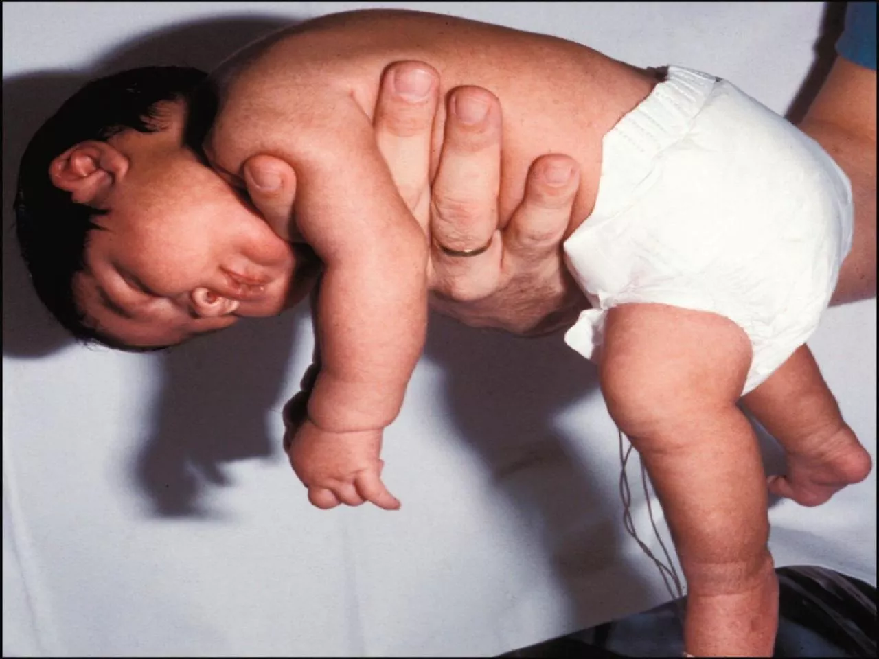
21. Neurologic examinationDetailed neurologic assessment- tone, strength and reflexesAssessment of tone- begin by examining posture and movement. A floppy infant often lies with limbs abducted and extended.
22. Evaluation of hypotoniaObservation of position at rest, Palpation of muscles,Passive movementsFurther evaluation of hypotonia by Amiel Tison method and 18o degree flip merhod.
23. Amiel-Tison methodAge(months)Adductor anglePopliteal angleDorsiflexion angleScarf sign0-340˚-80˚80˚-100˚60˚-70˚Elbow does not cross midline4-670˚-110˚90˚-120˚60˚-70˚Elbow crosses midline7-9110˚-140˚110˚-160˚60˚-70˚Elbow goes beyond axillary line10-12140˚-180˚150˚-170˚60˚-70˚-----
24. 180 degree flip methodA supine infant is pulled to sit, held vertical, held in ventral suspension and finally put in prone position, completing 180 degree flip.Supine- assess any preferred position, like external rotation of lower limb indicating weakness of floppiness on that side. Floppy baby may have frog like posture.
25. Traction responseThe presence of more than minimal head lag and failure to counter traction by flexion of the limbs in the term newborn is abnormal and indicate hypotonia.
26. Sitting- assess sitting ability(with or without support),rounded or straight back etc.
27. Vertical suspensionThe examiner places both the hands in the infant’s axillae and without grasping the thorax, lifts up. Normal response- Head erect in the mid line with flexion at the knee.hip and ankle joints.Hypotonic infant-suspends vertically, head falls forward, legs dangle and the infant may slip through examiner’s hand.
28. Horizontal suspensionBaby suspended in the prone position with the examiner’s palm underneath the chestNormal infant- keeps the head erect, maintains the back straight and flexes the elbow,hip,knee and ankle joints.Hypotonia- infants drape over the examiner’s hand legs hanging limply
29. Prone- place the child prone in bed and look for any arching.
30. Deep tendon reflexes(DTRs)DTRs often normal/hyperactive in central conditions.DTRs- normal/decreased/absent in peripheral disorders.
31. Other pertinent findingsDecreased resistance to flexion and extension of the extremities.Exaggerated hip abduction and ankle dorsiflectionOral-motor dysfunctionPoor respiratory effortsGastro esophageal refluxNote the distribution of weakness. Ex. Face is spared versus the trunk and extremities.
32. VariableCentral injuryCentral developmentalAnterior horn cellPeripheral nerveNM junctionMuscleStrengthNormal or slight weaknessNormal or slight weaknessweaknessweaknessweaknessweaknessDTRsNormal/increasednormaldecreaseddecreasedNormal/decreasedDecreased to absentBabinski’s sign+/-+/-absentabsentInfantile reflexpersistentPersistent/absentabsentabsentabsentabsentMuscle fasciculationabsentabsentprominentabsentabsentabsentMuscle massNormal/disuse atrophyNormal/disuse atrophyProminent atrophy(proximal)Distal atrophyNormal/decreasedProximal atrophy: increased or decreased pseudo hypertrophySensationnormalnormalnormalIncreased /decreasednormalnormalToneDecreased/evolving to increaseddecreaseddecreaseddecreasedDecreased/normaldecreasedLocalization of disorder producing hypotonia
33. Anterior horn cell disease versus NM junction disorderUsually spares extra ocular muscles, while diseases of the neuromuscular junction may be characterized by ptosis and extra ocular muscles weakness.
34. Clues to central hypotoniaDysmorphic features : Trisomy-21,Prader-willi syndrome, DiGeorge syndrome.Depressed level of consciousness or lethargyAbnormal eye movement or inability to track visuallyEarly onset seizure.ApneaExaggerated irregular breathing patterns .. Joubert syndromePredominant axial weaknessNormal strength with hypotoniaScissoring on vertical suspension- Cerebral palsyFisting of the handsHyperactive or normal reflexesMalformation of other organs
35. Characteristics of peripheral causes of hypotoniaAlert infant and appropriate response to surroundingsNormal sleep-wake patternsAssociated with profound weaknessHypotonia and hyporeflexia/ areflexiaOther features- muscle atrophy, presence of respiratory and feeding impairment.
36. Clues to anterior horn cell disorders(Infantile form of spinal muscular atrophy)HypotoniaGeneralized weaknessAbsent reflexesFeeding difficultiesFasciculation of tongueAlert faceProfound distal weakness
37. Clues to specific diagnosisHepatosplenomegaly-storage disorder, congenital infections.Hepatomegaly,retinitis pigmentosa-neonatal adrenoleukodystrophy.Renal cyst, high forehead, wide fontanel's- Zellweger’s syndrome.Congenital cataracts,glucoma-oculocerebrorenal syndrome.Abnormal odor- metabolic disorder.Hyperpigmentation,undescended testis- Prader willi
38. Peripheral causesDiagnosis mainly by history and clinical examinationMolecular genetics-CTG repeats, deletions in SMN geneNerve conduction studies and muscle biopsy(Depending on clinical situation, may be delayed until around 6 months of age as neonatal results are difficult to interpret)Creatine kinase-elevated in muscular dystrophy but not in spinal muscular atrophy or many other myopathiesSpecific DNA testing-for myotonic dystrophy and for spinal muscular atrophy(SMN gene)Electrophysiological studies- shows abnormalities in nerves , myopathies and disorders of NM junction.
39.
40.