
Epiretinal membrane can also be known by other names macular pucker preretinal membrane cellophane maculopathy surface wrinkling retinopathy and premacular How the eye works Light passes thr ID: 953434
Download Pdf The PPT/PDF document "Epiretinal membrane macular pucker" is the property of its rightful owner. Permission is granted to download and print the materials on this web site for personal, non-commercial use only, and to display it on your personal computer provided you do not modify the materials and that you retain all copyright notices contained in the materials. By downloading content from our website, you accept the terms of this agreement.


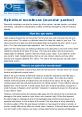
Epiretinal membrane (macular pucker) Epiretinal membrane can also be known by other names: macular pucker, pre-retinal membrane, cellophane maculopathy, surface wrinkling retinopathy, and pre-macular How the eye works Light passes through the cornea at the front of your eye, and is focused by the lens onto your retina. The retina is a delicate tissue that lines the inside of your eye. The retina converts the light into electrical signals that travel along the optic nerve to your brain. The brain interprets these signals to “see” the world around you. Light from the object you are looking at directly is focused onto a tiny area of the retina called the macula at the back of the eye. The macula is about 4mm across and is responsible for detailed central vision and most colour vision. It provides the vision you need to read, recognise faces, drive a car, see colours clearly, and any other activity that requires detailed, �ne vision. The rest of the retina gives you side vision What is an epiretinal membrane? An epiretinal membrane is scar tissue that has formed on the macula which can cause Most of the eye’s interior is �lled with vitreous, a jelly-like substan
ce that �lls about 80 percent of the eye and helps it maintain a round shape. The vitreous contains However, sometimes when the vitreous pulls away from the retina, there is microscopic damage to the retina’s surface (note: this is not a macular hole). When this happens, the retina begins a healing process to the damaged area and forms scar tissue, or an epiretinal membrane, on the surface of the retina. This scar tissue is �rmly attached to e the retina to wrinkle, or What causes epiretinal membrane? Most epiretinal membranes are related to vitreous detachment, which usually occurs in people over age 50. As we age, there is an increased risk of epiretinal membrane. Epiretinal membrane can also be triggered by certain eye diseases and disorders, people with diabetes sometimes develop an eye disease called diabetic retinopathy, which can cause epiretinal membrane. An epiretinal membrane can also form following What are the symptoms of epiretinal membrane? Vision loss from epiretinal membrane can vary from no loss to severe loss, although . They may have dif�culty in seeing �ne detail and reading small print. There may be a dull area in Epi
retinal membrane usually only affects one eye. Is a macular pucker similar to a macular hole? A macular pucker and a macular hole are different conditions, although they both similar symptoms - distorted and blurred vision. Also, a macular pucker will very rarely How is epiretinal membrane treated? blurriness are mild, and no treatment is necessary. People usually adjust to the mild visual distortion, since it does not affect activities of daily life, such as reading and Sometimes, vision deteriorates to the point where it affects daily routine activities. When this happens, surgery may be recommended. This procedure is called a vitrectomy, in which the vitreous gel is removed to prevent it from pulling on the retina and replaced with a salt solution. (Because the vitreous is mostly water, you will notice no change between the salt solution and the normal vitreous). Also, the scar tissue which causes the wrinkling is removed. A vitrectomy is usually performed under local anesthesia. After the surgery, you will be given eye drops to use for several weeks. Your surgeon will also provide further instructions regarding any restrictions on your activity in the How successful is this surgery
? Surgery to repair an epiretinal membrane is very delicate, and while vision improves What are the risks of surgery? vitrectomy. Other, less common complications are retinal detachment either during or after surgery, and infection after surgery. Also, the epiretinal membrane may grow Managing vision loss with you to assess your individual needs and determine which aids and technologies can help. There are many excellent solutions to help you live well with low vision. Fortunately it is very uncommon for vision loss from epiretinal membrane to be so Contact Macular Disease Foundation Australia to discuss your low vision needs and to Macular Disease Foundation Australia 1800 111 709 info@mdfoundation.com.au Web:www.mdfoundation.com.au Disclaimer: from a doctor. The Macular Disease Foundation Australia cannot be liable for any error or omission in this publication or for damages arising from its supply, performance or use, and makes no warranty of any kind, either expressed or implied July 2015 Macular Disease Foundation Australia Resources Macular Disease Foundation Australia has developed a comprehensive range of publications on macular degeneration, diabetic eye disease and other macula