
Danielle R Snowflack PhD wwwedvotekcom EDVOTEK The Biotechnology Education Company Celebrating OVER 30 years of science education Proteins are everywhere Proteins are diverse molecules that perform important functions within a cell ID: 1032629
Download Presentation The PPT/PDF document "Biotechnology Basics: Protein Electropho..." is the property of its rightful owner. Permission is granted to download and print the materials on this web site for personal, non-commercial use only, and to display it on your personal computer provided you do not modify the materials and that you retain all copyright notices contained in the materials. By downloading content from our website, you accept the terms of this agreement.


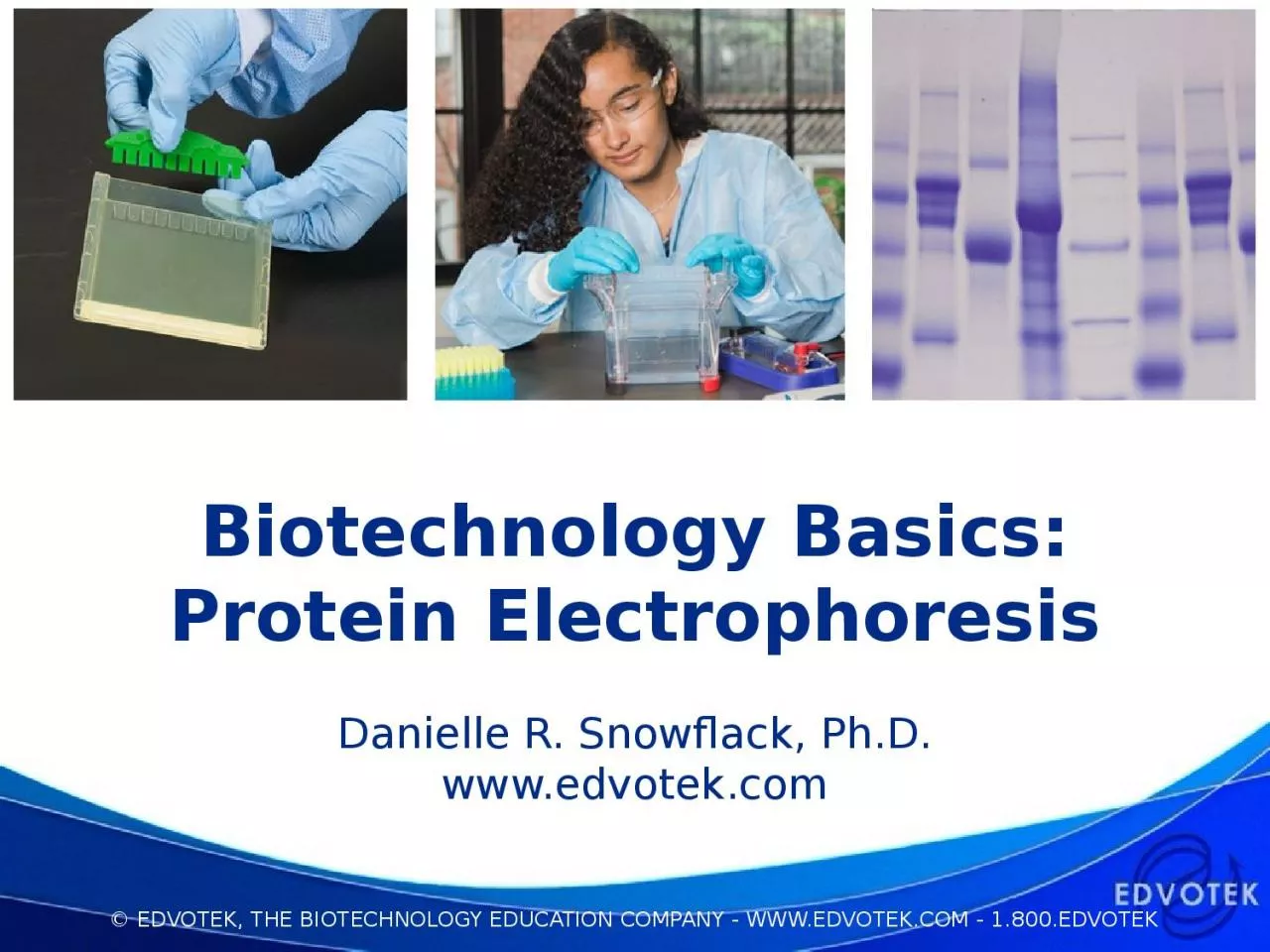
1. Biotechnology Basics:Protein ElectrophoresisDanielle R. Snowflack, Ph.D.www.edvotek.com
2. EDVOTEKThe Biotechnology Education CompanyCelebrating OVER 30 years of science education!
3. Proteins are everywhere!Proteins are diverse molecules that perform important functions within a cellenzyme catalysishomeostasisbinding and transport of small moleculesgene regulationimmunological defense cell structureThis Photo by Unknown Author is licensed under CC BY-SA-NC
4. Today’s Experiment: Protein ElectrophoresisThe amino acid sequence of a protein determines its size, shape, and charge. SDS Polyacrylamide Gel Electrophoresis can be used to determine the size of proteins.Today’s experiment: Determination of Protein Molecular Weight (Kit #153)
5. Summary of Protein Electrophoresis
6. Preparing for Protein Electrophoresis How to use precast 12% SDS-PAGE gelsRemove gel from packagingRemove tape at bottom of gel cassette (if present)Gently remove combInsert gel into tank and submerge in buffer
7. Vertical electrophoresis apparatusD.C. power sourceMicropipettePolyacrylamide gelsElectrophoresis BufferProtein samplesA way to visualize samplesWhat Do I Need to Perform SDS-PAGE Experiments?
8. Power Supplies Provide Current for ElectrophoresisCat. #5010QuadraSource™(10-300V)Cat. #509DuoSource™ 150(75/150 V)
9. The genetic code is translated to create proteinsA gene is a unit of heredity with a known nucleotide sequence Most genes code for proteinsThere are 20 standard amino acids found in proteinThe molecular weight is a function of the number and type of amino acids in the polypeptide chain. Scott Henry Maxwell, CC BY-SA 4.0 via Wikimedia Commons
10. Building a proteinA protein is a polymer built from amino acid monomersEach amino acid consists of a carbon molecule bonded to four groupsAmino group (N)Carboxyl group (C)Hydrogen atomR group (a chemically unique side chain)A peptide bond is formed between the N group of one amino acid and the C group of the next one
11. Protein 3D structureProteins exhibit different three-dimensional shapes and folding patterns which are determined by their amino acid sequences. Proteins can have spherical, elliptical or rod-like shapes. The precise three-dimensional configuration of a protein is critical to its function. A proteins can consist of a single polypeptide or several polypeptides specifically associated with each other. Kep17, CC BY-SA 4.0, via Wikimedia Commons
12. The Green Fluorescent Protein (GFP)First isolated from the jellyfish A. victoria in the 1970’s.Small protein (27kD) that absorbs blue light and emits green light in response.Once the DNA sequence was identified, genetic engineering techniques were used to introduce the protein into other organisms, like E. coli.
13. Point mutations in the GFP chromophore change its colorFrom “Fluorescent Protein Tracking and Detection:”Cold Spring Harbor Protocols 2009(12):pdb.top63Specific residues form ‘chromophore’, a special structure that is responsible for light production. Changing the amino acid sequence affects the patterns of light absorption and emissionThis creates a rainbow of fluorescent proteins. For their discovery and development of fluorescent proteins, Osamu Shimomura, Martin Chalfie and Roger Tsien were awarded the 2008 Nobel Prize in Chemistry.
14. Protein structure and human healthHemoglobin is a heterotetramerHetero – differentTetramer – four subunitsIn adults, two subunits are:Alpha hemoglobin (red), or HbABeta hemoglobin (blue), or HbBEach monomer has an iron-containing heme groupHeme transports oxygenZephyris at the English-language Wikipedia, CC BY-SA 3.0
15. Changes in a protein’s sequence can affect its functionSickle cell disease has been linked to DNA mutations that change the amino acid sequence of the beta hemoglobin proteinThe most common genetic change is a single point mutation is A to T which changes the amino acid sequence of the proteinGlutamic acid to ValineThe change from a negatively-charged amino acid to a hydrophobic on changes the protein structure
16. Summary of Protein Electrophoresis
17. Electrophoresis separates biomolecules by physical propertiesSamples are loaded into the gel, and an electrical current is passed through the gel. The current drives the negatively charged molecules through the gel towards the positive electrode, and positively charged molecules to the negative electrode.The molecules separate into distinct zones based on their charge.
18. Problem: Protein structure affects migration Proteins that are in their normal, biologically active forms are called native.A protein can have a net negative or net positive charge, depending on its amino acid composition and the pH of the solution. The three-dimensional shape of a protein influences how it travels through a gel/www.savemyexams.co.uk
19. SDS-PAGE Separates Proteins by SizeProtein structure is disrupted using reducing agents, detergent, and heatSDS-PAGE uses an electric field and a porous gel matrix to separate proteins by size.When an electrical current is passed through the gel, the current moves the denatured proteins towards the positive electrodeProteins will migrate through the polyacrylamide gel in a predictable manner
20. Staining agarose gels with FlashBlue™ ProteinThe proteins in this kit have been pre-stained, so they can be visualized right away!To enhance the results, use our new FlashBlue Protein Stain!Uses Ethanol and Vinegar – eliminate methanol!Fast – stain in 15 minutes, then destain in solutionVisualize on a white light boxLaunching soon in our protein experiments!
21. LaneSampleMolecular Weight (kDa)1Protein Standard Marker94, 67, 38, 30, 20, 142Sample 1683Sample 2584Sample 350Results and InterpretationCalculate protein molecular weight using a standard curve: https://youtu.be/ssUFYMQfSGE
22. Biotechnology Basics:Protein ElectrophoresisProteins are built from amino acidsThe amino acid sequence determines the 3D structure and protein functionHigher order structure makes proteins can be difficult to separate by electrophoresisDenatured proteins can be separated by size using SDS-PAGE
23. EDVOTEK, Inc.The Biotechnology Education CompanyTo Request this Presentation:https://forms.gle/pWxavaWiCpkp4C2QAFor Orders & Technical Service: Phone: 1-800-EDVOTEK (1-800-338-6538)Web site: www.edvotek.comEmail: info@edvotek.comCheck out our YouTube Channel:www.youtube.com/EdvotekIncFollow our social media for offers & updates: www.facebook.com/edvotekhttps://twitter.com/edvotekhttps://www.instagram.com/edvotek