
Polycythaemia vera PV Primary myelo31brosis MF Chronic myeloid leukaemia CML Less common types of MPNs include Chronic eosinophilic leukaemia CEL Chronic neutrophilic leukaemia CNL Your ID: 946970
Download Pdf The PPT/PDF document "Essential thrombocythaemia ET" is the property of its rightful owner. Permission is granted to download and print the materials on this web site for personal, non-commercial use only, and to display it on your personal computer provided you do not modify the materials and that you retain all copyright notices contained in the materials. By downloading content from our website, you accept the terms of this agreement.


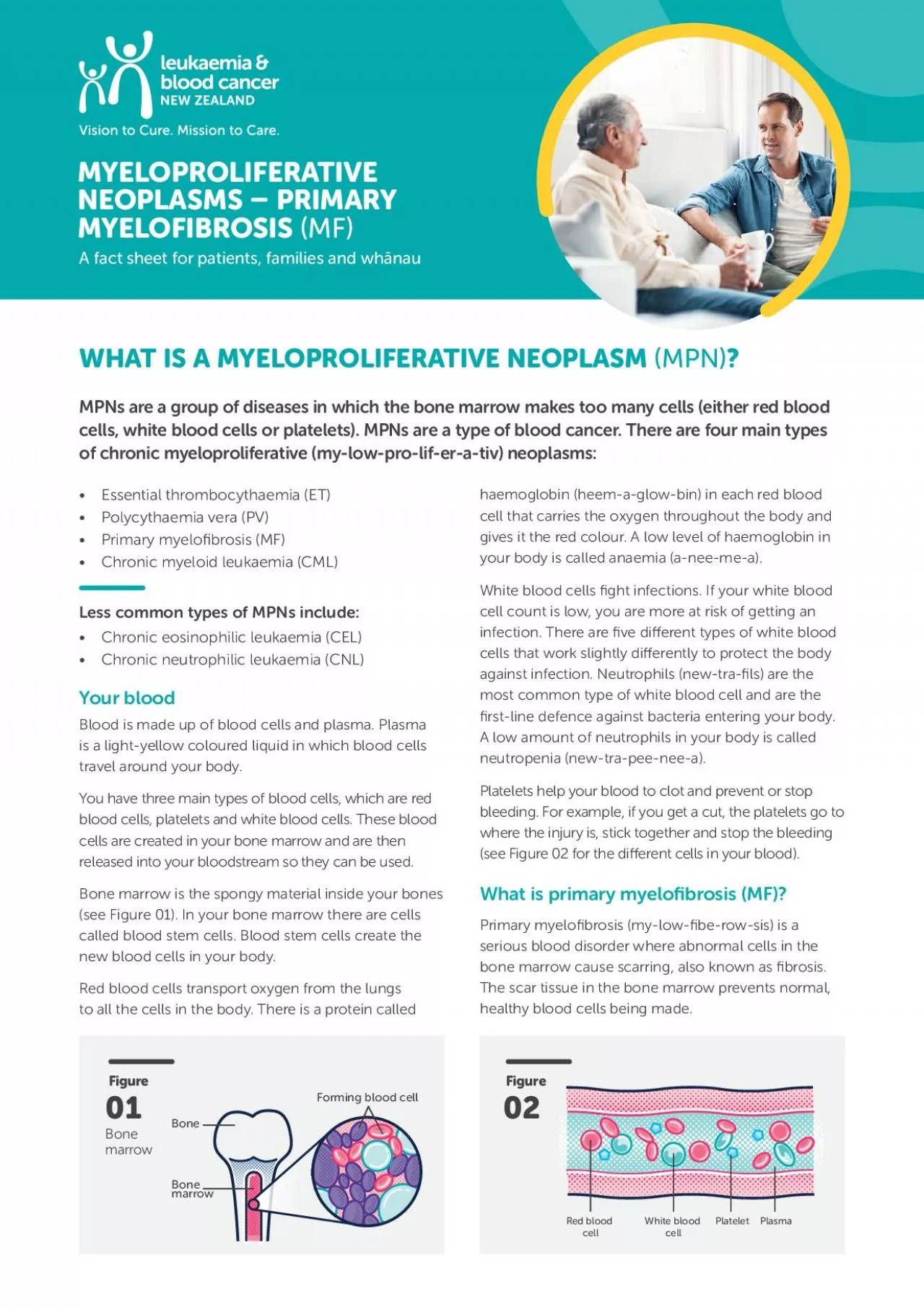
Essential thrombocythaemia (ET) Polycythaemia vera (PV) Primary myelo brosis (MF) Chronic myeloid leukaemia (CML) Less common types of MPNs include: Chronic eosinophilic leukaemia (CEL) Chronic neutrophilic leukaemia (CNL) Your blood Blood is made up of blood cells and plasma. Plasma is a light-yellow coloured liquid in which blood cells travel around your body. You have three main types of blood cells, which are red blood cells, platelets and white blood cells. These blood cells are created in your bone marrow and are then released into your bloodstream so they can be used. Bone marrow is the spongy material inside your bones (see Figure 01). In your bone marrow there are cells called blood stem cells. Blood stem cells create the new blood cells in your body. Red blood cells transport oxygen from the lungs to all the cells in the body. There is a protein called haemoglobin (heem-a-glow-bin) in each red blood cell that carries the oxygen throughout the body and gives it the red colour. A low level of haemoglobin in your body is called anaemia (a-nee-me-a). White blood cells ght infections. If your white blood cell count is low, you are more at risk of getting an infection. There are ve di erent types of white blood cells that work slightly di erently to protect the body against infection. Neutrophils (new-tra- ls) are the most common type of white blood cell and are the rst-line defence against bacteria entering your body. A low amount of neutrophils in your body is called neutropenia (new-tra-pee-nee-a). Platelets help your blood to clot and prevent or stop bleeding. For example, if you get a cut, the platelets go to where the injury is, stick together and stop the bleeding (see Figure 02 for the di erent cells in your blood). What is primary myelo brosis (MF)? Primary myelo brosis (my-low- be-row-sis) is a serious blood disorder where abnormal cells in the bone marrow cause scarring, also known as brosis. The scar tissue in the bone marrow prevents normal, healthy blood cells being made. WHAT IS A MYELOPROLIFERATIVE NEOPLASM MPN ? MPNs are a group of diseases in which the bone marrow makes too many cells (either red blood cells, white blood cells or platelets). MPNs are a type of blood cancer. There are four main types of chronic myeloproliferative (my-low-pro-lif-er-a-tiv) neoplasms: Bone marrow Red bood cell Whie bood cell PlteltPlasma Figure Figure 02 01 Forming bood ellBone marow Bone MYELOPROLIFERATIVE NEOPLASMS PRIMARY MYELOFIBROSIS MF A fact sheet for patients, families and wh nau Both adults and children can be diagnosed with MF but it is more commonly seen in people over the age of 60 years old. Approximately 40 people are diagnosed with MF each year in New Zealand. Primary myelo brosis may also be called myelosclerosis, myeloid metaplasia or idiopathic myelo brosis. What causes MF? The cause of MF is not fully understood but it is believed that mutations of particular genes lead to the production of scar tissue in the bone marrow. The most common gene that is a ected in MF is called JAK2. Other genes (like CALR, MPL and TN) may also be involved, which cause mutations. Below are the percentages of gene mutations for MF: JAK2 60% C
ALR 25-30% TN 10% MPL MF can arise on its own or as a progression from another MPN like essential thrombocythaemia (ET) or polycythaemia vera (PV). Risk factors associated with MF include: • Age: risk increases as you get older Environment: exposure to certain chemicals and ionising radiation may increase risk MF is not contagious. For most people the gene mutations that cause MF are not inherited and occur during a person’s lifetime. What are the symptoms of MF? Some people have no noticeable symptoms when they are rst diagnosed with MF and it might have only been picked up in a routine blood test. More signi cant symptoms can develop over time. Common signs of MF include: Enlarged spleen, which can cause abdominal pain, bloating and feeling full Low blood counts, which can cause bleeding, bruising (low platelets), infections (low white blood cells), fatigue and pale skin (low red blood cells) Unexplained weight loss and loss of muscle mass Unexplained fever Drenching night sweats Fatigue or tiredness •Bone pain The spleen is an organ that sits at the bottom of your left ribcage. Its main function is to lter the blood. When the bone marrow isn’t working properly this can cause the spleen to work harder and faster. The spleen can become enlarged when there are a lot of abnormal blood cells being ltered through it. Complications of MF If MF is not well managed with drugs and a balanced lifestyle, the scarring in the bone marrow can continue to get worse. The only known cure for this condition is allogeneic blood stem cell transplant (bone marrow/stem cells donated from another person) and is only an option for some people. A stem cell transplant might be o ered for someone with advanced disease and in healthier and younger people (the risk of treatment can be too high for some individuals). Medications can reduce some of the symptoms but won’t slow down the scarring in the bone marrow. Over time, less and less healthy blood cells are made, which can cause more symptoms and complications. If enough unhealthy cells accumulate then it can transform into acute myeloid leukaemia (20% of people in 10 years). Acute myeloid leukaemia is an aggressive bone marrow cancer. Increased risk of infection can be another complication of MF if someone has a low number of white blood cells. It is important to have good hand hygiene, check your body temperature if feeling hot or unwell, and be careful when other people have infections. It is important to let your health care team know if you have a high temperature or are showing signs of infection. How is MF diagnosed? There are some tests and examinations the doctor will do before con rming a diagnosis of MF. In most cases it is ruling out other blood conditions as well. The four main things a doctor will do before con rming MF are: Take a medical history Physical examination, which includes assessing the spleen and skin Blood tests including genetic tests to see if there is a mutation Bone marrow examination and biopsy Myeloproliferative neoplasms – primary myelofibrosis (MF) 2 Bone marrow biopsy is a test where the doctor takes a sample of your bone marrow to be examined under a microscope. The sample is usually taken from the ba
ck of your hip bone (iliac crest). The doctor might give you a drug to make you relaxed and sleepy. You might also have some pain relief. To do a bone marrow biopsy the doctor puts a long needle through the numbed skin into the bone, where they draw out some of the liquid bone marrow. We recommend you take someone with you for this procedure for support and also to drive you home as you might feel drowsy from the drugs. How is MF managed? Your doctor will regularly monitor your blood count so they can determine the stage of MF, and to ascertain if there is a potential cure with an allogeneic stem cell transplant in those who are young, otherwise healthy and have advanced disease. Unfortunately, the majority of people who are diagnosed with MF are over 60 years of age and often aren’t eligible for an allogeneic stem cell transplant. For those who are ineligible, treatment includes the best supportive management with the use of drugs and blood transfusions. Blood transfusion If your levels of red blood cells and platelets are dangerously low then a blood transfusion may be given to you at the hospital. This will boost the amount of red blood cells or platelets circulating in your bloodstream. Some people with MF will need regular blood transfusions to keep their blood levels at a functioning level. Drugs The most common drugs used in MF are: Hydroxyurea Interferon-alpha Ruxolitinib Hydroxyurea (Hydrea) Hydroxyurea is classed as a chemotherapy drug because it causes cell death. It works by suppressing the function of your bone marrow and controlling blood cell production. It interferes with the DNA of blood cells so instead of growing and maturing normally, they die. Common side e ects of hydroxyurea include symptoms of low blood counts like increased risk of infection, anaemia and bruising/bleeding. Other common side e ects include: Fatigue and extreme tiredness Diarrhoea or constipation Gout (pain and in ammation in joints) Less common side e ects that a ect less than 1% of people include: Nausea, vomiting, loss of appetite Itchy skin, ulcers, skin rashes Headache, dizziness or hallucinations Fever or chills Interferon-alpha Interferon-alpha is a drug that slows down the production of platelets, more speci cally the megakaryocyte clone in the bone marrow. Interferons are substances that are part of our immune system to help ght viruses and bacteria. Interferon-alpha may be used if people can’t have hydroxyurea. It has been shown to normalise high blood counts and reduce clotting. Common side e ects include: Flu-like symptoms (reduced appetite, fever, fatigue) Reduced white blood cells Headache Diarrhoea Hair loss or thinning Interferon is given as an injection and most people learn to do this themselves at home. Ruxolitinib (Jakavi) Ruxolitinib is now funded in New Zealand for people with MF with more advanced disease. It is a protein kinase inhibitor drug that helps control the number of blood cells. It works by interfering with the function of JAK2, which is often abnormal in people with MF. Interestingly, ruxolitinib works for all MF regardless of the mutation. The main e ect is to reduce the spleen size and the major symptoms. Unfortunately, despite blocking the JAK2 mutation,
it doesn’t appear to slow down the progression of the disease. Myeloproliferative neoplasms – primary myelofibrosis (MF) 3 Side e ects include: Low blood counts Bleeding Bruising Fever Tiredness Shortness of breath Infection Headache Increased cholesterol Increased risk of non-melanoma skin cancer Infection can escalate very quickly if you have low blood counts so it is important to contact your health care team if you suspect you have an infection. Signs of infection may be di erent for each person. Most likely you will have a high temperature (above 37.5°C) so it is important to check this with a thermometer. Other common signs of infection are: Skin feels hot to touch Feeling cold or shivery Aching muscles Feeling tired Headache Feeling confused or dizzy New pain in your body Cold-like symptoms (sore throat, coughing) Nausea, vomiting and/or diarrhoea Clinical trials Clinical trials are research studies that help determine whether a new treatment is safe, e ective and works better than the current treatment. Ask your haematologist if there are any clinical trials that you are eligible to be on. The bene ts of participating in a clinical trial are that you have access to the latest treatments or developments to current treatments. There may also be some risks involved, which depend on the type of clinical trial and your own health. MF and pregnancy In general, pregnancy increases a woman’s risk of blood clots so if you have MF as well then there is a greater risk. Many drugs used to treat MF should be avoided if pregnant due to the risk on the developing foetus. You should discuss the options with your haematologist if you are planning on getting pregnant in the future, and what the safest and most e ective treatments are. Future treatments There is ongoing research into developing a cure for MF and more e ective ways to manage di erent MPNs. For the latest information on speci c drugs it is best to ask your haematologist. Drugs that are publicly funded in New Zealand may be di erent to other countries. Looking after your health It is important to try and have a balanced lifestyle with a focus on quality sleep, good nutrition, adequate hydration and regular exercise. Drinking plenty of water each day is very important. It is also good to reduce stress in your life as much as possible. A history of smoking or high blood pressure can increase your risk of thrombosis even more. Your doctor may advise you on ways to stop smoking and/ or maintain a healthy weight and blood pressure. It can be hard to know how to make these changes so please ask your health care team or LBC Support Services Coordinator for more information. They may be able to refer you to other helpful organisations that can also support you. Leukaemia & Blood Cancer New Zealand Myeloproliferative neoplasms – primary myelofibrosis (MF) 4 If you would like to get in touch: Call: 0800 15 10 15 Email: info@leukaemia.org.nz Visit: www.leukaemia.org.nz Mail: PO Box 99182, Newmarket, Auckland 1149 Or visit one of our Support Services o ces in Auckland, Hamilton, Wellington, Christchurch or Dunedin. For more information and to download other fact sheets, see our website www.leukaemia.org.nz Important information available onlin