

acquired skin condition characterized by circumscribed depigmentation There is a complete loss of melanocytes from affected areas found in all races its prevalence may be as high as 1 its inheritance is ID: 780363
Download The PPT/PDF document "Vitiligo Definition :" is the property of its rightful owner. Permission is granted to download and print the materials on this web site for personal, non-commercial use only, and to display it on your personal computer provided you do not modify the materials and that you retain all copyright notices contained in the materials. By downloading content from our website, you accept the terms of this agreement.


Slide1
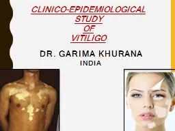
Vitiligo
Slide2Definition : acquired
skin condition characterized by
circumscribed depigmentation There is a complete loss of melanocytes from affected areas.found in all races; its prevalence may be as high as 1%; its inheritance is polygenicMale = female The patches of skin affected become white and usually have sharp margins. Typically both sides of the body are affected. Often the patches begin on areas of skin that are exposed to the sun.
Slide3causes
The etiology is unknown, but is thought to be multifactorial •Genetic predisposition •Autoimmune destruction of melanocytes •Oxidative stress (free radicals)•Intrinsic defects of melanocytes •May be triggered by stress or skin injury (e.g., sunburn)
Classification according to location
Generalized
(most common): widespread distribution of lesions, frequently with mucosal involvement
Localized
: isolated area affected (e.g.,
dermatomal
)
Universal
: Almost the entire body is affected.
Slide41)Generalized vitiligo
(non-segmental
vitiligo ) : More common .Usually starts at the second decade.There’s family history in 30% of patients .most frequent in those with autoimmune diseases such as diabetes, thyroid disorders and pernicious anemiaPathophysiology : It’s thought that , in this type , melanocytes are the target of a cell-mediated autoimmune attack or self destruct as a result of an inability to remove toxic melanin precursors.In Caucasoids the surrounding skin is sometimes partially depigmented or hyperpigmented (trichrome vitiligo).
Divided into two main patterns:
Slide5Clinical course : the sharply defined, usually symmetrical , white patches are especially common on the backs of the hands, wrists, fronts of knees, neck and around body orifices
. The
hair of the scalp and beard may depigment too.The course is unpredictable. Lesions may remain static or spread, sometimes following minor trauma (K¨obner phenomenon); occasionally, they repigment spontaneously from the hairfolliclesLeukotrichia: depigmented hair
Slide6Rare .
restricted to
one part of the body, but not necessarily to a dermatome.It occurs earlier in life than generalized vitiligo less likely to be associated with autoimmune diseases.Clinical course : The individual areas look like the generalized type but their segmental distribution is striking (unilateral ). it responds poorly to most treatments, although spontaneous repigmentation occurs more often in this type than in generalized vitiligo .*Note : Trauma and sun burn can precipitate both generalized and segmental vitiligo
.
2)Segmental
vitiligo
:
Slide7Slide8Differantial diagnosis :
Contact with
depigmenting chemicals, such as hydroquinones and substituted phenols in the rubber industry, should be excluded.Pityriasis alba: A common hypopigmented scaly patch seen in sun-exposed areas, esp. in children (resolves spontaneously or with topical steroids).Pityriasis versicolor must be considered; its fine scaling and less complete pigment loss separate it from vitiligo.Post-inflammatory depigmentation may look very like vitiligo but is less white and improves spontaneously.tropical diseases
that cause patchy hypopigmentation like leishmaniasis and
pinta
Piebaldism
are present at
birth….
rare autosomal dominant disorder of melanocyte development.
Leprosy
Cont segmental
vitiligo
:
Slide9Usually a clinical diagnosisIf diagnosis is uncertain:Wood's lamp examination: The
vitiligo
lesions appear as well defined blue-white areas.Dermoscopy: Vitiligo lesions have a characteristic perilesional hyperpigmentation and telangiectasia.Skin biopsy and histology: Melanocytes are absent, perilesional lymphocytes may be observed.Serological markers of autoimmune disease (e.g thyroid function tests and anti-thyroid antibodies) once vitiligo is confirmedDiagnosis
Slide10Treatment :
Slide11Treatment is
unsatisfactory
Sun avoidance White peopleBest left untreated in mostCamouflage preparations (dihydroxyacetone)Black patients with extensive vitiligo can be completely and irreversibly depigmented by creams containing the monobenzyl ether of hydroquinoneRecent patches may respond to a potent or very potent topical corticosteroidcalcineurin inhibitors (ointment )Psoralens Narrowband
UVB is also effective
antioxidant
Irradiating
skin
Where pigment is absent in hair follicles or in skin without hair follicles,
autologous skin grafts
can be performed.
Melanocyte and stem cell transplants
.
It’s
advice about suitable camouflage
preparations (tanning
lotions and makeups ) to cover unsightly patches should be given.
Slide12<<Thank you for listening >>