
Produce movement Maintain posture Stabilize joints Generate heat The Muscular System Muscles are responsible for all types of body movement Three basic muscle types are found in the body Skeletal muscle ID: 920255
Download Presentation The PPT/PDF document "The Muscular System Function of Muscles" is the property of its rightful owner. Permission is granted to download and print the materials on this web site for personal, non-commercial use only, and to display it on your personal computer provided you do not modify the materials and that you retain all copyright notices contained in the materials. By downloading content from our website, you accept the terms of this agreement.


Slide1
The Muscular System
Slide2Function of Muscles
Produce movement
Maintain postureStabilize jointsGenerate heat
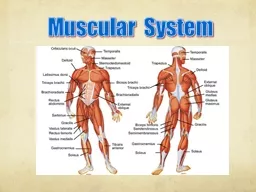
Slide3The Muscular System
Muscles are responsible for all types of body movement
Three basic muscle types are found in the bodySkeletal muscleCardiac muscle
Smooth muscle
Slide4Characteristics of Muscles
Muscle cells are elongated
(muscle cell = muscle fiber)Contraction of muscles is due to the movement of microfilaments
All muscles share some terminology
Prefix
myo
refers to muscle
Prefix
mys
refers to muscle
Prefix
sarco
refers to flesh
Slide5Skeletal Muscle Characteristics
Most are attached by tendons to bones
Cells are multinucleateStriated – have visible bandingVoluntary – subject to conscious controlCells are surrounded and bundled by connective tissue
Slide6Connective Tissue Wrappings of Skeletal Muscle
Endomysium – around single muscle fiber
Perimysium – around a fascicle (bundle) of fibers
Figure 6.1
Slide7Connective Tissue Wrappings of Skeletal Muscle
Epimysium – covers the entire skeletal muscle
Fascia – on the outside of the epimysium
Figure 6.1
Slide8Skeletal Muscle Attachments
Epimysium
blends into a connective tissue attachment
Tendon – cord-like structure
Aponeuroses
– sheet-like structure
Sites of muscle attachment
Bones
Cartilages
Connective tissue coverings
Slide9Smooth Muscle Characteristics
Has no striations
Spindle-shaped cellsSingle nucleus
Involuntary – no conscious control
Found mainly in the walls of hollow organs
Figure 6.2a
Slide10Cardiac Muscle Characteristics
Has striations
Usually has a single nucleusJoined to another muscle cell at an intercalated disc
Involuntary
Found only in the heart
Figure 6.2b
Slide11Naming of Skeletal Muscles
Direction of muscle fibers
Example: rectus (straight)
Relative size of the muscle
Example:
maximus
(largest)
Slide12Naming of Skeletal Muscles
Location of the muscle
Example: many muscles are named for bones (e.g., temporalis)
Number of origins
Example:
triceps
(three heads
)
Origin: attachment to bone that does NOT move
Insertion: attachment to bone that MOVES
Slide13Naming of Skeletal Muscles
Location of the muscle’s origin and insertion
Example: sterno
(on the sternum)
Shape of the muscle
Example:
deltoid
(triangular
)
Trapezius (trapezoid shaped)
Action of the muscle
Example:
flexor and extensor (flexes or extends a bone)
e
Slide14Microscopic Anatomy of Skeletal Muscle
Cells are multinucleate
Nuclei are just beneath the sarcolemma
Figure 6.3a
Slide15Microscopic Anatomy of Skeletal Muscle
Sarcolemma – specialized plasma membrane
Sarcoplasmic reticulum – specialized smooth endoplasmic reticulum
Figure 6.3a
Slide16Figure 6.3b
Microscopic Anatomy of Skeletal Muscle
Myofibril
Bundles of myofilaments
Myofibrils are aligned to give distinct bands
I band =
light band
A band =
dark band
Slide17Microscopic Anatomy of Skeletal Muscle
Sarcomere
Contractile unit of a muscle fiber
Figure 6.3b
Slide18Microscopic Anatomy of Skeletal Muscle
Organization of the sarcomere
Thick filaments = myosin filamentsComposed of the protein myosinHas ATPase enzymes
Figure 6.3c
Slide19Microscopic Anatomy of Skeletal Muscle
Organization of the sarcomere
Thin filaments = actin filamentsComposed of the protein actin
Figure 6.3c
Slide20Microscopic Anatomy of Skeletal Muscle
Myosin filaments have heads (extensions, or cross bridges)
Myosin and actin overlap somewhat
Figure 6.3d
Slide21Microscopic Anatomy of Skeletal Muscle
At rest, there is a bare zone that lacks actin filaments
Sarcoplasmic reticulum (SR) – for storage of
calcium
Figure 6.3d
Slide22Properties of Skeletal Muscle Activity
Irritability – ability to receive and respond to a stimulus
Contractility – ability to shorten when an adequate stimulus is received
Animation of muscle contraction
Slide23Nerve Stimulus to Muscles
Skeletal muscles must be stimulated by a nerve to contract
Motor unitOne neuronMuscle cells stimulated by that neuron
Figure 6.4a
Slide24Nerve Stimulus to Muscles
Neuromuscular junctions – association site of nerve and muscle
Figure 6.5b
Slide25Nerve Stimulus to Muscles
Synaptic cleft – gap between nerve and muscle
Nerve and muscle do not make contactArea between nerve and muscle is filled with interstitial fluid
Figure 6.5b
Slide26Transmission of Nerve Impulse to Muscle
Neurotransmitter – chemical released by nerve upon arrival of nerve impulse
The neurotransmitter for skeletal muscle is acetylcholineNeurotransmitter attaches to receptors on the sarcolemmaSarcolemma becomes permeable to sodium (Na
+
)
Slide27Transmission of Nerve Impulse to Muscle
Sodium rushing into the cell generates an action potential
Once started, muscle contraction cannot be stopped
Slide28The Sliding Filament Theory of Muscle Contraction
Activation by nerve causes myosin heads (crossbridges) to attach to binding sites on the thin filament
Myosin heads then bind to the next site of the thin filament
Figure 6.7
Slide29The Sliding Filament Theory of Muscle Contraction
This continued action causes a sliding of the myosin along the actin
The result is that the muscle is shortened (contracted)
Figure 6.7