

The production of murmurs is due to 3 main factors high blood flow rate through normal or abnormal orifices forward flow through a narrowed or irregular orifice into a dilated vessel ID: 946583
Download Pdf The PPT/PDF document "Heart murmurs" is the property of its rightful owner. Permission is granted to download and print the materials on this web site for personal, non-commercial use only, and to display it on your personal computer provided you do not modify the materials and that you retain all copyright notices contained in the materials. By downloading content from our website, you accept the terms of this agreement.

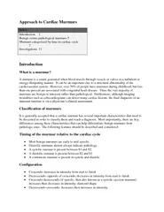


Heart murmurs ï The production of murmurs is due to 3 main factors: ï high blood flow rate through normal or abnormal orifices ï forward flow through a narrowed or irregular orifice into a dilated
vessel or chamber ï backward or regurgitant flow through an incompetent valve, septal defect, or patent ductus arteriosus Heart murmurs ï Intensity grading: ï 1 is so faint that it is heard only wi
th special effort ï 2 is soft but readily detected ï 3 is prominent but not loud ï 4 is loud (and usually palpable) ï 5 is very loud ï 6 murmur is loud enough to be heard with the stethoscope
just removed from contact with the chest wall Heart murmurs ï Systolic murmur begins with or after the first heart sound and ends at or before the subsequent second heart sound ï Diastolic murmur beg
ins with or after the second heart sound and ends before the subsequent first heart sound ï Continuous murmur begins in systole and continues without interruption through the timing of the second heart so
und into all or part of diastole Heart murmurs ï Most systolic heart murmurs do not signify cardiac disease, and many are related to physiological increases in blood flow velocity ï Diastolic and c
ontinuous murmurs virtually always represent pathological conditions and require further cardiac evaluation Aortic stenosis (AS) â etiology Aortic stenosis ï normal adult valve orifice is 3.0 t
o 4.0 cm 2 ï aortic valve area must be reduced to ¼ its normal size before significant changes in the circulation occur ï Classification ï mild: area � 1.5cm 2 ï moderate: area 1.0 to 1
.5 cm 2 ï severe: area cm 2 ï In severe stenosis the mean transvalvular pressure gradient is generally �50 mmHg Aortic stenosis Aortic stenosis AS - pathophysiology AS â c
linical manifestations ï a long latent, asymptomatic period ï cardinal symptoms: angina pectoris, syncope, heart failure ï the arterial pulse rises slowly and is small and sustained (pulsus parvus et
tardus) ï t he systolic murmur of AS ï late - peaking ï heard best at the base of the heart ï often well transmitted along the carotid vessels and to the apex AS â natural history ï
asymptomatic patients have an excellent prognosis ⢠once patients with AS become symptomatic with angina or syncope, the average survival is 2 to 3 years, whereas with congestive heart failure it i
s 1.5 years AS - treatment ï aortic valve replacement ï transcatheter aortic valve implantation ï b alloon a ortic v alvuloplasty AS - treatment Aortic Regurgitation (AR) - eti
ology ï Valvular disease ï Rheumatic fever ï Concomitant with aortic stenosis ï Infective endocarditis ï Bicuspid valve ï Aortic root disease ï Age related aortic dilatation ï Marfa
n syndrome ï Aortic dissection Clinical manifestations â h istory of AR ï m ost patients remain asymptomatic ï e xertional dyspnea ï o rthopnea and paroxysmal nocturnal dyspnea ï a ng
ina pectoris â the late sign ï âUncomfortable awarness of the heartbeatâ Clinical manifestations â physical examination ï systolic blood pressure ï diastolic blood pressure ï
hyperdynamic, diffuse apical impulse ï diastolic murmur Clinical manifestations â physical examination ï De Musset sign - head nodding in time with the heart beat ï Corrigan pulse (water
- hammer) - rapid upstroke and collapse of the carotid artery pulse ï Muller sign - pulsations of uvula AR - workup Prognosis ï Relatively good prognosis (asymptomatic patients with mode
rate severe AR): ï 75% survive 5 years ï 50% survive 10 years ï In symptomatic patients ï Angina pectoris â expected survival: 4 years ï Heart failure â expected survival: 2 years T
reatment ï Aortic valve replacement ï Correction of dilated aortic root Mitral stenosis (MS) â etiology ï RHEUMATIC FEVER !!! ï Congenital MS â Patophysiology ï Normal mitra
l orifice: 4 - 6cm 2 ï 2cm 2 â mild MS ï 1cm 2 â critical MS - � pressure of 25mmHg in left atrium is required to maintain normal cardiac output exertional dyspnea RR in pulmonary v
eins RR in LA pulmonary hypertension signs of RV failure MS â history ï exertional dyspnea ï risk of pulmonary edema ï hemoptysis ï angina - like c hest pain ï s ystemic embolisat
ion (mainly in patients with atrial fibrillation) MS â physical examination ï m itral facies (pinkish - purple patches on the cheeks) ï a ccentation of S1 ï d iastolic murmur, best heard at th
e apex, radiating to the left axilla ï RV failure Treatment ï m edical â anticoagulant therapy ï v alvotomy ï Surgical ï Baloon mitral valvuloplasty ï m itral valve replacement
Treatment â anticoagulant therapy Anticoagulant therapy with a target INR in the upper half of the range 2 to 3 is indicated inpatients with either permanent or paroxysmal AF . In patients with sinu
s rhythm, anticoagulation is indicated when there has been prior embolism, or a thrombus is present in the left atrium (recommendation class I, level of evidence C) and should also be considered when TOE shows
dense spontaneous echo contrast or an enlarged left atrium (M - mode diameter 50 mm or LA volume 60 ml/m2 (recommendation class IIa, level of evidence C) Mitral regurgitation (MR) - etiology ï chro
nic rheumatic heart disease ï secondary to dilation of LV ï degenerative calcification of the mitral valves ï dysfunction of the papillary muscles MR â history ï chronic weakness and fatig
ue ( secondary to a low cardiac output ) are more prominent features in MR ï History is like in MS, but less dramatic ï acute pulmonary edema occurs less frequently ï hemoptysis and systemic emb
olisation are less common ï t he development of atrial fibrillation affects the course adversely but not as dramatically as it does in MS MR â physical examination ï holosystolic murmur: ï
usually constant in intensity ï b lowing ï loudest at the apex with radiation to the axilla MR â Treatment ï the reconstructive procedure s ï annuloplasty (with the use of a rigid or
a flexible prosthetic ring ) ï reconstruction of the valve ï repair of the subvalvular apparatus: replacement, reimplantation, elongation or shortening of chordae tendineae, splitting of the papillary
muscle ï mitral valve replacement Mitral valve prolapse (MVP) ï Affects 3 to 5% of the population ï Usually a primary condition ï A large majority of patients are asymptomatic ï Patients ma
y complain of syncope, presyncope, palpitations, chest discomfort ï The auscultatory finding : systolic click MVP ï Echocardiography confirms the diagnosis ï Sometimes coexist mild MR ï Progres
sive MR is the most frequent serious complication (10 - 15%) ï Asymptomatic patients without evidence of MR have an excellent prognosis ï Patients with MVP and severe MR should be treated as are