/
Arsenic-Induces Dysfunction in Poly(ADP-Ribose) Polymerase-1: Arsenic-Induces Dysfunction in Poly(ADP-Ribose) Polymerase-1:

Arsenic-Induces Dysfunction in Poly(ADP-Ribose) Polymerase-1: - PowerPoint Presentation

holly
holly . @holly
Follow
64 views
Uploaded On 2024-01-03

Arsenic-Induces Dysfunction in Poly(ADP-Ribose) Polymerase-1: - PPT Presentation

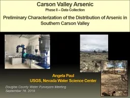
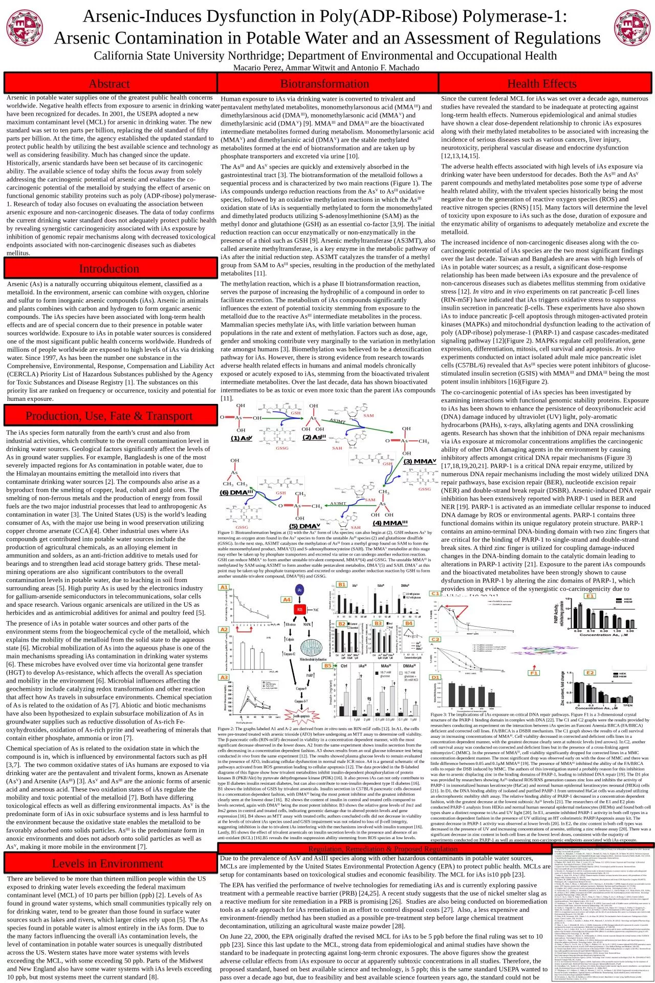
Arsenic Contamination in Potable Water and an Assessment of Regulations California State University Northridge Department of Environmental and Occupational Health Macario Perez Ammar Witwit and Antonio F ID: 1037663

arsenic ias amp water ias arsenic water amp parp levels exposure dna species cells cell repair health drinking asiii

Share:

Link:

Embed:

Download Presentation from below link

Download Presentation The PPT/PDF document "Arsenic-Induces Dysfunction in Poly(ADP-..." is the property of its rightful owner. Permission is granted to download and print the materials on this web site for personal, non-commercial use only, and to display it on your personal computer provided you do not modify the materials and that you retain all copyright notices contained in the materials. By downloading content from our website, you accept the terms of this agreement.


Presentation Transcript

1. Arsenic-Induces Dysfunction in Poly(ADP-Ribose) Polymerase-1: Arsenic Contamination in Potable Water and an Assessment of RegulationsCalifornia State University Northridge; Department of Environmental and Occupational Health Macario Perez, Ammar Witwit and Antonio F. Machado Abstract Biotransformation Regulation, Remediation & Proposed Regulation Introduction Production, Use, Fate & Transport Levels in Environment Health Effects ReferencesHuman exposure to iAs via drinking water is converted to trivalent and pentavalent methylated metabolites, monomethylarsonous acid (MMAIII) and dimethylarsinous acid (DMAIII), monomethylarsonic acid (MMAV) and dimethylarsinic acid (DMAV) [9]. MMAIII and DMAIII are the bioactivated intermediate metabolites formed during metabolism. Monomethylarsonic acid (MMAV) and dimethylarsinic acid (DMAV) are the stable methylated metabolites formed at the end of biotransformation and are taken up by phosphate transporters and excreted via urine [10]. The AsIII and AsV species are quickly and extensively absorbed in the gastrointestinal tract [3]. The biotransformation of the metalloid follows a sequential process and is characterized by two main reactions (Figure 1). The iAs compounds undergo reduction reactions from the AsV to AsIII oxidative species, followed by an oxidative methylation reactions in which the AsIII oxidation state of iAs is sequentially methylated to form the monomethylated and dimethylated products utilizing S-adenosylmethionine (SAM) as the methyl donor and glutathione (GSH) as an essential co-factor [3,9]. The initial reduction reaction can occur enzymatically or non-enzymatically in the presence of a thiol such as GSH [9]. Arsenic methyltransferase (AS3MT), also called arsenite methyltransferase, is a key enzyme in the metabolic pathway of iAs after the initial reduction step. AS3MT catalyzes the transfer of a methyl group from SAM to AsIII species, resulting in the production of the methylated metabolites [11]. The methylation reaction, which is a phase II biotransformation reaction, serves the purpose of increasing the hydrophilic of a compound in order to facilitate excretion. The metabolism of iAs compounds significantly influences the extent of potential toxicity stemming from exposure to the metalloid due to the reactive AsIII intermediate metabolites in the process. Mammalian species methylate iAs, with little variation between human populations in the rate and extent of methylation. Factors such as dose, age, gender and smoking contribute very marginally to the variation in methylation rate amongst humans [3]. Biomethylation was believed to be a detoxification pathway for iAs. However, there is strong evidence from research towards adverse health related effects in humans and animal models chronically exposed or acutely exposed to iAs, stemming from the bioactivated trivalent intermediate metabolites. Over the last decade, data has shown bioactivated intermediates to be as toxic or even more toxic than the parent iAs compounds [11]. 1. Agency for Toxic Substances and Disease Registry. (2014). CERCLA Priority List of Hazardous Substances for 2013. Retrieved from http://www.atsdr.cdc.gov/spl/2. Gardner, R., Hamadani, J., Grander, M., Tofail, F., Nermell, B., Palm, B., Kippler, M., & Vahter, M. (2011). Persistent exposure to arsenic via drinking water in rural Bangladesh despite major mitigation efforts. American Journal of Public Health, 101, S333-8. 3. World Health Organization. (2001). Arsenic and Arsenic Compounds. Retrieved from http://www.inchem.org/documents/ehc/ehc/ehc224.htm.4. Hughes, M.F., Beck, B.D., Chen, Y., Lewis, A.S., & Thomas, D.J. (2011). Arsenic Exposure and Toxicology: A Historical Perspective. Toxicological Sciences, 123(2), 305-332.5. Agency for Toxic Substances and Disease Registry. (2014). Toxicological profile for arsenic. Retrieved from http://www.atsdr.cdc.gov/ToxProfiles/tp2.pdf6. Davolos, D., Pietrangeli, B. (2013). A molecular study on bacterial resistance to arsenic-toxicity in surface and underground waters of Latium (Italy). Ecotoxicology and Environmental Safety, 96, 1-9. 7. Sarkar, A., Kazy, S.K., & Sar, P. (2012). Characterization of arsenic resistant bacteria from arsenic rich groundwater of West Bengal, India. Ecotoxicology, 22, 363-376. 8. Tiemann, M. (2008). Arsenic in drinking water: Regulatory developments and issues (CRS Report for Congress RS20672). Washington, DC: Congressional Research Service.9. Nollen, M., Ebert, F., Moser, J., Mullenders, L.H.F., Hartwig, A., & Scherdtle, T. (2009). Impact of arsenic on nucleotide excision repair: XPC function, protein level, and gene expression. Molecular Nutrition and Food Research, 53, 572-582.10. Hughes, M.F. (2002). Arsenic toxicity and potential mechanisms of action. Toxicological Letters, 133, 1-16.11. Valenzuela, O.L., Drobna, Z., Hernandez-Castellanos, E., Sanchez-Pena, L.C., Garcia-Vargas, G.G., Borja-Aburto, V.H., Styblo, M., & Razo, L.M.D. (2009). Association of AS3MT polymorphisms and the risk of premalignant arsenic skin lesions. Toxicology and Applied Pharmacology, 239(2), 200-207. 12. Lu, T., Su, C., Chen, Y., Yang, C., Wu, C., Hung, D., Chen, C., Cheng, P., Liu, S., & Huang, C. (2011). Arsenic induces pancreatic β-cell apoptosis via the oxidative stress-regulated mitochondria-dependent and endoplasmic reticulum stress-triggered signaling pathways. Toxicological Letters, 201, 15-26.13. Steinmaus, C., Yuan, Y., Bates, M.N., & Smith, A.H. (2003). Case-control study of bladder cancer and drinking water arsenic in the western United States. American Journal of Epidemiology, 158, 1193-1201.14. Surdu, S., Fitzgerald, E.F., Bloom, M.S., Boscoe, F.P., Carpenter, D.O., Haase, R.F., Gurzau, E., Rudnai, P., Koppova, K., Vahter, M., Leonardi, G., Goessler, W., Kumar, R., & Fletcher, T. (2014). Polymorphisms in DNA repair genes XRCC1 and XRCC3, occupational exposure to arsenic and sunlight, and the risk of non-melanoma skin cancer in a European case-control study. Environmental Research, 134, 382-389.15. Hunt, K.M., Srivastava, R.K., Elmets, C.A., & Athar, M. (2014). The mechanistic basis of arsenicosis: Pathogenesis of skin cancer. Cancer Letters, 354, 211-219.16. Douillet, C., Currier, J., Saunder, J., Bodnar, W.M., Matousek, T., & Styblo, M. (2012). Toxicology and Applied Pharmacology, 267(1), 11-15. 17. Shen, S., Lee, J., Weinfeld, M., & Le, C. (2008). Attenuation of DNA damage-induced p53 expression by arsenic: A possible mechanism for arsenic co-carcinogenesis. Molecular Carcinogenesis, 47, 508-518.18. Shen, S., Lee, J., Cullen, W.R., Le, X.C., & Weinfeld, M. (2009). Arsenite and its mono- and dimethylated trivalent metabolites enhance the formation of benzo[a]pyrene diol epoxide-DNA adducts in xeroderma pigmentosum complementation group A cells. Chemical Research in Toxicology, 22, 382-390.19. Peremarti, J., Ramos, F., Marcos, R., & Hernandez, A. (2014). Arsenic exposure disrupts the normal function of the FA/BRCA repair pathway. Toxicological Sciences, 142(1), 93-104.20. Cooper, K.L., Yager, J.W., & Hudson, L.G. (2014). Melanocytes and keratinocytes have distinct and shared responses to ultraviolet radiation and arsenic. Toxicology Letters, 224, 407-415.21. Wang, F., Zhou, X., Liu, W., Sun, X., Chen, C., Hudson, L.G., & Liu, K.J. (2013). Arsenite-induced ROS/RNS generation causes zinc loss and inhibits the activity of poly (ADP-ribose) polymerase-1. Free Radical Biology and Medicine, 249-256.22. Protein Data Bank. (2012). Crystal structure of the human PARP-1 DNA binding domain in complex with DNA. Retrieved from http://pdb.org/pdb/results/results.do?qrid=6B3341F8&tabtoshow=Current23. United States Environmental Protection Agency. (2014). Arsenic Rule. Retrieved from http://water.epa.gov/lawsregs/rulesregs/sdwa/arsenic/regulations.cfm. 24. U.S. Environmental Protection Agency. (2014). Technology brief: Arsenic treatment technologies (Pub. No. EPA/600/S-07/007). Research Triangle Park, NC: Author.25. U.S. Environmental Protection Agency. (2014). Application of the permeable reactive barrier technology for the treatment of Arsenic in ground water. Retrieved from http://www.epa.gov /ada/gw/pdfs/research_07.pdf 26. Chowdhury, S.R., Yanful, E.K., & Pratt, A.R. (2014). Recycling of nickel smelter slag for arsenic remediation—an experimental study. Environmental Science and Pollution Research, 21, 10096-10107.27. Villadangos, A.F., Ordonez, E., Pedre, B., Messens, J., Gil, J.A., & Mateos, L.M. (2014). Engineered coryneform bacteria as a bio-tool for arsenic remediation. Applied Genetics and Molecular Biotechnology, (Epub ahead of print), retrieved from http://www.ncbi.nlm.nih.gov/pubmed/25208910.28. Srivastava, S., Raj, K.R., & Kardam, A. (2013). Efficient arsenic depollution in water using modified maize powder. Environmental Chemistry Letters, 11, 47-53. Figure 1: Biotransformation begins at (1) with the AsV form of iAs species; can also begin at (2). GSH reduces AsV by removing an oxygen atom found in the AsV species to form the unstable AsIII species (2) and glutathione disulfide (GSSG). In the next step, AS3MT catalyzes the methylation of AsIII from a methyl group found on SAM to form the stable monomethylated product, MMAV(3) and S-adenosylhomocysteine (SAH). The MMAV metabolite at this stage may either be taken up by phosphate transporters and excreted via urine or can undergo another reduction reaction. GSH can reduce MMAV to form another unstable trivalent compound, MMAIII(4) and GSSG. The unstable MMAIII is methylated by SAM using AS3MT to form another stable pentavalent metabolite, DMAV(5) and SAH. DMAV at this point may be taken up by phosphate transporters and excreted or undergo another reduction reaction by GSH to form another unstable trivalent compound, DMAIII(6) and GSSG. Arsenic (As) is a naturally occurring ubiquitous element, classified as a metalloid. In the environment, arsenic can combine with oxygen, chlorine and sulfur to form inorganic arsenic compounds (iAs). Arsenic in animals and plants combines with carbon and hydrogen to form organic arsenic compounds. The iAs species have been associated with long-term health effects and are of special concern due to their presence in potable water sources worldwide. Exposure to iAs in potable water sources is considered one of the most significant public health concerns worldwide. Hundreds of millions of people worldwide are exposed to high levels of iAs via drinking water. Since 1997, As has been the number one substance in the Comprehensive, Environmental, Response, Compensation and Liability Act (CERCLA) Priority List of Hazardous Substances published by the Agency for Toxic Substances and Disease Registry [1]. The substances on this priority list are ranked on frequency or occurrence, toxicity and potential for human exposure.The iAs species form naturally from the earth’s crust and also from industrial activities, which contribute to the overall contamination level in drinking water sources. Geological factors significantly affect the levels of As in ground water supplies. For example, Bangladesh is one of the most severely impacted regions for As contamination in potable water, due to the Himalayan mountains emitting the metalloid into rivers that contaminate drinking water sources [2]. The compounds also arise as a byproduct from the smelting of copper, lead, cobalt and gold ores. The smelting of non-ferrous metals and the production of energy from fossil fuels are the two major industrial processes that lead to anthropogenic As contamination in water [3]. The United States (US) is the world’s leading consumer of As, with the major use being in wood preservation utilizing copper chrome arsenate (CCA)[4]. Other industrial uses where iAs compounds get contributed into potable water sources include the production of agricultural chemicals, as an alloying element in ammunition and solders, as an anti-friction additive to metals used for bearings and to strengthen lead acid storage battery grids. These metal-mining operations are also significant contributors to the overall contamination levels in potable water, due to leaching in soil from surrounding areas [5]. High purity As is used by the electronics industry for gallium-arsenide semiconductors in telecommunications, solar cells and space research. Various organic arsenicals are utilized in the US as herbicides and as antimicrobial additives for animal and poultry feed [5]. The presence of iAs in potable water sources and other parts of the environment stems from the biogeochemical cycle of the metalloid, which explains the mobility of the metalloid from the solid state to the aqueous state [6]. Microbial mobilization of As into the aqueous phase is one of the main mechanisms spreading iAs contamination in drinking water systems [6]. These microbes have evolved over time via horizontal gene transfer (HGT) to develop As-resistance, which affects the overall As speciation and mobility in the environment [6]. Microbial influences affecting the geochemistry include catalyzing redox transformation and other reaction that affect how As travels in subsurface environments. Chemical speciation of As is related to the oxidation of As [7]. Abiotic and biotic mechanisms have also been hypothesized to explain subsurface mobilization of As in groundwater supplies such as reductive dissolution of As-rich Fe-oxyhydroxides, oxidation of As-rich pyrite and weathering of minerals that contain either phosphate, ammonia or iron [7]. Chemical speciation of As is related to the oxidation state in which the compound is in, which is influenced by environmental factors such as pH [3,7]. The two common oxidative states of iAs humans are exposed to via drinking water are the pentavalent and trivalent forms, known as Arsenate (AsV) and Arsenite (AsIII) [3]. AsV and AsIII are the anionic forms of arsenic acid and arsenous acid. These two oxidation states of iAs regulate the mobility and toxic potential of the metalloid [7]. Both have differing toxicological effects as well as differing environmental impacts. AsV is the predominate form of iAs in oxic subsurface systems and is less harmful to the environment because the oxidative state enables the metalloid to be favorably adsorbed onto solids particles. AsIII is the predominate form in anoxic environments and does not adsorb onto solid particles as well as AsV, making it more mobile in the environment [7]. Arsenic in potable water supplies one of the greatest public health concerns worldwide. Negative health effects from exposure to arsenic in drinking water have been recognized for decades. In 2001, the USEPA adopted a new maximum contaminant level (MCL) for arsenic in drinking water. The new standard was set to ten parts per billion, replacing the old standard of fifty parts per billion. At the time, the agency established the updated standard to protect public health by utilizing the best available science and technology as well as considering feasibility. Much has changed since the update. Historically, arsenic standards have been set because of its carcinogenic ability. The available science of today shifts the focus away from solely addressing the carcinogenic potential of arsenic and evaluates the co-carcinogenic potential of the metalloid by studying the effect of arsenic on functional genomic stability proteins such as poly (ADP-ribose) polymerase-1. Research of today also focuses on evaluating the association between arsenic exposure and non-carcinogenic diseases. The data of today confirms the current drinking water standard does not adequately protect public health by revealing synergistic carcinogenicity associated with iAs exposure by inhibition of genomic repair mechanisms along with decreased toxicological endpoints associated with non-carcinogenic diseases such as diabetes mellitus. There are believed to be more than thirteen million people within the US exposed to drinking water levels exceeding the federal maximum contaminant level (MCL) of 10 parts per billion (ppb) [2]. Levels of As found in ground water systems, which small communities typically rely on for drinking water, tend to be greater than those found in surface water sources such as lakes and rivers, which larger cities rely upon [5]. The As species found in potable water is almost entirely in the iAs form. Due to the many factors influencing the overall iAs contamination levels, the level of contamination in potable water sources is unequally distributed across the US. Western states have more water systems with levels exceeding the MCL, with some exceeding 50 ppb. Parts of the Midwest and New England also have some water systems with iAs levels exceeding 10 ppb, but most systems meet the current standard [8]. Due to the prevalence of AsV and AsIII species along with other hazardous contaminants in potable water sources, MCLs are implemented by the United States Environmental Protection Agency (EPA) to protect public health. MCLs are setup for contaminants based on toxicological studies and economic feasibility. The MCL for iAs is10 ppb [23].The EPA has verified the performance of twelve technologies for remediating iAs and is currently exploring passive treatment with a permeable reactive barrier (PRB) [24,25]. A recent study suggests that the use of nickel smelter slag as a reactive medium for site remediation in a PRB is promising [26]. Studies are also being conducted on bioremediation tools as a safe approach for iAs remediation in an effort to control disposal costs [27]. Also, a less expensive and environment-friendly method has been studied as a possible pre-treatment step before large chemical treatment decontamination, utilizing an agricultural waste maize powder [28].On June 22, 2000, the EPA originally drafted the revised MCL for iAs to be 5 ppb before the final ruling was set to 10 ppb [23]. Since this last update to the MCL, strong data from epidemiological and animal studies have shown the standard to be inadequate in protecting against long-term chronic exposures. The above figures show the greatest adverse cellular effects from iAs exposure to occur at apparently subtoxic concentrations in all studies. Therefore, the proposed standard, based on best available science and technology, is 5 ppb; this is the same standard USEPA wanted to pass over a decade ago but, due to feasibility and best available science fourteen years ago, the standard could not be met. Since the current federal MCL for iAs was set over a decade ago, numerous studies have revealed the standard to be inadequate at protecting against long-term health effects. Numerous epidemiological and animal studies have shown a clear dose-dependent relationship to chronic iAs exposures along with their methylated metabolites to be associated with increasing the incidence of serious diseases such as various cancers, liver injury, neurotoxicity, peripheral vascular disease and endocrine dysfunction [12,13,14,15].The adverse health effects associated with high levels of iAs exposure via drinking water have been understood for decades. Both the AsIII and AsV parent compounds and methylated metabolites pose some type of adverse health related ability, with the trivalent species historically being the most negative due to the generation of reactive oxygen species (ROS) and reactive nitrogen species (RNS) [15]. Many factors will determine the level of toxicity upon exposure to iAs such as the dose, duration of exposure and the enzymatic ability of organisms to adequately metabolize and excrete the metalloid. The increased incidence of non-carcinogenic diseases along with the co-carcinogenic potential of iAs species are the two most significant findings over the last decade. Taiwan and Bangladesh are areas with high levels of iAs in potable water sources; as a result, a significant dose-response relationship has been made between iAs exposure and the prevalence of non-cancerous diseases such as diabetes mellitus stemming from oxidative stress [12]. In vitro and in vivo experiments on rat pancreatic β-cell lines (RIN-m5F) have indicated that iAs triggers oxidative stress to suppress insulin secretion in pancreatic β-cells. These experiments have also shown iAs to induce pancreatic β-cell apoptosis through mitogen-activated protein kinases (MAPKs) and mitochondrial dysfunction leading to the activation of poly (ADP-ribose) polymerase-1 (PARP-1) and caspase cascades-meditated signaling pathway [12](Figure 2). MAPKs regulate cell proliferation, gene expression, differentiation, mitosis, cell survival and apoptosis. In vivo experiments conducted on intact isolated adult male mice pancreatic islet cells (C57BL/6) revealed that AsIII species were potent inhibitors of glucose-stimulated insulin secretion (GSIS) with MMAIII and DMAIII being the most potent insulin inhibitors [16](Figure 2).The co-carcinogenic potential of iAs species has been investigated by examining interactions with functional genomic stability proteins. Exposure to iAs has been shown to enhance the persistence of deoxyribonucleic acid (DNA) damage induced by ultraviolet (UV) light, poly-aromatic hydrocarbons (PAHs), x-rays, alkylating agents and DNA crosslinking agents. Research has shown that the inhibition of DNA repair mechanisms via iAs exposure at micromolar concentrations amplifies the carcinogenic ability of other DNA damaging agents in the environment by causing inhibitory affects amongst critical DNA repair mechanisms (Figure 3) [17,18,19,20,21]. PARP-1 is a critical DNA repair enzyme, utilized by numerous DNA repair mechanisms including the most widely utilized DNA repair pathways, base excision repair (BER), nucleotide excision repair (NER) and double-strand break repair (DSBR). Arsenic-induced DNA repair inhibition has been extensively reported with PARP-1 used in BER and NER [19]. PARP-1 is activated as an immediate cellular response to induced DNA damage by ROS or environmental agents. PARP-1 contains three functional domains within its unique regulatory protein structure. PARP-1 contains an amino-terminal DNA-binding domain with two zinc fingers that are critical for the binding of PARP-1 to single-strand and double-strand break sites. A third zinc finger is utilized for coupling damage-induced changes in the DNA-binding domain to the catalytic domain leading to alterations in PARP-1 activity [21]. Exposure to the parent iAs compounds and the bioactivated metabolites have been strongly shown to cause dysfunction in PARP-1 by altering the zinc domains of PARP-1, which provides strong evidence of the synergistic co-carcinogenicity due to inhibition [19,20,21]. Figure 2: The graphs labeled A1 and A-2 are derived from in vitro tests on RIN-m5F cells [12]. In A1, the cells were pre-treated treated with arsenic trioxide (ATO) before undergoing an MTT assay to determine cell viability. The β-pancreatic cells (RIN-m5F) decreased in viability in a concentration dependent manner, with the most significant decrease observed in the lower doses. A2 from the same experiment shows insulin secretion from the cells decreasing in a concentration dependent fashion. A3 shows results from an oral glucose tolerance test being conducted in vivo from the same experiment [12]. The results showed plasma glucose levels to remain evaluated in the presence of ATO, indicating cellular dysfunction in normal male ICR mice. A4 is a general schematic of the pathways activated from ROS generation leading to cellular apoptosis [12]. The data provided in the B-labeled diagrams of this figure show how trivalent metabolites inhibit insulin-dependent phosphorylation of protein kinases B (PKB/Akt) by pyruvate dehydrogenase kinase (PDK) [16]. It also proves iAs can not only contribute to type II diabetes insulin resistant diabetes, but can also contribute to non-insulin dependent type II diabetes [16]. B1 shows the inhibition of GSIS by trivalent arsenicals. Insulin secretion in C57BL/6 pancreatic cells decreased in a concentration dependent fashion, with DMAIII being the most potent inhibitor and the greatest inhibition clearly seen at the lowest dose [16]. B2 shows the content of insulin in control and treated cells compared to levels secreted, again with DMAIII being the most potent inhibitor. B3 shows the relative gene levels of Ins1 and Ins2 genes in control and treated cells, indicating genomic damage due to altered protein levels and gene expression [16]. B4 shows an MTT assay with treated cells; authors concluded cells did not decrease in viability at the levels of trivalent iAs species used and GSIS impairment was not related to loss of β-cell integrity, suggesting inhibition is due to trivalent iAs interfering with the mechanisms involved with insulin transport [16]. Lastly, B5 shows the effect of trivalent arsenicals on insulin secretion levels in the presence and absence of an anti-oxidant (KCL) [16].B5 reveals the insulin suppression ability stemming from ROS species at low levels. Figure 3: The implications of iAs exposure on critical DNA repair pathways. Figure F1 is a 3-dimensional crystal structure of the PARP-1 binding domain in complex with DNA [22]. The C1 and C2 graphs were the results provided by researchers conducting an experiment on the interaction between iAs species an Fanconi Anemia BRCA (FA/BRCA) deficient and corrected cell lines. FA/BRCA is a DSBR mechanism. The C1 graph shows the results of a cell survival assay in increasing concentrations of MMAIII. Cell viability decreased in corrected and deficient cells lines in a concentration dependent manner, with the greatest decrease clearly seen at subtoxic levels (red arrows). In C2, another cell survival assay was conducted on corrected and deficient lines but in the presence of a cross-linking agent mitomycin-C (MMC). In the presence of MMAIII, cell viability significantly dropped for corrected lines in a MMC concentration dependent manner. The most significant drop was observed early on with the dose of MMC and there was little difference between 0.05 and 0.1μM MMAIII [19]. The presence of MMAIII inhibited the ability of the FA/BRCA cells to repair the DSB induced by MMC. The authors of this investigation stated a probably reason for this inhibition was due to arsenic displacing zinc in the binding domains of PARP-1, leading to inhibited DNA repair [19]. The D1 plot was provided by researchers showing AsIII-induced ROS/RNS generation causes zinc loss and inhibits the activity of PARP-1 in immortalized human keratinocyte (HaCat) and normal human epidermal keratinocytes neonatal (HEKn) cells [21]. In D1, the DNA binding ability of isolated and purified PARP-1 from untreated HaCat cells was analyzed utilizing an electrophoretic mobility shift assay. The relative binding ability of PARP-1 decreased in a concentration dependent fashion, with the greatest decrease at the lowest subtoxic AsIII levels [21]. The researchers of the E1 and E2 plots conducted PARP-1 analysis from HEKn and normal human neonatal epidermal melanocytes (HEMn) and found both cell types share a distinct response to iAs and UV light [20]. In E1, arsenite inhibited PARP-1 activity in both cell types in a concentration dependent fashion in the presence of UV utilizing an HT colorimetric PARP/Apoptosis assay kit. The greatest decrease in PARP-1 activity was observed at lower levels [20]. In E2, the zinc content in both cell types was decreased in the presence of UV and increasing concentrations of arsenite, utilizing a zinc release assay [20]. There was a significant decrease in zinc content in both cell lines at the lowest level doses, consistent with the majority of experiments conducted on PARP-1 as well as assessing non-carcinogenic endpoints associated with iAs exposure.