
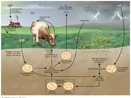
Approximately 75 are reutilized The excess nitrogen forms urea Proteins represent 1015 of total energy supply Digestion and Absorption of Proteins The α amino group of many amino acids is transferred to ID: 908444
Download Presentation The PPT/PDF document "Amino Acid Metabolism The continuous deg..." is the property of its rightful owner. Permission is granted to download and print the materials on this web site for personal, non-commercial use only, and to display it on your personal computer provided you do not modify the materials and that you retain all copyright notices contained in the materials. By downloading content from our website, you accept the terms of this agreement.


Slide1
Slide2Slide3Amino Acid Metabolism
The continuous degradation and synthesis of cellular proteins occur in all forms of life. Each day humans turn over 1–2% of their total body protein, principally muscle protein.
Approximately 75% are reutilized.
The excess nitrogen forms urea.
Proteins represent 10-15 % of total energy supply.
Slide4Digestion and Absorption of Proteins.
Slide5The
α
-amino group of many amino acids is transferred to
α
-
ketoglutarate
to form
glutamate, which is then
oxidatively deaminated to yield ammonium ion (NH4+).
Slide6Slide7Transamination
Slide8All the protein amino acids except
lysine,
threonine
,
proline
, and
hydroxyproline
participate in
transamination
.
Transamination
is readily reversible, and
aminotransferases
also function in amino acid biosynthesis.
The coenzyme
pyridoxal
phosphate
(PLP)
is present at the catalytic site of
aminotransferases
.
Slide9Aminotransferases
Aspartate aminotransferase(AST), one of the most important of these enzymes, catalyzes the transfer of the amino group of
aspartate to
α
-
ketoglutarate
Alanine aminotransferase(ALT) catalyzes the transfer of the amino group of alanine to
α
-
ketoglutarate
.
Slide10Glucose -
Alanine
Cycle
Blood
glucose
Blood
alanine
Alanine
serves as a carrier of ammonia and of the carbon skeleton of
pyruvate
from skeletal muscle to liver. The ammonia is excreted and the
pyruvate
is used to produce glucose, which is returned to the muscle
.
UREA
Slide11Oxidative
deamination
This reaction is catalyzed by
glutamate
dehydrogenase
. This enzyme is unusual in being
able to utilize either NAD+ or NADP+.
Slide12Peripheral Tissues Transport Nitrogen to the Liver
Nitrogen can also be transported as glutamine.
Glutamine
synthetase
catalyzes the synthesis of glutamine from glutamate and NH4 + in an ATP-dependent reaction:
The
nitrogens
of glutamine can be converted into urea in the liver.
Slide13Fates of the Carbon Skeletons of Amino Acids
Glucogenic
amino acids are shaded red, and
ketogenic
amino acids are shaded yellow. Most amino acids are both
glucogenic
and
ketogenic
.
Slide14Slide15Ammonia
Ammonia (NH
3
) is a relatively strong base,
and at physiological pH values it is mainly present in the form of the
ammonium ion NH4+ .
NH
3
and NH
4
+ are toxic, and at higher concentrations cause brain damage in particular. Ammonia therefore has to be
effectively inactivated and excreted. This can be carried out in various ways.
Slide16If liver function is compromised, as in
cirrhosis
or
hepatitis
, elevated blood ammonia levels generate clinical signs and symptoms which may lead to coma
“hepatic coma”.
Rare metabolic disorders involve each of the five urea cycle enzymes.
Only traces of ammonia (10–20μg/
dL
) normally are present in peripheral blood.
Slide17Formation & Secretion of Ammonia
Maintains Acid-Base Balance
Excretion into urine of ammonia produced by renal tubular cells facilitates
cation
conservation and regulation of acid-base balance. Ammonia production from intracellular renal amino acids, especially glutamine, increases in
metabolic acidosis and decreases in metabolic alkalosis
.
Slide18Aquatic animals can excrete
NH4+
directly. For example, fish excrete NH4+ via the gills (
ammonotelic
animals
).
Terrestrial vertebrates, including
humans, hardly excrete any NH3, and instead, most ammonia is converted into
urea
before excretion (
ureotelic
animals
).
Birds and reptiles,
form
uric acid
, which is
mainly excreted as a solid in order to save water (
uricotelic
animals
).
Slide19Fish
Birds
US
Slide20Slide21Urea Cycle
Slide22Urea Cycle
Slide23Urea Cycle
[1] In the first step,
carbamoyl
phosphate is
formed in the mitochondria from hydrogen
carbonate (HCO3–) and NH4+, with two ATP
molecules being consumed. In this compound,
the
carbamoyl residue (–O–CO–NH2)
is at a high chemical potential. In hepatic
mitochondria, enzyme [1] makes up about
20% of the matrix proteins.
Slide24Carbamoyl
phosphate
synthase
I
, the rate-limiting enzyme of the urea cycle, is active only in the presence of its
allosteric
activator
N-
acetylglutamate, which enhances the affinityof the
synthase
for ATP.
Major changes in diet can increase the concentrations of individual urea cycle enzymes 10-fold to 20-fold.
Starvation
, for example, elevates enzyme levels to cope with the increased production of ammonia that accompanies enhanced protein degradation.
Slide25[2] In the next step, the
carbamoyl
residue
is transferred to the non-
proteinogenic
amino acid
ornithine
, converting it into
citrulline, which is also non-proteinogenic. This is passed into the cytoplasm via a transporter.
Slide26[3] The second
NH2
group of the later urea
molecule is provided by
aspartate
, which
condenses with
citrulline
into argininosuccinate.ATP is cleaved into AMP and diphosphate
(
PPi
) for this
endergonic
reaction. To shift the equilibrium of the reaction to the side of the product,
diphosphate
is removed from the equilibrium by hydrolysis.
Slide27[4] Cleavage of
fumarate
from
argininosuccinate
leads to the
proteinogenic
amino acid
arginine
, which is synthesized in this way in animal metabolism.
[5] In the final step, urea is released from the
guanidinium
group of the
arginine
by hydrolysis , and is immediately rearranged into
urea. In addition,
ornithine
is regenerated and returns via the
ornithine
transporter into the mitochondria, where it becomes available for the cycle once again.
Slide28The rate of urea formation is mainly controlled
by reaction [1].
N-
acetyl glutamate
, as
an
allosteric
effector, activates carbamoylphosphatesynthase. In turn, the concentration
of acetyl
glutamate depends on
arginine
and ATP levels, as
well as other factors.
Slide29Krebs Bi-cycles
Slide30Inherited Defects of the Urea Cycle Cause
Hyperammonemia
and Can Lead to
Brain Damage
All defects in the urea cycle lead to an elevated level of NH4+ in the blood (
hyperammonemia
). Some of these genetic defects become evident a day or two after birth, when the affected infant
becomes lethargic and vomits periodically.
Coma and irreversible brain damage may soon follow.
Slide31Symptoms of
Ammonia Intoxication
This include tremor, slurred speech, blurred vision,
coma, and ultimately death.
Ammonia may be toxic to the brain in part because it reacts with α-
ketoglutarate
to form glutamate. The resulting depleted levels of α-
ketoglutarate
then impair function of the
tricarboxylic
acid (TCA) cycle in neurons.
Slide32END