
1 2 Gel Electrophoresis Principle Types and Applications Description of Module Subject Name Paper Name Module NameTitle Gel Electrophoresis Principle Types and Applications Dr Vijaya Khader Dr ID: 958431
Download Pdf The PPT/PDF document "Gel Electrophoresis Principle Types and ..." is the property of its rightful owner. Permission is granted to download and print the materials on this web site for personal, non-commercial use only, and to display it on your personal computer provided you do not modify the materials and that you retain all copyright notices contained in the materials. By downloading content from our website, you accept the terms of this agreement.


1 Gel Electrophoresis, Principle, Types and Applications 2 Gel Electrophoresis, Principle, Types and Applications Description of Module Subject Name Paper Name Module Name/Title Gel Electrophoresis, Principle, Types and Applications Dr. Vijaya Khader Dr. MC Varadaraj 3 Gel Electrophoresis, Principle, Types and Applications 1. Objectives 1. To understand gel electrophoresis Principle 2. Types of Electrophoresis(for DNA, RNA, Proteins) 3. Chemistry of electrophoresis 4. Applications 2. Lay Out 4 Gel Electrophoresis, Principle, Types and Applications
Gel Electrophoresis Agarose SDS - PAGE 2D - E 5 Gel Electrophoresis, Principle, Types and Applications 3. 1 Description ï· When charged particles move in an electric field ï· E lectrophoresis is most commonly used for biomolecule separation such as DNA, RNA or protein ï· May be used as a preparative technique prior to use of other methods such as RFLP, PCR, cloning, DNA sequencing, or blotting 3.2 History Father of electrophoresis : Arne Tiselius (Nobel Prize in 1948) 3 . 3 Principle When we place any charged molecules in an electric field, they move toward the positive or negative pole according t
o the charge they are having . Proteins do not have any net charge whereas nucleic aci ds have a negative charge so they move towards the anode when electric field is applied . 3.4 Factors Affecting gel Electrohoresis Electrophoretic velocity depends on: Inherent Factors ï· How much charge the particles have ï· What is the m olecular weight ï· Secondary structures (i.e., its shape). External Environment ï· pH of s olution ï· Electric field ï· Solution viscosity ï· Temperature 6 Gel Electrophoresis, Principle, Types and Applications 3.5 Gel Proteins and nucleic acids are electrophoresed ( movement
under the effect of electric current) in a gel . Usually the gel is polymerized in the shape of a thin slab and have wells to load sample. Agarose ï· Agarose is a polysaccharide extracted from seaweed (Gelidium) and contains many agarobiose subunits ï· During solidification , agarose form a network of polymers and its pore sizes can be determined by its concentration ï· It is usually used at concentrations of 0.5 to 2%. ï· Stiffer gel means the agarose concentration is higher ï· Heat agarose with buffer to prepare gel and after it cools down it is poured in to the tray called as casting tray ï· These gels are not toxic unlike acry
lamide gels ï· Range of separation in agarose gels is higher but resolving power is low. ï· By varying the con centration of agarose, we can separate 500 t0 4000 bp of DNA ï· 3.6 Stainin g Ethidium Bromide ï· Intercalation between base pairs of nucleic acids results in very strong binding ï· When EtBr is exposed to uv light, electrons in the aromatic r ing of the ethidium molecule get activated, which release s energy in the form of light ï· Stock prepared : 10mg/ml Working concentration : 0.5 µg/mL. 7 Gel Electrophoresis, Principle, Types and Applications Precaution: EtBr is carcinogenic since it intercalates b
etween base pairs of nucleic acids so not to touch with bare hands. Always use gloves while handling stain and stained gels. Other Available safe options for staining agarose gels: There are other stains for DNA available in the market for agarose gels . Some of these stains comes under name SYBR Gold, SYBR green, Crystal Violet and Methyl Blue. Methyl Blue and Crystal Violet do not require exposure of the gel to uv light to see DNA bands, therefore reducing the chancesn of mutation. But than they are less sensitive than EtBr. SYBR gold and SYBR green have good sensitivity. These are ultra - violet light dependent dyes and are less toxic than EtBr, but th
ese are not very economic . T he other dyes do not give up to the mark results, when they are added directly to the gel, and it is required to be stained after electrophoresis is over. Ethidium bromide is still widely used because i t is easy to use, it is economic, and more sensitive tha other dyes. Though its disposal will always be a matter of concern because of its carcinogenic nature. Still if students and staff are properly instructed for its handling it will not be a matter or concern. Loading Dye Loading dyes which are used in gel electrophoresis have three main roles . T hey add weight to the sample, so it gets settled down in the well proper
ly. T hey provide color and are visible so its easy to load the sample in the well. T he dye s move at steady rate in the gel, so we can get estimation about how far DNA fragments have move in the gel. Ficoll & Orange G (6x) (10ml) 1.5g Ficoll 400 Orange G dye Double distilled water The dye is s tore d in small aliquots at 4°C in a regrigerator . Sucrose & xylene cyanol / bromophenol blue (6x) (10ml) S ucrose 4g 25mg bromophen ol blue or xylene cyanol Double distilled water 8 Gel Electrophoresis, Principle, Types and Applications This dye is s tore d at 4°C to avoid any contamination . Glycerol & bromophenol blue (6x) (10ml) 3ml glycerol
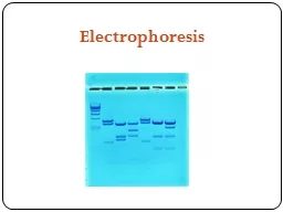
(30%) 25mg bromophenol blue (0.25%) Double distilled water 3.7 Applications: No work of molecular biology is possible without agarose gel electrophoresis DNA EXPERIMENTS ï· To visualize Products of animal, plant or bacterial DNA Extractions ï· To study RFLP FORENSIC SCIENCE ï· Paternity test ï· DNA Fingerprinting HUMAN HEALTH & DISEASE ï· To study Human Genomics & Bioinformatics ï· Restriction Enzyme Mapping ï· Diagnostics (Human and plant pathogens) ï· DNA Profiles of âInfectious Diseasesâ TRANSFORMATION & BACTERIAL CLONING ï· To screen cloned products POLYMERASE CHAIN REACTION 9 Gel Elect
rophoresis, Principle, Types and Applications ï· To check amplified DNA 4. Sodium d odecyl sulphate poly acryl amide gel electrophoresis ( SDS PAGE) Polyacrylamide: Fig.1 Formation of polyacrylamide ï· Polyacrylamide is formed by is a l inking of acrylamide molecules ï· T he concentration of acrylamide is used between 3.5 and 20%. ï· Acrylamide is a neurotoxin and needs careful handling 10 Gel Electrophoresis, Principle, Types and Applications ï· Polyacrylamide is non - toxic, but polyacrylamide gels should not be touched bare handed because of there is still possibi lity that acrylamide is present in fr
ee state. ï· P olyacrylamide gels shows narrow separation range , but their power of resolving two close sizes of proteins very high ï· If we want to separate DNA fragments less than 500 bp, polyacrylamide gel is used ï· DNA fragments differing in length by even a single base pair can also be resolved. ï· The polymerization reaction starts by formation of free radical with ammonium per sulfate (APS) as which acts as a initiator and N, N, Nâ, Nâ - tetramethylene dia mine (TEMED) which acts as a catalyst. SDS (sodium dodecy l sulfate) is a detergent which can dissolve hydrophobic molecules and has a negative
charge attached to it. I f a cell is incubated with SDS, the me mbranes will get dissolved, and proteins will be denatured by the detergent also all the proteins will imparted with negative charges. 11 Gel Electrophoresis, Principle, Types and Applications Fig. 2 Role of SDS in PAGE Fig.3 Cross section of polyacrlamide gel Role of Ammonium persulphate and TEMED ï· Ammonium Persulfate (APS) (NH4 )2S2O8 is an oxidizing agent which is used in combination with TEMED to catalyze the polymerization of acrylamide and bisacrylamide ï· Ammonium persulfate forms free radicals when it is d issolved in water and it initiate
polymerization of acrylamide soluti ons. Percentage of gel is made according to the expected molecular weight of protein we want to study Table.1 Ingredients of SDS PAGE (30 ml) 12 Gel Electrophoresis, Principle, Types and Applications Table 2. Sample/Running Buffer 13 Gel Electrophoresis, Principle, Types and Applications Gel Making 14 Gel Electrophoresis, Principle, Types and Applications Fig.4 Diagrammatic view of gel making process Fig.5 Diagrammatic view of gel making process Contd. 15 Gel Electrophoresis, Principle, Types and Applications
Electrophoresis Fig. 6. Apparatus set up for SDS PAGE The different pH buffer system and the stacking gel A buffer is required for electrophoresis. Usually discontinuous buffer is used that means the pH of buffer in the gel is different than that of pH of buffer in the gel tank. In stacking gel 6.8 pH is used and running gel 8.8pH buffer is used. . 16 Gel Electrophoresis, Principle, Types and Applications Fig.7 D ifferent pH levels used during running of SDS PAGE So you must be wondering that why this different pH for single gel? We all know that glycine can exist i n three different states of charge depending on pH. positivity, neutr
al or negativity As soon as electrophoresis starts , the glycine ions which have negative charge in the pH 8.3 electrode buffer are forced to enter the stacking gel, where the pH is 6.8. Now glycine acts as zwitte rionic (neutrally charged) . Because of this loss of charge they mov e very slowly in applied electric field. T he Cl - ions (from Tris - HCl) , move much fast in the electric field and they moves faster than the glycine. The separa tion of Cl - from the Tris - ion lead to the formation of very small zone with voltage gradient whi ch pulls the glycine behind it, results in two small separated fronts of migrating ions; the highly mobile Cl - fron
t, followed by the slower, mostly neutral glycine front . 4.1 Gel Staining Gel staining: Coomassie blue Chemical reagents required: ï· Brilliant Blue R - 250 (BBR) ï· Sterile distilled water Solutions: 17 Gel Electrophoresis, Principle, Types and Applications ï· Fixing solution: It is prepared by mixing different ratios of methanol(50), acetic acid(10) and water(40) ï· Destaining solution: It is prepared by mixing different ratios of methano(45)l, ace tic acid(10) and water(45). ï· Coomassie concentrated stain solution: It has 12.0 g BBR to which 300 mL Methanol is added then acetic acid (60ml) is added. After mixing all c
omponents it is stirred properly. ï· Coomassie Working solution: To this 500 mL Meth anol we add 30 ml of Coomassie stain solution and 400ml of water plus 100 ml of acetic acid. After mixing it is filter sterilized using a syringe filter ï· Silver Staining ï· The stain is more sensitive and able to detect protein concentrations from 1 ng to 1 mg. ï· If gel is not stained properly it can be destained and stained again which is not possible in coomassie staining. Silver Staining Protocol 18 Gel Electrophoresis, Principle, Types and Applications Table 3. Silver staining Protocol Destaining Protocol ï· Des
tain until no band is visible ï· Gel is w ash ed 3 - 5 times for 10 min in a distilled water which should be sterile also till all the stain is removed ï· Stain the gel gain using same staining protocol 19 Gel Electrophoresis, Principle, Types and Applications 4.2 Applications of SDS PAGE ï· Molecular weight of the proteins can be determined ï· To perform western blotting SDSPAGE is required 5. 2D - Gel electrophoresis ï· It is a type of gel electrophoresis where prote ins are separated in two dimensions ï· It is the only method which is available and is capable of simultaneously separating thousands of p rot
eins. Basis of separation ï· Proteins are se parated on the basis of two properties. ï· F irst ly they are separated on the basis of their net charge. ï· After that, PAGE separates the proteins on the basis of their mass. ï· Using this method s mall changes in charge a nd mass can be detected , as it is not possible that two different proteins will resolve to the same place in both dimensions Isoelectric Point ï· There is a pH at which ther e is no net charge on a protein and this point is called isoelectric point (p I). ï· A protein has a net negative charge above its isoelectric point, and it migrates toward the anode in an electri
cal field. ï· T he protein is positive b elow its isoelectric point, and it migrates toward the cathode. Isoelectric Focusing: ï· When proteins are separated by isoelectric points it is called isoelectric focusing (IEF). ï· Therefore , a gradient of pH is applied to a gel and an electric potential is applied across the gel 20 Gel Electrophoresis, Principle, Types and Applications ï· proteins will have charge a t all pH values other than their isoe lectric point, ï· Proteins move towards the negative end of the gel If they are positively charged and if they are negatively charged they will move to the positive end of the gel. ï
· In the first dimension proteins will move along the gel and will accumul ate at their isoelectric point 5.1 First Dimension Electrophoresis ï· C ommercially available Immobilized pH Gradient (IPG) strips are used for f ocusing ï· IPG strips are solid surfaces with coat of dehydrated polyacrylamide ï· These strips are av ailable in a range of pH gradients such as 5 - 7, 3 - 8, 6 - 12 etc. Fig.8 IPG Strips ï· Rehydration of strips is done with rehydration buffer before protein loading ï· For rehydration of IPG strips these IPG strips are kept in rehydration solution for 10 - 15 hours . ï· The foc using is carried out on an equipment
which supplies g radient electric current to the IPG strips. ï· IPG strip has two sides, a base of plastic and the ano ther side which have gel on it. ï· After this IPG strip is then placed into the lane in a way that the surface with gel faces downwards and is in direct contact with the solution around it. 21 Gel Electrophoresis, Principle, Types and Applications ï· This is left on the working bench for 15 hours. ï· After 15 hours t he IPG strips are r emoved from the rehydration solution carefully using forceps ï· This IPG strip is then placed in the fresh tray, with the gel side facing upwards Fig.9 Isoelectr
ic focusing Isoelectric Focusing ï· The strips are put in isoelectric focusing unit ï· The voltage g radients and time intervals depends on the type of strip that we are using to run the samples . ï· The separation can be monitored directly ï· After focusing the strips are taken out and can be used in next step 22 Gel Electrophoresis, Principle, Types and Applications Fig.10. Isoelectric focusing contd 5.2 Second Dimension Electrophoresis Equilibrium of Strips ï· We need to equilibrate the strips before separating them in second dimension ï· It involves the denaturation of proteins, for separation on the basis of
molecular weight during SDS - PAGE. Equilibration 23 Gel Electrophoresis, Principle, Types and Applications Fig11. Second dimension electrophoresis 5.3 Result and Image Analysis ï· After the proteins gets separa ted in 2DE, each dot on gel is a unique protein which is specific to pI and molecular weight. ï· We can not do p rotein expression profiling manually as the number of spots are very large and manual interpretation can always lead to false result ï· We can use commercially available software for analysis 24 Gel Electrophoresis, Principle, Types and Applications Fig.12. Gel Image af