
By Louann W Lawrence Larry A Broussard Introduction Porphyrins hemoglobin amp myoglobin all contain porphyrin ring 4 pyrrole groups bonded by methene bridges Porphyrins can chelate metals ID: 915935
Download Presentation The PPT/PDF document "Chapter 19: Porphyrins and Hemoglobin" is the property of its rightful owner. Permission is granted to download and print the materials on this web site for personal, non-commercial use only, and to display it on your personal computer provided you do not modify the materials and that you retain all copyright notices contained in the materials. By downloading content from our website, you accept the terms of this agreement.


Slide1
Chapter 19: Porphyrins and Hemoglobin
By Louann W. Lawrence, Larry A. Broussard
Slide2Introduction
Porphyrins, hemoglobin, & myoglobin all contain porphyrin ring: 4 pyrrole groups bonded by methene bridges.
Porphyrins can chelate metals
to form functional groups that participate in oxidative metabolism.
Hemoglobin designed to bind, deliver, and release oxygen.
Porphyrias
:
disturbances heme synthesis
Hemoglobinopathies:
disorders of
qualitative defects
in
hemoglobin
molecule
(example: Sickle Cell Anemia)
Thalassemias:
disorders of
quantitative defects
in production of normal hemoglobin molecules
(examples: alpha and beta thalassemia)
Slide3Introduction
Basic structure of porphyrins
Slide4Introduction
Slide5Porphyrins
Porphyrins
(in the body):
Act as chemical intermediates in synthesis of hemoglobin, myoglobin
, & other respiratory pigments called cytochromes
P
art of peroxidase & catalase enzymes, which contribute to efficiency of internal respiration
Chelate iron to form
heme
Used to diagnose
porphyrias
(result from
heme
disturbances)
Slide6Heme Synthesis
Main synthesis sites are the bone marrow and liver
Synthesis of
heme
:
Delta-
aminolevulinic
acid
Porphobilinogen
Uroporphyrinogen
Coproporphyrinogen
Protoporphyrin
Heme
Slide7Slide8Porphyrins
Chemistry of Porphyrins
Porphyrins are organic compounds found
in nature
.
Pigment Chlorophyl is a magnesium porphyrin
Four basic isomers may exist for every porphyrin compound (I-IV) however, only types I & III, which differ in side chain arrangement, occur in nature.
Only Type III isomers form heme
Porphyrins are stable & red-violet to red-brown and fluoresce red when excited by light near 400 nm.
Porphyrinogens:
reduced form of of porphyrins, functional forms that must be used in heme synthesis; unstable, colorless, do not fluoresce
Slide9Porphyrins
3 porphyrin compounds clinically significant in humans:
Protoporphyrin (PROTO) – primarily excreted in feces
Uroporphyrin (URO) – primarily excreted in urine
Coprophorphyrin (COPRO) – can be both
Excess of the 3 in biological fluids is a sign of abnormal heme synthesis.
Again – porphyrinogens are the functional form of the compound that MUST be used in heme synthesis.
Slide10Porphyrins
Porphyrin Synthesis
All cells contain hemoproteins & can synthesize heme, but bone marrow & liver are main sites.
Rate of heme synthesis in cells of liver is achieved largely through regulation of enzyme
-aminolevulinic acid (ALA) synthase.
Increase in heme causes decrease in ALA synthase.
Decrease in heme causes increase in ALA synthase.
Rate of heme synthesis is flexible & can change rapidly in response to many external stimuli.
Slide11Porphyrins
Clinical Significance and Disease Correlation
Porphyrias: inherited or acquired enzyme deficiencies that result in overproduction of heme precursors in bone marrow or liver
Diagnosis is made by combination of history & physical & laboratory findings.
Types associated with neuropsychiatric symptoms (these 3 conditions are associated with excess of early precursors (ALA, phorphobilinogen or both)
ALA dehydratase (ALAD)
Deficiency porphyria (ADP)
Acute intermittent porphyria (AIP)
Slide12Porphyrins
Clinical Significance and Disease Correlation
Types associated with cutaneous symptoms (photosensitivity, blisters, excess facial hair, and hyperpigmentation). The following are due to excess later intermediates:
Porphyria cutanea tarda (PCT)
**increased fragility to light-exposed skin
Hepatoerythropoietic porphyria (HEP)
Erythropoietic porphyria (EP)
Congenital erythropoietic porphyria (CEP)
Types associated with neurocutaneous symptoms – due to excess in both early and later intermediates.
Hereditary coproporphyria (HCP)
Variegate porphyria (VP)
Slide13Summary of
Porphyrias
Porphyria
Enzyme Deficiency
Symptoms
Plumboporphyria
(PP)
ALA
dehydratase
Neuropsychiatric
Acute intermittent
porphyria
(AIP)
Porphobilinogen
deaminase
Neuropsychiatric
Porphyria
cutanea
tarda
(PCT)
Uroporphyrinogen
decarboxylase
Cutaneous
Hepatoerythropoietic
porphyria
(HEP)
Uroporphyrinogen
decarboxylase
Cutaneous
Erythropoietic
porphyria
(EP)
Ferrochelatase
Cutaneous
Congenital
erythropoietic
porphyria
(CEP)
Uroprophyrinogen
III
cosynthase
Cutaneous
Slide14Porphyrins
Methods of Analyzing Porphyrins
Tests for urinary PBG (porphobilinogen) & ALA
Watson-Schwartz
Hoesch
Hoesch sometimes used to confirm results of Watson-Schwarts due to Hoesch not having interference with uronbilinogen
Bo
th of above use
Ehrlich’s reagent
. When PBGs mixed with Ehrlich’s – form a red-orange color.
Tests for porphyrins
Enhanced fluorescence of compounds in acidic solution
Chromatic separation & quantitation with spectrophotometry or fluorometry
Molecular diagnostic techniques
Slide15Hemoglobin
Role in Body: transports oxygen to tissue & CO
2
to lungs
Structure
A large, spherical, complex protein molecule (molecular weight is 64,000)
Comprises heme (3%) & globin proteins (97%)
Contains 4 heme groups attached to 4 globin chains
Each globin chain consists of 141 or more amino acids & has 4-fold structure.
Majority of hemoglobin in normal adults is hemoglobin A
(or A
1
).
Hemoglobinopathies:
diseases related to defects in hemoglobin structure
Slide16Hemoglobin (cont’d)
Hemoglobin A: structure of the hemoglobin molecule
Slide17Hemoglobin
Synthesis and Degradation of Hemoglobin
Synthesis occurs in immature RBCs in bone marrow:
65% in nucleated cells
35% in reticulocytes
Normal synthesis depends on adequate iron supply & heme & protein synthesis to form globin portion.
Two pathways degrade hemoglobin
:
Extravascular
(80–90%): occurs outside circulatory system within
phagocytic cells of spleen
, liver, & bone marrow
Intravascular
(10–20%): released directly into bloodstream &
dissociated into
& dimers
Slide18Hemoglobin
Clinical Significance and Disease Correlation
Hemoglobin Qualitative Defects: The Hemoglobinopathies
Hemoglobin S
Amino acid defect is at 6th position on
-chain – glutamic acid substituted by valine.
Most common hemoglobinopathy in U.S
.
HbAS (heterozygous = trait) and HbSS (homozygous = disease)
Heterozygous state offers protection from parasites (plasmodium falciparum)
Hemoglobin C
Glutamic acid in 6th position of
-chain is replaced by lysine, resulting in net positive charge
.
Found in West Africa (North Ghana) in 17–28% of population & in 2–3% of African Americans
Slide19Hemoglobin
Clinical Significance and Disease Correlation
Hemoglobin Qualitative Defects: The Hemoglobinopathies
Hemoglobin SC
Most common mixed hemoglobinopathy
-genes code for -S chains & -C chains, leaving no normal chains to produce hemoglobin A.
Less severe than HbSS disease but still has similar clinical symptoms to HbSS disease
.
Hemoglobin E
An amino acid substitution of
lysine for glutamic acid in 26th position of
chain
, resulting in net positive charge
Found in Asia in about 20 million people, 80% of whom live in Southeast Asia
Slide20Hemoglobin
Clinical Significance and Disease Correlation
Hemoglobin Qualitative Defects: The Hemoglobinopathies
Hemoglobin D
Variant with electrophoretic mobility on cellulose acetate, like that of hemoglobin S but with negative solubility test
Hemoglobin Quantitative Defects: The Thalassemias
Thalassemias:
group of diseases in which defect causes reduced synthesis of 1 or more of hemoglobin chains
-Thalassemias – defective production of alpha chains
-Thalassemias – defective production of beta chains
Slide21Hemoglobin
-
Thalassemias
2 alpha genes located on each chromosome 16, yielding a total of 4
genes.
Hydrops
fetalis
– (4 gene deletion) total absence of alpha chain synthesis
Make hemoglobin
Barts
(four gamma chains)
Die before birth or upon birth
Hemoglobin H disease (3 gene deletion)
Hgb
H = beta chain tetramers (due to excess beta chains and lack of alpha chains).
-Thalassemia minor (2 gene deletion)
Mild, microcytic, hypochromic anemia.
Silent carrier (1 gene deletion) – sufficient alpha chains for normal amounts of hemoglobin
Often discovered by expression of 1 to 2%
Hgb
Barts
Slide22Hemoglobin
-
Thalassemias
(point mutations vs. gene deletions)
Many possible expressions of this condition/disease
-Thalassemia Major – aka Cooley’s anemia (severe anemia present)
In -Thalassemia, there is only one gene inherited from each parent – however – there can be a gene expression – which correlates to varying degree of severity
0
= no beta chain production
+
= decreased beta chain production
= normal beta chain production
Clinical findings of Beta Thalassemia
Increased
Hb
F (2 alpha/2 gamma)
Varied increases
in HbA
2
(2 alpha/2 delta)
Slide23Hemoglobin
Methodology
Solubility test (screening test for sickling hemoglobins)
Based on principle that sickling hemoglobin, in deoxygenated state, is relatively insoluble & forms precipitate in solution
If sickling hemoglobin present – can’t see through.
If No sickling hemoglobin present – see through
.
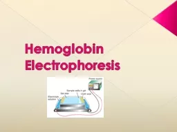
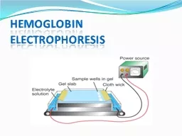
Cellulose acetate (alkaline) hemoglobin electrophoresis
Fresh hemolysate made from packed RBC sample is applied to a cellulose acetate plate using a buffer of alkaline pH (8.4–8.6); electrophoresis is performed.
Citrate agar (acidic) electrophoresis
Performed at acid pH (6.0–6.2) after abnormal hemoglobin is detected on cellulose-acetate electrophoresis
Slide24Hemoglobin
Methodology
Hemoglobin A
2
quantitation
(2 alpha / 2 delta)
Best accomplished by microcolumn chromatography or high-performance liquid chromatography
(HPLC)
Acid elution stain for hemoglobin F
(2 alpha / 2 gamma)
Distinguishes erythrocytes containing increased amount of hemoglobin F from normal adult cells
Hemoglobin F quantitation
Based on principle that fetal hemoglobin is resistant to alkali denaturation in 1.25 mol/L NaOH for 2 minutes
Slide25Hemoglobin (cont’d)
DNA Technology
Definitive diagnosis of some hemoglobinopathies & thalassemias that involve combinations of genetic defects may require DNA analysis.
DNA sequence of interest may be easily analyzed from whole blood or spots of dried blood on filter paper.
Advantages:
provides definitive info on genotype of individuals tested & sometimes direct detection of molecular lesions
Disadvantages:
higher cost & lack of availability in many labs
Especially useful in prenatal diagnosis of thalassemia major & sickle cell anemia
Slide26Myoglobin
Structure and Role in the Body
A simple heme protein found in skeletal & cardiac muscle
Can reversibly bind oxygen, similar to hemoglobin molecule
Unable to release oxygen, except under low oxygen tension
Acts as oxygen carrier in cytoplasm of muscle cell
Serves as an extra reserve of oxygen to sustain activity in exercising muscle
Slide27Myoglobin
Clinical Significance
Elevated levels in serum & urine often
indicate muscle damage.
Combination of high serum myoglobin & low clearance rate indicates high risk for acute renal failure.
Primary use of serum myoglobin is investigation of chest pain to rule out acute myocardial infarction
.
Also has been investigated to aid in diagnosis & differentiation of types of hereditary progressive muscular dystrophy
Slide28Myoglobin
Methodology
Several immunoassay methods are used to measure & identify myoglobin (fluorescence, chemiluminescence, immunochromic).
Procedures incorporate binding of specific antibodies to myoglobin.
Resulting chemical or physical change can be measured & correlated to myoglobin concentration.
Methods have been adapted to point-of-care devices for rapid assessment of chest pain.