/
European Journal of Molecular  Clinical Medicine European Journal of Molecular  Clinical Medicine

European Journal of Molecular Clinical Medicine - PDF document

isabella2
isabella2 . @isabella2
Follow
352 views
Uploaded On 2022-08-24

European Journal of Molecular Clinical Medicine - PPT Presentation

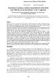
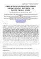
ISSN 2515 8260 Volume 08 Issue 03 2021 939 UDC 6167282 0016 0531 08 0535 6 RESULTS OF SURGICAL TREATMENT OF CONGENITAL HIP DISLOCATION Ibragimov Sadullo Yusupovich Samarkand Sta ID: 941191

hip patients years treatment patients hip treatment years congenital results dislocation radiography patient months issue open reduction operated volume

Share:

Link:

Embed:

Download Presentation from below link

Download Pdf The PPT/PDF document "European Journal of Molecular Clinical ..." is the property of its rightful owner. Permission is granted to download and print the materials on this web site for personal, non-commercial use only, and to display it on your personal computer provided you do not modify the materials and that you retain all copyright notices contained in the materials. By downloading content from our website, you accept the terms of this agreement.


Presentation Transcript

European Journal of Molecular & Clinical Medicine ISSN 2515 - 8260 Volume 08, Issue 03 , 2021 939 UDC 616.728.2 - 001.6 - 053.1 - 08 - 053.5 / 6 RESULTS OF SURGICAL TREATMENT OF CONGENITAL HIP DISLOCATION Ibragimov Sadullo Yusupovich Samarkand State Medical Institute Uzbekistan Urinboev Paizullo Urinboevich Samarkand State Medical Institute Uzbekistan Kudratova Gulsara Najmiddinovna Samarkand State Medical Institute Uzbekistan Saloxiy Otabek Ikromovich Samarkand State Medical Institute Uzbekistan Muxtorov Zokir Safarovich Samarkand State Medical Institute Uzbekistan Kuvatov Dilshod Xolmurodovich Samarka nd State Medical Institute Uzbekistan Ashirov Mavlon Umurzokovich Samarkand State Medical Institute Uzbekistan Kayumov Rustam Shuxratovich Samarkand State Medical Institute Uzbekistan Abstract. Relevance. At present, although significant progress has been made in the field of early detection of congenital malformations and conservative treatment, a large number of patients with this pathology remain untreated in a timely manner. In 10 - 15% of patients treat ed conservatively with congenital malformations of the thighs, the need for surgical treatment arises. P urpose of the study. It consists of studying the causes of complications observed during long periods of treatment of patients treated surgically on con genital malformations of the thighs and looking for measures to prevent them. In such cases, depending on the age of the patients and the severity of the discharge, surgical procedures are performed, ranging from simple open placement to repair and opening of the proximal

part of the femur and the roof of the joint. This article presents the results of surgical treatment performed for 111 children with congenital hip dislocation. After 30 years, the long term results of the intervention were studied in 76 (68.5%) patients. According to observation, 22(28.9%) patients and 31(40.8%) patients demonstrated good and satisfactory long term results respectively. While results of surgical treatment for 23(30.3%) patients estimated as unsatisfactory. Identified t he causes of unsatisfactory results and preventive measures were recommended. Keywords: congenital hip dislocation, reconstruction, contracture, coxarthrosis, ankylosis. Relevance. Congenital hip dislocation ranks first in terms of frequency and number of published scientific papers among orthopedic diseases, and is one of the most pressing problems of pediatric orthopedics [1,3,7]. European Journal of Molecular & Clinical Medicine ISSN 2515 - 8260 Volume 08, Issue 03 , 2021 940 Currently, despite significant progress in the ear ly detection of congenital malformations and conservative treatment, a large number of patients with this pathology remain without timely treatment. In 10 - 15% of patients with congenital hip dislocation treated conservatively, there is a need for surgical treatment [2]. In the literature of recent years, a group of congenital hip dislocation that does not respond to conservative treatment has been described. According to the literature, the frequency of such dislocations ranges from 1 - 5% to 10 - 20% [6]. In s uch cases, depending on th

e degree of congenital hip dislocation, operations are performed from simple open reduction to open reduction with corrective osteotomy of the femur and acetabular plasty. There is a lot of data in the literature about the immedia te results of surgical treatment of congenital hip dislocation, but long - term results are very poorly covered. Purpose of the study. Study of long - term results (30 years) of treatment of patients treated with surgery with congenital hip dislocation, study of complications and ways to prevent complications. Research material. The number of patients treated for congenital hip dislocation in 1991 in the department of pediatric orthopedics of the Samarkand regional hospital of orthopedics and the consequences o f trauma was 310 patients, which amounted to 47.8% of the total number of patients. 111 (17.1%) patients underwent surgical treatment. A retrospective analysis of these patients was performed. Among sick boys there were 10 (9%), girls were 101 (91%). The p atients were distributed by age as follows: from 2 to 5 years - 27 (24.3%) patients, from 5 to 8 years - 73 (65.7%) patients, patients over 8 years old - 11 (9.9%) patients. In 39 (35.3%) patients, the pathology was right - sided, in 55 (49.5%) left - sided, a nd in 17 (15.3%) patients, bilateral. All patients were examined by a pediatrician before and after surgery. After preliminary preparation, the patients underwent the following operations under intubation anesthesia: simple open reduction of the femoral he ad - in 5 (4.5%) patients, open reduction with subtrochanteric corrective osteotomy of the femur - in 58 (52.2%) patients, open reduction with subtrochanteric corrective osteotomy o

f the femur and plastic of the acetabular roof - in 34 (30.6%) patients, ex tra - articular operations - in 8 (7.2%) patients. After the operation, depending on the complexity of the operation, a 1.5 coxite plaster cast was applied from 4 weeks to 6 - 7 weeks. No early postoperative complications were observed in the patients (shock, secondary bleeding, pneumonia, relaxation, hematoma suppuration). All postoperative wounds healed by primary intention. After removing the plaster cast, physiotherapeutic treatment was carried out aimed at restoring the range of motion in the operated hip joint. After 2.5 - 3 months, walking was allowed with loading of a healthy limb using crutches. The load on the operated limb was allowed one year after the operation. The immediate results after the operations were assessed as good. After the end of treatme nt, the parents were given the following recommendations: until the end of growth, be under the European Journal of Molecular & Clinical Medicine ISSN 2515 - 8260 Volume 08, Issue 03 , 2021 941 supervision of a pediatric orthopedist, exemption from physical education, limitation of work performed while standing, when choosing a profession, prefer seden tary professions, girls limit themselves to two pregnancies. Scaglietti O., Calandriello B. wrote in 1961: “Open reduction of hip dislocation is not a guarantee of a good result. During the operation, the femoral head and the acetabulum are matched. It is necessary to create conditions for the correct development of the pathologically altered femoral head and acetabul

um ”. Treatment Results: Results were evaluated 30 years after the end of treatment. The results of treatment were studied in 76 (68.5%) patie nts. At the time of the examination, the patients were 35 - 42 years old. Of these, 68 (89.5%) were women, 8 (10.5%) were men. Patients underwent clinical, radiological and CT examinations according to indications. The treatment results were rated as good, s atisfactory and unsatisfactory. In 22 (28.9%) patients, the results were assessed as good. These patients do not present complaints, walk independently and correctly, movements in the hip joints are not limited, the length of the lower limbs is the same. T he development and volume of the pelvic and femoral muscles is the same. X - ray indicators: the femoral heads are spherical, the heads are centered in the acetabulum, the cervical - shaft angles are within 120 - 1250. Shenton's line is not broken. The Viberg an gle is within 20 - 250. Mild subchondral sclerosis is determined in the femoral head and acetabulum. These patients were operated on at the age of 3 - 5 years, were performed open reduction with subtrochanteric corrective osteotomy of the femur, plastic of the roof of the acetabulum. Patients in the postoperative period were observed by an orthopedist, followed the doctor's instructions flawlessly. Here's an example. Patient B., 35 years old, at 3 years and 6 months was operated on for congenital dislocation of the right hip. Figure # 1. Patient B., 3 years and 6 months. Radiography in direct projection. Diagnostic radiography. European Journal of Molecular & Clinical Medicine

ISSN 2515 - 8260 Volume 08, Issue 03 , 2021 942 The operation “Open reduction of the right thigh with corrective subtrochanteric osteotomy, Salter osteotomy of the pelvis” was performed. After six months, the metal retainers were removed. Figure 2. Patient B., 3 years and 6 months. Radiography in direct projection. Result one month after surgery (Patient in a plaster cast). Fi g. 6. Patient B., 4 years old. Radiography in direct projection. Result 6 months after surgery (before removing the metal retainers). European Journal of Molecular & Clinical Medicine ISSN 2515 - 8260 Volume 08, Issue 03 , 2021 943 Fig. 4. Patient B., 5 years 4 months. Radiography in direct projection. Result in 1 year and 8 months. The treatment res ults were assessed as good. Figure 5. Patient B., 35 years old. Radiography in direct projection. Result 30 years after surgery In 31 (40.8%) patients, the results were assessed as satisfactory. These patients get tired at the end of the day, there is mild lameness and pain in the hip joints. The muscles on the operated limb are atrophied (1 - 1.5 cm), internal rotation and abduction in the hip joints are limited within European Journal of Molecular & Clinical Medicine ISSN 2515 - 8260 Volume 08, Issue 03 , 2021 944 15 - 200. Patients work by profession. X - ray indicators: the heads of the femo ral bones are spherical, the heads are centered in

the acetabulum, the Shenton's line is broken in 6 patients, the I degree of coxarthrosis is determined in all patients, the Viberg angle is within 18 - 200. These patients were operated on at the age of 5 - 8 years, 5 patients underwent open reduction, 18 patients underwent open reduction with subtrochanteric corrective osteotomy of the femur. In the postoperative period, these patients did not always come for an orthopedic examination, did not follow the docto r's instructions. In 23 (30.3%) patients, the results were assessed as unsatisfactory. These patients indicate persistent pain in the operated joint, all patients have limited movement in the operated joint. In 4 patients, the absence of movement in the jo int (ankylosis) is determined. In 8 patients, II and III degrees of coxarthrosis are determined. 7 sick invalids of the II group. 4 patients underwent hip arthroplasty. 6 patients were offered arthroplasty. These patients were operated on at the age of ove r 8 years; coxarthrosis was determined in 12 patients before the operation. In the postoperative period, they did not receive rehabilitation, they worked in hard work. 6 women had 3 - 5 pregnancies. Here's an example. Patient A.L., 39 years old, was operated on at the age of 8 for right - sided congenital dislocation of the hip, left - sided subluxation of the hip. Fig. 6. Patient A.L., 8 years old. Radiography in direct projection. Diagnostic radiography. The operation "Open reduction of the right femur with c orrective subtrochanteric osteotomy, plastic of the acetabular roof according to Pemberton" was performed. Six months later, the operation "Corrective subtrochanteric osteotomy of the left femur, plastic of the acetabula

r roof according to Pemberton" was p erformed. After another six months, the metal retainers were removed, European Journal of Molecular & Clinical Medicine ISSN 2515 - 8260 Volume 08, Issue 03 , 2021 945 photo # 7. Patient A.L., 8 years 6 months. Radiography in direct projection. Control X - ray 6 months after surgery. Patient A.L., 8 years old. Radiography in direct projection. Contro l radiography 1 year after surgery. European Journal of Molecular & Clinical Medicine ISSN 2515 - 8260 Volume 08, Issue 03 , 2021 946 Fig. 9. Patient A.L., 38 years old. Radiography in direct projection. Control radiography 30 years after surgery. "Bilateral dysplastic coxarthrosis, right grade III, left grade II." The patient was offered endoprosth etics. Conclusions: Congenital hip dislocation is a widespread disease of the musculoskeletal system. According to our data, this pathology accounts for 47.8% of inpatients. Studies have shown that clinically good long - term results are observed in 22 (28.9%) patients. This o nce again indicates that it is advisable to begin treatment of congenital hip dislocation from the hospital. With constant dispensary observation of these patients until the end of growth, positive results can be achieved. References. 1. Baindurashvili AG, Chukhraeva I. Yu. On the issue of early diagnosis of pathology of the musculoskeletal system in children. Topical issues of pediatric traumatology an

d orthopedics: scientific and practical materials. conf. children orthopedic traumatologists of Russia. SP b .; Syktyvkar, 2009; 8 - 10. 2. Bakhteeva N.Kh., Vinokurov V.A., Norkin I.A., Persova E.A. Conservative treatment of children with congenital hip dislocation. Bulletin of Traumatology and Orthopedics. N.N. Priorov. - 2003. - No. 4. - S. 34 - 37. 3. Gribova I. V. Surgical treatment of congenital dislocation and residual hip subluxation in children after three years. Abstract of doctoral dissertation. M., 2002 4. Pozdnikin I.Yu. Surgical treatment of young children with hip dysplasia and congenital hip dislocatio n. Abstract of the thesis for Candidate of Medical Sciences. St. Petersburg. 2006. European Journal of Molecular & Clinical Medicine ISSN 2515 - 8260 Volume 08, Issue 03 , 2021 947 5. Pozdnikin I.Yu., Baskov VE, Voloshin S.Yu., et al. Errors in diagnosis and initiation of conservative treatment of children with congenital hip dislocation // Pediatric T raumatology, Orthopedics and Reconstructive Surgery. - 2017. - T. 5. - Issue. 2. - P. 42 – 51. doi: 10.17816 / PTORS5242 - 51. 6. Kamosko M.M., Poznovich M.S. Conservative treatment of hip dysplasia. Pediatric Traumatology, Orthopedics and Reconstructive Surge ry. T. II. Issue 4. 2014 p. 2309 - 3994. 7. Lazovaya Yu.I. Assessment of the dynamics of the development of the hip joint in children under conditions of a persisting pathological process (Congenital dislocation of the hip: diagnosis and treatment). Abstract of the thesis for Candidate of Medical Sciences. Moscow,