
Meissner EG Bennett JE Qvarnstrom Y da Silva A Chu EY Tsokos M et al Disseminated Microsporidiosis in an Immunosuppressed Patient Emerg Infect Dis 201218711551158 httpsdoiorg103201eid1807120047 ID: 1009557
Download Presentation The PPT/PDF document "Figure 1 Figure 1. . . . . Microsporidiu..." is the property of its rightful owner. Permission is granted to download and print the materials on this web site for personal, non-commercial use only, and to display it on your personal computer provided you do not modify the materials and that you retain all copyright notices contained in the materials. By downloading content from our website, you accept the terms of this agreement.


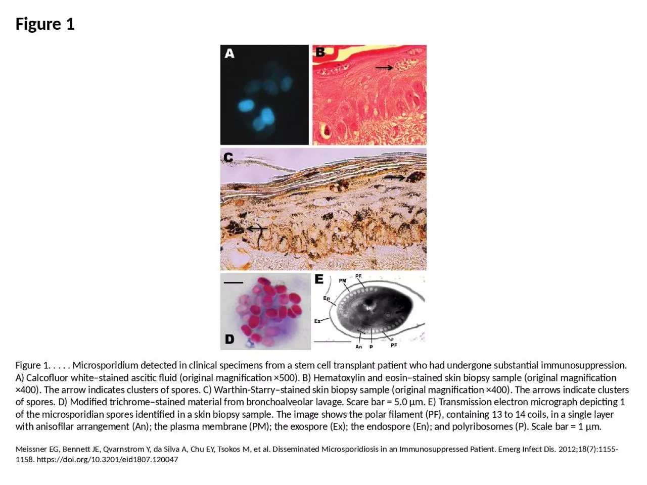
1. Figure 1Figure 1. . . . . Microsporidium detected in clinical specimens from a stem cell transplant patient who had undergone substantial immunosuppression. A) Calcofluor white–stained ascitic fluid (original magnification ×500). B) Hematoxylin and eosin–stained skin biopsy sample (original magnification ×400). The arrow indicates clusters of spores. C) Warthin-Starry–stained skin biopsy sample (original magnification ×400). The arrows indicate clusters of spores. D) Modified trichrome–stained material from bronchoalveolar lavage. Scare bar = 5.0 μm. E) Transmission electron micrograph depicting 1 of the microsporidian spores identified in a skin biopsy sample. The image shows the polar filament (PF), containing 13 to 14 coils, in a single layer with anisofilar arrangement (An); the plasma membrane (PM); the exospore (Ex); the endospore (En); and polyribosomes (P). Scale bar = 1 μm.Meissner EG, Bennett JE, Qvarnstrom Y, da Silva A, Chu EY, Tsokos M, et al. Disseminated Microsporidiosis in an Immunosuppressed Patient. Emerg Infect Dis. 2012;18(7):1155-1158. https://doi.org/10.3201/eid1807.120047