
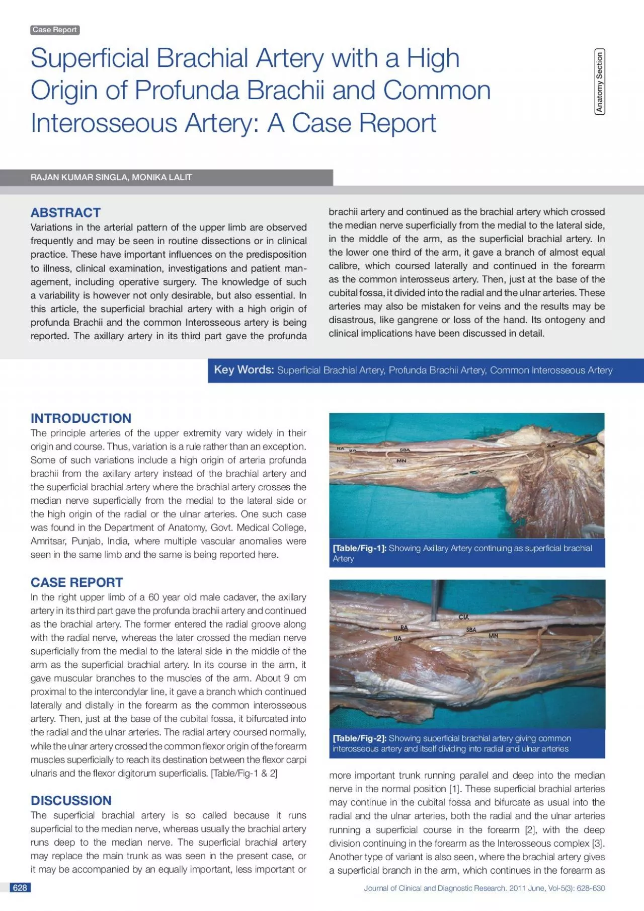
628 628 The principle arteries of the upper extremity vary widely in their origin and course Thus variation is a rule rather than an exception Some of such variations include a high origin of arter ID: 938140
Download Pdf The PPT/PDF document "Journal of Clinical and Diagnostic Resea..." is the property of its rightful owner. Permission is granted to download and print the materials on this web site for personal, non-commercial use only, and to display it on your personal computer provided you do not modify the materials and that you retain all copyright notices contained in the materials. By downloading content from our website, you accept the terms of this agreement.

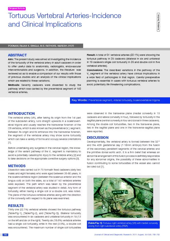


Journal of Clinical and Diagnostic Research. 2011 June, Vol-5(3): 628-630 628 628 The principle arteries of the upper extremity vary widely in their origin and course. Thus, variation is a rule rather than an exception. Some of such variations include a high origin of arteria profunda brachii from the axillary artery instead of the brachial artery and the super cial brachial artery where the brachial artery crosses the median nerve super cially from the medial to the lateral side or the high origin of the radial or the ulnar arteries. One such case RAJAN KUMA Key Words: Case Report A [Table/Fig-1]: Showing Axillary Artery continuing as super cial brachial [Table/Fig-2]: www.jcdr.netRajan Kumar Singla and Monika Lalit, super cial brachial artery with a high origin of profunda Brachii and the common Interosseous arteryJournal of Clinical and Diagnostic Research. 2011 June, Vol-5(3): 628-630 629 629 Key Words:Super cial Brachial Artery, Profunda Brachii Artery, Common Interosseous Arterythe common interosseous artery, while the brachial artery divides into the radial and the ulnar arteries at the normal position [4]. Our case was partially so, in that the common interosseous artery came from the super cial brachial artery, about 9 cm proximal to the intercondylar line. However, there was no deep component and the super cial brachial artery itself terminated into the radial and the ulnar arteries, both running a super cial course in the forearm. The incidence of the super cial brachial artery is reported to vary between 0.2 and 19.7% (see [Table/Fig-3])Manners Smith [16] opined that many of the variations which are noted in man, represent a retention or the reappearance of primitive patterns and this is in consonance with the view that ontogeny repeats phylogeny. Arey [16] [17] is of view that the anomalous The choice of unusual paths in the primitive vascular plexusesThe persistence of vessels which are normally obliteratedThe disappearance of vessels which are normally retainedFusions and absorption of the parts which are usually distinctOntogeny of the super cial brachial artery was rst described by Senior in 192618 and this was later on modi ed by Baeza et al in 1995 [15]. According to Baeza et al [15], the arteries of the upper limb develop as follows: [See Table/Fig-4]• Thevessel that plays an important role in the normal arterial • Themedial one which is a super cial antebrachial artery and a lateral one which continues in the forearm as a part of the the • Thebranches- the median and the ulnar arteries [7],[18],[19]. Each of these branches anastomoses with a corresponding branch of the primitive axial artery, which are trunks in the origin of the median and the ulnar arteries respectively. Gradually, the trunks of deep origin attain a haemodynamic predominance and the super cial antebrachial artery, together with a preanastomotic segment of its terminal branch, retrogresses. Therefore, two segments can be distinguished in both the median and the ulnar arteries; a proximal or deep one which corresponds to the trunks of origin in the primitive axial artery and another distal or super cial one which represents the postanastomotic segment of the terminal branch of the super cial antebrachial artery.• Theand the ulnar arteries. Thus, the lateral terminal branch of the super cial brachial artery anastomoses with a trunk for a deep origin of the radial artery in the primitive axial artery. A deep, haemodynamic predominance determines the regression of those super cial arterial segments which are located proximal to the anastomosis, while the distal segments persist as a part of the radial artery. This statement is in agreement with that which was given by Senior and Singer [18],[20]. However, they de ned the super cial branch of the anastomosis as the p
roper super cial brachial artery and not as its lateral, terminal branch. This difference may be justi ed by the fact that once the anastomosis between the trunk for the deep origin of the radial artery and the lateral branch of the super cial brachial artery is made, the de nitive patterns of the median and ulnar arteries have already been established [18], [20].This explanation stands a good stead as far as the origin of the radial and the ulnar arteries from the super cial brachial artery are concerned. However, an origin of the common interosseous artery from the super cial brachial artery can not be explained on the basis of this. For this, a slight modi cation in Baeza et al’s [15] model of development of the human brachio antebrachial system is recommended as follows [See Table/Fig-5]:The super cial brachial artery, before dividing into its two terminal branches (Stage II of Baeza et al ,1995), gives a branch (n in Fig II) which joins the axial artery, distal to the origin of the deep component of the radial and the ulnar arteries. Usually, this component also disappears, as the haemodynamic preference goes for the deep component of the axial artery. But in the present case, this component persisted and the deep component disappeared, leading to an origin of the common interosseous artery from the super cial brachial artery.The presence of the large, common interosseus artery provides enough blood supply to the upper limb to prevent any ischaemia in YearPercentagecentage18440.22.Gruber [6]18480.43.Muller [7]19031.04.Degaris and Swartley [8]19289.05.Miller [9]19393.06.Treves and Rogers [4]eves and Rogers [4]19535.758.Skopakoff [11]Lanz and Wachsmuth [12]achsmuth [12]196112.3 or 3.611.Fuss et al [13]198517.012.Lippert and Pabst [14]198522.013.Baeza et al [15]199511.914.Present Case[Table/Fig-3]: [Table/Fig-4]:Baeza et al 1995,s modi cations in development of Human Brachio antebrachial system Trunk of deep origin of radial arteryTrunk of deep origin of ulnar arteryTrunk of deep origin of median arteryCommon interosseous artey Rajan Kumar Singla and Monika Lalit, super cial brachial artery with a high origin of profunda Brachii and the common Interosseous arterywww.jcdr.netJournal of Clinical and Diagnostic Research. 2011 June, Vol-5(3): 628-630 630 an event of occlusion of the super cial brachial artery. The super cial brachial artery, as well as the super cial position of the ulnar and the radial arteries not only makes them more vulnerable to trauma and thus to bleeding, but also makes them more accessible to cannulation, if needed. These arteries may also be mistaken for a vein. If certain drugs are injected into these vessels, the results may be disastrous, like gangrene or loss of the hand [21]. Moreover, if the super cial brachial artery is retained, it is usually associated with a retarded development of the palmar arch [22],[23],[24].ch [22],[23],[24]. Adachi B. Arteriensystem des japaner. Kyoto 1928; 1: 205-210. Cited by Keen JA. A study of arterial variations in limbs with special reference to symmetry of vascular patterns. ns. Keen JA. A study of arterial variations in limbs with special reference to symmetry of vascular patterns. ns. Vare A M and Bansal P C. A case of anomalous brachial artery and other associated vascular anomalies in a single upper limb. J Anat Soc c Treves FB and Rogers L. Surgical applied Anatomy. In: The upper limb11th Ed. London, Toronto, Melbourne and Sydney: Cassell & Co Ltd.; ; Quains. The Anatomy of Arteries of Human Body. London: Taylor and Walton; 1844. Cited by McCormack L J et al. Brachial and antebrachial arterial patterns. Lateral branch of Super- cial brachial Trunk of deep origin of radial arteryMedian branch of Super- cial ante- Trunk of deep origin of
ulnar arteryTrunk of deep origin of median arteryTrunk of deep origin of common Interosseous artey Trunk of super cial origin of common Interosseous Artery [Table/Fig-5]:Development of Super cial Brachial Artery as in the present case Gruber W. 1848. Cited by McCormack L J et al. Brachial and antebrachial arterial patterns. ns. Muller.Die Arm – Arteries des Menschen Anat Hefte 1903; 22: 379. Cited by Keibel F and Mall F.P. Manual of human embryology. In: Development of blood vascular system – The Arteries. Edited by Minot C.S; Evans H.M; Tandler J and Sabin F.R. 1st Edition. J.B Lippincot Lippincot DeGaris CF and Swartley WB. The axillary artery in white and negro o Miller RA. Observations upon the arrangement of axillary artery and and McCormack LJ, Cauldwell EW and Anson BJ. Brachial and antebrachial arterial patterns. ns. Skopakoff C. Ober die variabilitat der ab und Verzweigung der A brachialis super cialis. Anat Anz 1959; 106: 356-368. Cited by Keen JA. A study of arterial variations in limbs with special reference to symmetry of vascular patterns. ns. Lanz T and Wachsmuth W. Praktische Anat;omie Arm Springer Berlin 1959; 1(3): 124-125. Cited by Baeza et al. An Anatomical study and ontogenic explanation of 23 cases with variations in main pattern of of Fuss FK, Matula CHW, Tschabhcher M. Die Arteria brachialis super cialis. Anat Anz 1985; 160: 285-294. Cited by Baeza et al. An Anatomical study and ontogenic explanation of 23 cases with variations in main pattern of brachio antebrachial arteries. J AnatAnat Lippert H and Pabst R. Arterial variations in man, Springer, New York 1985: 68-73. Cited by Baeza et al. An Anatomical study and ontogenic explanation of 23 cases with variations in main pattern of of Baeza R.A, Nebot J, Ferreira B, Reina F, Perez J, Saundo J.R . et al. An Anatomical study and ontogenic explanation of 23 cases with variations in main pattern of brachio antebrachial arteries. J Anat 1995; 1995; Manners Smith T. The limb arteries of primates. J Anat Physiol Physiol Arey L.B. Developmental Anatomy. In: Development of the arteries6th Edition. W.B Saunders Co, Philadelphia; 1957: 375-377..B Saunders Co, Philadelphia; 1957: 375-377. Senior H D. A note on development of radial artery. 1926; 32: 220. . Vancov V. Une variete extremement complexe des arteres du member superieur chez un foetus humain. Anat Anz 1961; 109: 400-404. Cited by Baeza et al. An Anatomical study and ontogenic explanation of 23 cases with variations in main pattern of brachio antebrachial arteries. Singer E. Embryological patterns persisting in arteries of the arm. Anat t Jurjus A, Sfeir R and Bezirdjian R. Unusual variation of arterial pattern of Breme G. Kasuistischer Beitrag zur kenntnis der Anomalien der Armarterien. Zcchr.f.Morph.u.Anthrop.Bd.1. S 1899; 1: 483-494. Cited by Singer E. Embryological patterns persisting in arteries of the arm. . Dubreuil-Chambradrel L. L’Artere mediane. Gazette Medieale du centre. 1906. Cited by Singer E. Embryological patterns persisting in arteries of f Schwalbe E. Beitrag zur kenntris der arterien vrietaten des menschlichen armes. Morph Arbeiten V.G. Schwalbe 1898; 8: 1-47. Cited by Singer E. Embryological patterns persisting in arteries of the arm. Anat Rec 1933; 1. Dr. Rajan Kumar Singla 2. Dr. Monika LalitATT1. Department of Anatomy, Government Medical College Amritsar-143001, Punjab, India.2. Department of Anatomy, Chintpurni Medical College Dr. Monika Lalit24, Lane-5, Gopal Nagar, Majitha Road Amritsar-143001, Punjab, INDIAATIONNo competing Interests. Date of Submission: Date of Peer Review: Date of Acceptance: Date of Publishing