
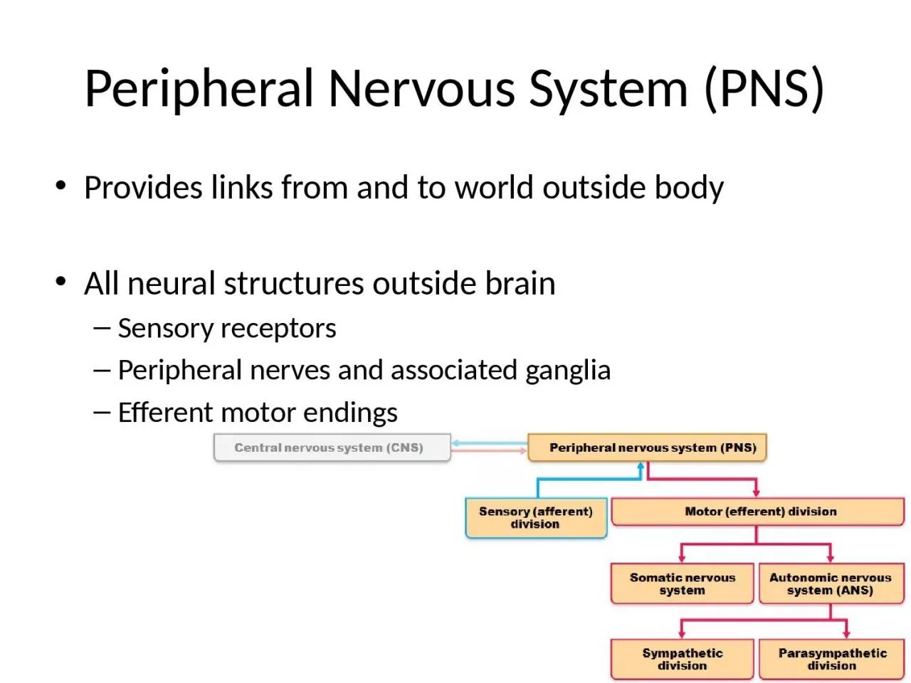
Provides links from and to world outside body All neural structures outside brain Sensory receptors Peripheral nerves and associated ganglia Efferent motor endings Sensory Receptors Specialized to respond to changes in environment ID: 915466
Download Presentation The PPT/PDF document "Peripheral Nervous System (PNS)" is the property of its rightful owner. Permission is granted to download and print the materials on this web site for personal, non-commercial use only, and to display it on your personal computer provided you do not modify the materials and that you retain all copyright notices contained in the materials. By downloading content from our website, you accept the terms of this agreement.


Slide1
Peripheral Nervous System (PNS)
Provides links from and to world outside bodyAll neural structures outside brainSensory receptorsPeripheral nerves and associated gangliaEfferent motor endings
Slide2Sensory Receptors
Specialized to respond to changes in environment (stimuli)Activation results in graded potentials that trigger nerve impulsesSensation
awareness
of
stimulus
Perception interpretation of meaning of stimulus
Both take place
in brain
Slide3Classification of Receptors
Based onType of stimulus they detectLocation in bodyStructural complexity
Slide4Classification by
Stimulus TypeMechanoreceptorsrespond to touch, pressure, vibration, and stretchThermoreceptors
sensitive
to changes in temperature
Photoreceptors
respond to light energy (e.g., retina)
Chemoreceptorsrespond to chemicals (e.g., smell, taste, changes in blood chemistry)
Nociceptors
sensitive
to pain-causing stimuli
extreme
heat or cold, excessive pressure, inflammatory
chemicals
Slide5Classification by
LocationExteroceptorsRespond to stimuli arising from outside bodyReceptors in skin for touch, pressure, pain, and temperature
Most
special sense
organs are
exteroceptors
Slide6Classification by
LocationInteroceptors (visceroceptors)Respond to stimuli arising in internal viscera and blood vesselsSensitive to chemical changes, tissue stretch, and temperature changes
Sometimes
cause discomfort
We are usually
unaware of their workings
Slide7Classification by
LocationProprioceptorsRespond to stretch in skeletal muscles, tendons, joints, ligaments, and connective tissue coverings of bones and musclesInform brain of one's movements
Slide8Classification by Receptor Structure
Simple receptors for general sensesTactile sensations touch, pressure, stretch, vibrationTemperature
Pain
muscle
sense
Modified dendritic endings of sensory neurons Receptors for special senses
Vision, hearing, equilibrium, smell, and taste (Chapter 15)
Slide9Simple
Receptors, NonencapsulatedNonencapsulated (free) nerve endingsAbundant in epithelia and connective tissuesAssociated with most
nonmyelinated
fibers ,
small-diameter group C
fibers (pain fibers) Respond mostly to temperature
and pain; pressure-induced tissue movementitch
Slide10Simple Receptors,
Nonencapsulated Free Nerve EndingsThermoreceptorsCold receptors in superficial dermis Heat receptors
in
deeper dermis
Outside those temperature ranges nociceptors activated pain
Slide11Simple Receptors,
Nonencapsulated Free Nerve EndingsNociceptorsRespond to:Pinchingchemicals from damaged
tissue
capsaicin
Slide12Simple,
NonencapsulatedLight touch receptorsTactile (Merkel) discsSensitive to light pressureHair follicle
receptors
Sensitive to hair movement
Slide13Encapsulated Dendritic Endings
Most mechanoreceptors in connective tissue capsuleTactile (Meissner's
)
corpuscles
discriminative touch
Found in nipples, external genitalia, finger tips, soles of the feet, and eyelids Lamellar
(Pacinian) corpusclesdeep
pressure and
vibration
Found in fingers, soles of feet, external genitalia, nipples
Bulbous
corpuscles
(
Ruffini
endings
)
deep
continuous
pressure
Found in dermis, hypodermis, and joint capsules
Muscle spindles
muscle
stretch
Tendon organs
stretch
in tendons
Joint
kinesthetic
receptors
joint
position and motion
Slide14From Sensation to Perception
Survival depends upon sensation and perceptionSensation the awareness of changes in the internal and external environmentPerception
the
conscious interpretation of those stimuli
Slide15Sensory Integration
Somatosensory system part of sensory system serving body wall and limbsReceives inputs fromExteroceptors, proprioceptors, and
interoceptors
Input
relayed toward head, but processed along way
Slide16Sensory Integration
Levels of neural integration in sensory systems:Receptor levelsensory receptorsCircuit level
processing
in ascending pathways
Perceptual level
processing in cortical sensory areas
Slide17Adaptation of Sensory Receptors
Adaptation is change in sensitivity in the presence of constant stimulusReceptor membranes become less responsive
Receptor
potentials decline in frequency or stop
Slide18Adaptation of Sensory Receptors
Phasic (fast-adapting) receptors signal beginning or end of stimulusreceptors for pressure, touch, and smell Tonic receptors adapt slowly or not at all
nociceptors
and most proprioceptors
Slide19Processing at the Circuit Level
Pathways of three neurons conduct sensory impulses upward to appropriate cortical regionsFirst-order sensory neuronsConduct impulses from receptor level to spinal reflexes or second-order neurons in CNSSecond-order sensory neuronsTransmit impulses to third-order sensory neuronsThird-order sensory neurons
Conduct impulses from thalamus to the somatosensory cortex (perceptual level)
Slide20Processing at the Perceptual Level
Interpretation of sensory input depends on specific location of target neurons in sensory cortexAspects of sensory perception:Perceptual detectionability
to detect a stimulus (requires summation of impulses)
Magnitude estimation
intensity
coded in frequency of impulses
Spatial discriminationidentifying site or pattern of stimulus (studied by two-point discrimination test)
Slide21Main Aspects of Sensory Perception
Feature abstractionidentification of more complex aspects and several stimulus propertiesQuality discriminationability
to identify
submodalities
of a sensation
sweet or sour tastesPattern recognition
recognition of familiar or significant patterns in stimuli melody in piece of
music
Slide22Perception of Pain
Warns of actual or impending tissue damage protective actionStimuli include extreme pressure
extreme temperature
Histamine
K
+ATPAcidsbradykinin
Some pain impulses are blocked by inhibitory endogenous opioids (e.g., endorphins)
Slide23Pain Tolerance
All perceive pain at same stimulus intensityPain tolerance varies"Sensitive to pain" means low pain tolerance, not low pain thresholdGenes
help determine pain tolerance, response to pain
medications
Slide24Homeostatic Imbalance
Long-lasting/intense pain leads to hyperalgesia (pain amplification)
chronic pain
phantom
limb
painfelt in limb no longer presentNow use epidural anesthesia to reduce
Early pain management critical to
prevent pain amplification responses
Slide25Visceral
PainStimulation of visceral organ receptorsFelt as vague aching, gnawing, burningActivated by tissue stretchingIschemia
Chemicals
muscle spasms
Slide26Referred Pain
Referred pain
Pain from one body region perceived from different region Visceral and somatic pain fibers travel in same nerves; brain assumes stimulus from common (somatic) regionE.g., left arm pain during heart attack
Slide27Structure of a Nerve
Cordlike organ of the Peripheral Nervous SystemBundle of myelinated and unmyelinated peripheral axons enclosed by connective tissue
Slide28Structure of a Nerve
Connective tissue coverings includeEndoneuriumloose connective tissue that encloses axons and their myelin sheathsPerineurium
coarse
connective tissue that bundles fibers into
fascicles
Epineuriumtough fibrous sheath around a nerve
Slide29Classification of Nerves
Most nerves are mixtures of afferent and efferent fibers and somatic and autonomic (visceral) fibersClassified according to direction transmit impulsesMixed nerves
both
sensory and motor
fibers
impulses both to and from CNSSensory (afferent) nerves
impulses only toward CNSMotor (efferent) nerves impulses only away from CNS
Slide30Classification of Nerves
Pure sensory (afferent) or motor (efferent) nerves are rare; most mixedTypes of fibers in mixed nerves:Somatic afferentSomatic efferentVisceral afferentVisceral efferent
Peripheral
nerves classified as cranial or spinal nerves
Slide31Ganglia
Contain neuron cell bodies associated with nerves in PNSGanglia associated with afferent nerve fibers contain cell bodies of sensory neuronsDorsal root
ganglia
Ganglia associated with
efferent
nerve fibers contain autonomic motor neuronsAutonomic ganglia
Slide32Regeneration of Nerve Fibers
Mature neurons are amitotic if soma of damaged nerve is intact, peripheral axon may regenerateIf peripheral axon damagedMacrophages
clean dead axon; myelin sheath intact
Axon filaments grow through regeneration tube
Axon regenerates; new myelin sheath forms
Greater distance between severed ends-less chance of regeneration
Slide33Regeneration of Nerve Fibers
Most CNS fibers never regenerateCNS oligodendrocytes bear growth-inhibiting proteins that prevent CNS fiber regenerationAstrocytes at injury site form scar tissue
that
blocks axonal
regrowth
Slide34Endoneurium
Schwann cells
Droplets
of myelin
Fragmented
axon
Site of nerve damage
The axon
becomes
fragmented at
the injury site.
1
Figure 13.5 Regeneration of a nerve fiber in a peripheral nerve. (1 of 4)
Slide352
Schwann cell
Macrophage
Macrophages
clean out the dead
axon distal to the
injury.
Figure 13.5 Regeneration of a nerve fiber in a peripheral nerve. (2 of 4)
Slide36Aligning Schwann cells
form regeneration tube
Fine axon sprouts
or filaments
Axon sprouts,
or filaments, grow
through a
regeneration tube
formed by
Schwann cells.
3
Figure 13.5 Regeneration of a nerve fiber in a peripheral nerve. (3 of 4)
Slide37Figure 13.5 Regeneration of a nerve fiber in a peripheral nerve. (4 of 4)
Schwann cell
New myelin
sheath forming
Single enlarging
axon filament
The axon
regenerates and a
new myelin sheath
forms.
4
Slide38Cranial Nerves
Twelve pairs of nerves associated with brainTwo attach to forebrain; rest with brain stem Most mixed nerves; two pairs purely sensoryEach numbered with Roman Numerals
Slide39I: The Olfactory Nerves
Sensory nerves of smell (sensory only)Receptors: nasal mucosaPurely sensory (olfactory) function
Slide40PathwayReceptors in olfactory epithelium in nasal cavity
Pass through cribriform
plate, roof of nasal cavity to olfactory bulb. Synapse in olfactory bulbOlfactory tract runs beneath frontal lobe to primary olfactory cortexI: The Olfactory Nerves
Slide41Homeostatic ImbalanceAnosmia
Partial or total loss of smell
I: The Olfactory Nerves
Slide42II: The Optic Nerves
Receptors: in retinas Purely Sensory PathwayPhotoreceptors in retinaPass through optic
canals as Optic Nerve
converge
and partially cross over at optic
chiasmaOptic tracts continue to thalamuswhere they synapse
Optic radiation fibers run to visual cortex in the occipital lobe
Slide43Homeostatic Imbalance
Damage done to optic nerve: blindness in affected eye
Damage done after optic chiasmaPartial visual lossClinical TestsAcuityPeripheral visionOphthalmascopic evaluation to view optic disc and blood vessels
II: The Optic Nerves
Slide44III: The
Oculomotor NervesOrigin: gray matter of midbrainPathway: gray matter of midbrain, near pons enters bony orbit through superior orbital fissure eye
Function:
Motor Nerve with some proprioception
Function in raising eyelid, directing eyeball
Voluntary Effectors: 4 of 6 extrinsic eye musclesInferior Oblique
Superior RectusMedial RectusInferior RectusAnd upper eyelid muscleLevator
Palpebra
Superioris
Slide45Involuntary Effectors
IrisPupillary constriction
Ciliary muscleChange in lens shape for focusingAutonomic Fibers: Parasympathetic Afferent FibersSensory from the four extrinsic eye muscles for proprioception
III: The
Oculomotor
Nerves
Slide46Clinical Tests
Pupillary reflex (pupils constrict)Convergence (Medial Rectus and Lens)
H or Z pattern for extrinsic eye muscle movementHomeostatic ImbalanceOculomotor nerve paralysis: External Strabismuseye rotates laterally at rest (Lateral Rectus muscle is not affected by CN III. PtosisDrooping eyelid (
Levator
Palpebra
Superioris loses function)Difficulty focusing (Lens)III: The
Oculomotor Nerves
Slide47IV: The
Trochlear NervesOrigin: midbrainPathway: from midbrain through superior orbital fissure to Superior Oblique musclePrimarily motor nerve that directs eyeballSome afferent fibers from proprioceptors
Slide48Clinical Testing H or Z pattern of eye movements
Looking down and laterallyDamage to
Trochlear NerveDouble vision and the inability to look down and laterallyIV: The Trochlear Nerves
Slide49Sensory and Motor: Mixed
Largest cranial nerveOrigins for motor fibers: Pons
Sensory Receptors locatedScalp, upper eyelid, nose, nasal cavity, corneal, lacrimal gland Ophthalmic divisionPalate, upper teeth, skin of cheek, upper lip, lower eyelidMaxillary divisionAnterior tongue (not taste), lower teeth, chin, temples
Mandibular
division
Cell bodies for sensory fibers located in Trigeminal Ganglion
Pathway: Runs in three branches to the faceV: The Trigeminal Nerves
Slide50V: The Trigeminal Nerves
Three divisionsOphthalmic divisionMaxillary division
Mandibular
division
Muscles of mastication
sensory
Motor
and sensory
Slide51Clinical Tests
Ophthalmic divisionCorneal reflex: check sensory to cornea by making light contact. Normal response: blinking
Maxillary divisionCheck pain/temperature responses. Hot/cold and sharp/dullMandibular divisionMotor: clench teeth, open mouth against resistance, move jaw side to sideTests muscles of mastication
V: The Trigeminal Nerves
Slide52Homeostatic Imbalance
Trigeminal neuralgiaTic
douloureux (painful twitch)Possibly vascular compression of CN VMild sensory stimuli (a breeze) can cause excruciating pain that last for seconds to minutesCan happen a hundred times a dayTreatmentNSAIDS/Analgesics are partially effective
Surgical destruction of the nerve
Loss of sensation to face
V: The Trigeminal Nerves
Slide53VI: The
Abducens NervesFibers from inferior pons enter orbits via superior orbital fissuresPrimarily a motor nerveinnervates
L
ateral Rectus muscle
Some sensory:
Proprioception from Lateral Rectus muscle
Slide54Homeostatic ImbalanceDamage causes an Internal Strabismus
Lateral Rectus muscle can not pull the eye laterally, so it rotates medially
Clinical Tests, along with III and IVH or Z pattern for eye movementVI: The Abducens Nerves
Slide55VII: The Facial Nerves
Pathway: from pons through internal acoustic meatus
emerge
through
stylomastoid
foramina to lateral aspect of faceChief motor nerves of face with 5 major branchesIncludes parasympathetic fibers
Motor functions include facial expression
Lacrimal
glands
Nasal and palatine glands
salivary glands
Submandibular
Sublingual
Somatic Motor, Voluntary
Autonomic Motor, Involuntary
Slide56Sensory function from anterior two-thirds of tongue
Taste Clinical Tests
Test for sweet/salty, etc to see if sensory portion intactClose eyes (oribicularis oculi), smile (facial muscles) to observe facial expressionsEncourage tear production (Lacrimal glands) Ammonia fumes
VII: The Facial Nerves
Slide57Homeostatic imbalance
Bells PalsyRapid onsetMay be related to Herpes Simplex 1 Virus
Paralysis of facial muscles on the affected sidePartial loss of taste sensationDrooping lower eyelid(upper eyelid controlled by CN III)Mouth corners sag (facial muscles impacted)Speaking and eating become difficultContinual tearing (loss of Lacrimal Gland control)
Eye can not close completely
May result in dry eye syndrome
VII: The Facial Nerves
Slide58VIII: The
Vestibulocochlear NervesAfferent fibers from hearing receptors (cochlear division) and equilibrium receptors (vestibular division) pass from inner ear through internal acoustic meatuses, and enter brain stem at pons-medulla borderMostly sensory function; small motor component for adjustment of sensitivity of receptors
Formerly
auditory nerve
Slide59Vestibulo
:Vestibule is related to balance and equilibriumCochlear
Cochlea houses the hearing receptors Mostly sensoryHearing and balanceMinor motor componentAdjusts sensitivity of the sensory receptorsVIII: The Vestibulocochlear Nerves
Slide60Origin: Receptors in the vestibule (balance) and cochlea (hearing).
Vestibular Nerve (cell bodies in the Vestibular Ganglion) joins Cochlear nerve (cell bodies in the Spiral Ganglion) to pass through the internal acoustic meatus to go to brain stem
VIII: The Vestibulocochlear Nerves
Slide61Clinical Tests
Evaluate air conduction/bone conduction with tuning forkCheck for hearing acuityHomeostatic Imbalance
Damage to cochlear nerve: central deafness (nerve deafness)Damage to vestibular nerve: DizzinessRapid involuntary eye movements (Nystagmus)Loss of balanceNausea and vomiting
VIII: The
Vestibulocochlear
Nerves
Slide62IX: The
Glossopharyngeal NervesOrigen/Pathway: from medulla leave skull via jugular foramen and run to throat
Motor
functions
innervates
part of tongue and pharynx
swallowingParotid salivary glands
Sensory functions
taste from pharynx and posterior tongue
general sensory (touch, pressure, pain)
impulses from pharynx and posterior
tongue
impulses
from carotid
chemoreceptors
and
baroreceptors
Chemoreceptor: sensitive to changes in blood O
2
and CO
2
levels
Baroreceptors
: sensitive to changes in blood pressure
SNS; Voluntary, skeletal
ANS; Parasympathetic
involuntary
Slide63Clinical TestingView uvula while mouth is open, saying “
aah”Use cotton-tipped swab to test for gag reflex
Check posterior tongue for ability to tasteHomeostatic ImbalanceDamage to CN IX result in impaired ability to swallow and tasteIX: The Glossopharyngeal Nerves
Slide64X: The
Vagus NervesVagus: “Vague” or wanderingOnly cranial nerves that extend beyond head and neck regionOrigin: from medulla through jugular foramen
neck, thorax, and abdomen
Most
motor fibers are parasympathetic fibers
regulate activities of
HeartLungsAbdominal viscera
Slide65Sensory fibers carry impulses from thoracic and abdominal viscera
Baroreceptors
Chemoreceptorstaste buds of posterior tongue and pharynx proprioceptive fibers from muscles of larynx and pharynx X: The Vagus Nerves
Slide66Clinical Testing
Similar to the testing for CN IX since their areas overlapHomeostatic Imbalance
Can lead to hoarseness or loss of voice if CN X is damagedDifficulty swallowingChanges in gastrointestinal motilityX: The Vagus Nerves
Slide67XI: The Accessory Nerves
Origin differs from the other cranial nervesAre spinal rootlets from the upper cervical regions instead of cranial rootlets Do
not
originate from the brain
Rootlets
pass into cranium through the foramen magnum
Slide68Mostly Motor Innervates
Trapezius muscles and Sternocleidomastoid
musclesMinor sensoryProprioceptive input from the skeletal musclesFormerly spinal accessory nerveXI: The Accessory Nerves
Slide69Clinical Testing
Muscle test the Traps and SCMs against resistanceAsk them to rotate head and shrug shoulders “I
dunno” movementHomeostatic ImbalanceHead turns toward side of injury due to SCM paralysisElevation of shoulder (shrugging) becomes difficultXI: The Accessory Nerves
Slide70XII: The Hypoglossal Nerves
Origin: Fibers from medulla exit skull via hypoglossal canal to target the tongueMixed nerve, mostly motorMotor function Innervate extrinsic
and intrinsic muscles of tongue that
contribute to swallowing and
speech
Sensory functionProprioceptive input from the skeletal muscles
Slide71Clinical TestingAsk subjects to protrude and retract tongue
(stick out their tongues)Check for deviation to one side
Homeostatic ImbalanceDifficulty with speech and swallowing Decreased movement/paralysis of tongue and eventual atrophyXII: The Hypoglossal Nerves
Slide72Spinal Nerves
31 pairs of mixed nerves named for point of issue from spinal cordSupply all body parts but head and part of neck8 cervical (C1–C8)
12 thoracic (T
1
–T
12)5 Lumbar (L1–L5)
5 Sacral (S1–S5)1 Coccygeal
(C
0
)
Slide73Spinal Nerves
Only 7 cervical vertebrae, yet 8 pairs cervical spinal nerves7 exit vertebral canal superior to vertebrae for which named1 exits canal inferior to C7Other spinal nerves exit
inferior to vertebra for which named
Slide74Figure 13.7 Spinal nerves.
Cervical plexus
Brachial plexus
Cervical
enlargement
Intercostal
nerves
Lumbar
enlargement
Lumbar plexus
Sacral plexus
Cauda equina
Cervical
nerves
C
1
– C
8
Thoracic
nerves
T
1
– T
12
Lumbar
nerves
L
1
– L
5
Sacral
nerves
S
1
– S
5
Coccygeal
nerve
Co
1
Slide75Spinal Nerves: Roots
Each spinal nerve connects to spinal cord via two rootsVentral rootsContain motor (efferent) fibers from ventral horn motor neuronsFibers
innervate skeletal muscles
Slide76Spinal Nerves: Roots
Dorsal rootsContain sensory (afferent) fibers from sensory neurons in dorsal root ganglia and conduct impulses from peripheral receptorsDorsal and ventral roots unite to form
spinal
nerves
emerge
from vertebral column via intervertebral foramina
Slide77Figure 13.8a Formation of spinal nerves and
rami
distribution.
Gray matter
White matter
Dorsal root
Dorsal and ventral
rootlets of spinal
nerve
Dorsal root
ganglion
Dorsal ramus
of spinal nerve
Ventral ramus
of spinal nerve
Spinal nerve
Sympathetic trunk
ganglion
Anterior view showing spinal cord, associated nerves, and vertebrae.
The dorsal and ventral roots arise medially as rootlets and join laterally to
form the spinal nerve.
Rami communicantes
Ventral root
Slide78Spinal Nerves: Rami
Spinal nerves quite short (~1-2 cm)Each branches into mixed ramiDorsal ramusVentral
ramus
larger
Meningeal branch tiny
reenters vertebral canalinnervates meninges and blood vessels
Rami
communicantes
(
autonomic pathways) join ventral
rami
in thoracic region
Slide79Dorsal ramus
Ventral ramus
Spinal nerve
Rami communicantes
Sympathetic trunk
ganglion
Dorsal root ganglion
Dorsal root
Ventral root
Branches of intercostal nerve
Lateral cutaneous
Anterior cutaneous
Sternum
Intercostal nerve
Cross section of thorax showing the main roots and branches of a spinal nerve.
Figure 13.8b Formation of spinal nerves and rami distribution.
Slide80Spinal Nerves: Rami
All ventral rami except T2–T12 form interlacing nerve networks
called
nerve plexuses
cervical, brachial, lumbar, and sacralThe back is innervated by dorsal
rami via several branchesVentral rami
supply
muscles of
ribs
anterolateral
thorax
abdominal
wall
Spinal
roots longer as move inferiorly in cord
Lumbar and sacral roots extend as
cauda
equina
Slide81Typical Spinal Nerve
Please note that due to differing operating systems, some animations will not appear until the presentation is viewed in Presentation Mode (Slide Show view). You may see blank slides in the “Normal” or “Slide Sorter” views. All animations will appear after viewing in Presentation Mode and playing each animation. Most animations will require the latest version of the Flash Player, which is available at http://get.adobe.com/flashplayer.
Slide82Spinal Nerves: Plexuses
Within plexus fibers criss-crossEach branch contains fibers from several spinal nervesFibers from ventral ramus go to body periphery via several routes
Each limb muscle innervated by more than one spinal nerve
Damage to
one spinal nerve
does not cause paralysis
Slide83Cervical Plexus and the Neck
Formed by ventral rami of C1–C4Most branches form
cutaneous
nerves
Innervate skin of neck, ear, back of head, and shoulders
Other branches innervate neck musclesPhrenic nerve
Major motor and sensory nerve of diaphragm receives fibers from C
3
–C
5
Irritation
hiccups
Slide84Brachial Plexus and Upper Limb
Formed by ventral rami of C5–C8 and T1
Gives rise to nerves that innervate upper limb
Major branches of this plexus:
Roots
five ventral rami
(C5–T1
Trunks
upper
, middle, and
lower
Divisions
anterior
and
posterior
Cords
lateral
, medial, and posterior
Slide85Anterior
divisions
Roots (ventral
rami
):
Posterior
divisions
Trunks
Roots
Posterior
divisions
Lateral
Posterior
Medial
Upper
Middle
Lower
C
4
C
5
C
6
C
7
C
8
T
1
Trunks
Cords
Figure 13.10a The brachial plexus.
Slide86Brachial Plexus: Five Important Nerves
Axillaryinnervates deltoid, teres minor, and skin and joint capsule of shoulderMusculocutaneous
innervates
biceps
brachii
and brachialis, coracobrachialis
, and skin of lateral forearmMedianinnervates skin
,
most flexors
, forearm
pronators
, wrist and finger flexors, thumb opposition muscles
Ulnar
supplies
flexor
carpi
ulnaris
, part of flexor
digitorum
profundus
,
most intrinsic hand muscles
, skin of medial aspect of hand, wrist/finger flexion
Radial
innervates
essentially
all extensor muscles
,
supinators
, and posterior skin of limb
Slide87Lumbar Plexus
Arises from L1–L4Innervates thigh, abdominal wall, and psoas muscleFemoral
nerve
innervates
quadriceps and skin of anterior thigh and medial surface of leg
Obturator nervepasses through
obturator foramen to innervate adductor muscles
Slide88Sacral Plexus
Arises from L4–S4Serves the buttock, lower limb, pelvic structures, and perineumSciatic nerveLongest and thickest nerve of body
Innervates hamstring muscles, adductor
magnus
, and most muscles in leg and foot
Composed of two nerves: tibial
common fibular
Slide89Anterolateral Thorax and Abdominal Wall
Ventral rami in thoracic wall run in a simple segmental patternForm intercostal nerves that supply intercostal muscles, muscle and
skin
Give off
cutaneous
branches to skinDorsal rami innervate posterior body trunk
Slide90Innervation of Skin: Dermatomes
Dermatome area of skin innervated by cutaneous branches of single spinal nerveAll
spinal nerves except C
1
participate in dermatomes
Extent of spinal cord injuries ascertained by affected dermatomesMost dermatomes overlap, so destruction of a single spinal nerve will not cause complete numbness
Slide91Peripheral Motor Endings
PNS elements that activate effectors by releasing neurotransmitters
Slide92Review of Innervation of Skeletal Muscle
Takes place at neuromuscular junctionNeurotransmitter acetylcholine (ACh) released when nerve impulse reaches axon terminal
ACh
binds to receptors, resulting in:
Movement of Na+ and K+ across membraneDepolarization of muscle cell
An end plate potential, which triggers an action potential muscle contraction
Slide93Figure 9.8 When a nerve impulse reaches a neuromuscular junction, acetylcholine (ACh) is released.
Slide 2
Synaptic vesicle
containing ACh
Synaptic cleft
Junctional
folds of
sarcolemma
Sarcoplasm of
muscle fiber
Action potential arrives at axon terminal of motor neuron.
1
Slide94Figure 9.8 When a nerve impulse reaches a neuromuscular junction, acetylcholine (ACh) is released.
Slide 3
Synaptic vesicle
containing ACh
Synaptic cleft
Junctional
folds of
sarcolemma
Sarcoplasm of
muscle fiber
Voltage-gated Ca
2+
channels open. Ca
2+
enters the axon terminal moving down its
electochemical
gradient.
2
Slide95Figure 9.8 When a nerve impulse reaches a neuromuscular junction, acetylcholine (ACh) is released.
Slide 4
Synaptic vesicle
containing ACh
Synaptic cleft
ACh
Junctional
folds of
sarcolemma
Sarcoplasm of
muscle fiber
Ca
2+
entry causes
ACh
(a
neurotransmitter) to be released
by
exocytosis
.
3
Slide96Figure 9.8 When a nerve impulse reaches a neuromuscular junction, acetylcholine (ACh) is released.
Slide 5
Synaptic vesicle
containing ACh
Synaptic cleft
Junctional
folds of
sarcolemma
Sarcoplasm of
muscle fiber
ACh
diffuses across the synaptic cleft and binds to its receptors on
the
sarcolemma
.
4
Slide97Figure 9.8 When a nerve impulse reaches a neuromuscular junction, acetylcholine (ACh) is released.
Slide 8
Action
potential (AP)
Myelinated axon
of motor neuron
Axon terminal of
neuromuscular
junction
Sarcolemma of
the muscle fiber
Synaptic vesicle
containing ACh
Synaptic
cleft
Junctional
folds of
sarcolemma
Sarcoplasm of
muscle fiber
Postsynaptic
membrane
ion channel opens;
ions pass.
Ion channel closes;
ions cannot pass.
ACh
binding opens ion
channels in the receptors that
allow simultaneous passage of
Na
+
into the muscle fiber and K
+
out of the muscle fiber. More Na
+
ions enter than K
+
ions exit,
which produces a local change
in the membrane potential called
the end plate potential.
Degraded ACh
ACh
Acetylcho-
linesterase
5
Slide98Figure 9.8 When a nerve impulse reaches a neuromuscular junction, acetylcholine (ACh) is released.
Slide 8
Action
potential (AP)
Myelinated axon
of motor neuron
Axon terminal of
neuromuscular
junction
Sarcolemma of
the muscle fiber
Junctional
folds of
sarcolemma
Ion channel closes;
ions cannot pass.
ACh
effects are terminated by
its breakdown in the synaptic
cleft by
acetylcholinesterase
and
diffusion away from the junction.
ACh
Acetylcho-
linesterase
6
Slide99Review of Innervation of Visceral Muscle and Glands
Autonomic motor endings and visceral effectors are simpler than somatic junctionsBranches form synapses using varicosities
Visceral
motor responses slower than somatic responses
Slide100Reflexes
Inborn (intrinsic) reflex - rapid, involuntary, predictable motor response to stimulusExample – maintain posture, control visceral activitiesCan be modified by learning and conscious effortLearned (acquired) reflexes result from practice or repetition, Example – driving skills
Slide101Reflex Arc
Components of a reflex arc (neural path)Receptorsite of stimulus action
Sensory neuron
transmits
afferent impulses to CNS
Integration center
either monosynaptic or polysynaptic region within CNSMotor neuron
conducts
efferent impulses from integration center to effector organ
Effector
muscle
fiber or gland cell that responds to efferent impulses by contracting or secreting
Slide102Figure 13.15 The five basic components of all reflex arcs.
Stimulus
Skin
Receptor
1
Slide103Figure 13.15 The five basic components of all reflex arcs.
Stimulus
Skin
Receptor
Sensory neuron
1
2
Slide104Figure 13.15 The five basic components of all reflex arcs.
Stimulus
Skin
Receptor
Sensory neuron
Integration center
1
2
3
Interneuron
Slide105Figure 13.15 The five basic components of all reflex arcs.
Stimulus
Skin
Receptor
Sensory neuron
Integration center
Motor neuron
1
2
3
4
Slide106Figure 13.15 The five basic components of all reflex arcs.
Stimulus
Skin
Receptor
Sensory neuron
Integration center
Motor neuron
Effector
1
2
3
4
5
Slide107Figure 13.15 The five basic components of all reflex arcs.
Stimulus
Skin
Receptor
Sensory neuron
Integration center
Motor neuron
Effector
1
2
3
4
5
Interneuron
Spinal cord
(in cross scetion)
Slide108Reflex Arc
Please note that due to differing operating systems, some animations will not appear until the presentation is viewed in Presentation Mode (Slide Show view). You may see blank slides in the “Normal” or “Slide Sorter” views. All animations will appear after viewing in Presentation Mode and playing each animation. Most animations will require the latest version of the Flash Player, which is available at http://get.adobe.com/flashplayer.
Slide109Reflexes
Functional classificationSomatic reflexesActivate skeletal muscleAutonomic (visceral) reflexesActivate visceral effectors
smooth
or cardiac muscle or
glands
Slide110Spinal Reflexes
Spinal somatic reflexesIntegration center in spinal cordEffectors are skeletal muscleTesting of somatic reflexes important clinically to assess condition of nervous systemIf exaggerated, distorted, or absent
degeneration/pathology of specific nervous system regions
Slide111Stretch and Tendon Reflexes
To smoothly coordinate skeletal muscle nervous system must receive proprioceptor input regardingLength of muscleFrom muscle spindlesAmount of tension in muscleFrom tendon organs
Slide112Functional Anatomy of Muscle Spindles
Composed of 3–10 modified skeletal muscle fibers intrafusal muscle fibers wrapped
in connective tissue capsule
Effector
fibers extrafusal muscle fibers
Slide113Intrafusal Fibers
Noncontractile in central regions lack myofilaments) Two types of afferent endings
Anulospiral
endings
primary sensory endingsEndings wrap around spindle; stimulated by rate
and degree of stretchFlower spray endings
secondary
sensory
endings
Small axons at spindle ends; respond to
stretch
Slide114Muscle Spindles
Excited in two waysExternal stretch of muscle and muscle spindleInternal stretch of muscle spindle
Activating
motor neurons stimulates ends to contract, thereby stretching spindle
Stretch causes increased rate of impulses to spinal cord
Slide115Slide116Muscle Spindles
Contracting muscle reduces tension on muscle spindleSensitivity lost unless muscle spindle shortened
Slide117The Stretch Reflex
Maintains muscle tone in large postural muscles, and adjusts it reflexivelyCauses muscle contraction in response to increased muscle length (stretch)
Slide118Stretch Reflexes
How stretch reflex worksStretch activates muscle spindleSensory neurons synapse directly with motor neurons in spinal cord motor neurons cause stretched muscle to contract
All
stretch reflexes are
monosynaptic
and ipsilateral
Slide119Stretch Reflexes
Reciprocal inhibition also occursSpecific fibers synapse with interneurons that inhibit motor neurons of antagonistic musclesExample
In
patellar
reflex:
stretched muscle (quadriceps) contracts antagonists (hamstrings) relax
Slide120Slide121Stretch Reflexes
Positive reflex reactions indicateSensory and motor connections between muscle and spinal cord intactStrength of response indicates degree of spinal cord excitability
Hypoactive
or absent
reflexes indicate
peripheral nerve damage ventral
horn injuryHyperactive responses indicatelesions of
corticospinal
tract
Slide122The Tendon Reflex
Polysynaptic reflexesHelps prevent damage due to excessive stretch Important for smooth onset and termination of muscle contraction
Slide123The Tendon Reflex
Produces muscle relaxation (lengthening) in response to tensionContraction or passive stretch activates tendon reflex Afferent impulses transmitted to spinal cord Contracting muscle relaxes; antagonist contracts (reciprocal activation)
Information
transmitted simultaneously to cerebellum and used to adjust muscle tension
Slide124Slide 2
Figure 13.19 The tendon reflex.
Quadriceps strongly contracts.
Tendon organs are activated.
Spinal cord
Quadriceps
(extensors)
Tendon organ
Hamstrings
(flexors)
+
+
+
–
+ Excitatory synapse
– Inhibitory synapse
1
Slide125Slide 3
Figure 13.19 The tendon reflex.
Afferent fibers synapse with
interneurons
in the spinal cord.
Interneurons
Spinal cord
Quadriceps
(extensors)
Tendon organ
Hamstrings
(flexors)
+
+
+
–
+ Excitatory synapse
– Inhibitory synapse
2
Slide126Slide 4
Figure 13.19 The tendon reflex.
Spinal cord
Quadriceps
(extensors)
Tendon organ
Hamstrings
(flexors)
Efferent impulses
to
muscle
with stretched
tendon
are damped.
Muscle relaxes,
reducing
tension.
+
+
+
–
+ Excitatory synapse
– Inhibitory synapse
3a
Slide127Slide 5
Figure 13.19 The tendon reflex.
Spinal cord
Quadriceps
(extensors)
Tendon organ
Hamstrings
(flexors)
Efferent impulses
to
antagonist
muscle
cause
it to contract.
3b
+
+
+
–
+ Excitatory synapse
– Inhibitory synapse
Slide128Tendon Reflex Responses
Example of a scale used for deep tendon reflexes:
The usual gradation for deep tendon reflexes ranges from 0 to 4 0 Absent reflex1 Feeble reflex2 Normal reflex3 Brisk reflex4 Brisk reflex with clonussometimes a fifth possibility is listed:
5 Brisk reflex with sustained
clonus
Slide129Tendon Reflex Repsones
Clonus
A series of short rhythmic contractions and relaxationsAssociated with hyperexcitability and upper motor neuron damageCommon with stroke and spinal cord injury Can last from several seconds to several minutesSustained clonus: always pathological
More than 4 beats
Slide130The Flexor and Crossed-Extensor Reflexes
Flexor (withdrawal) reflexInitiated by painful stimulusCauses automatic withdrawal of threatened body partIpsilateral
and polysynaptic
Protective; important
Brain can override
E.g., finger stick for blood test
Slide131Reflex Arc
Please note that due to differing operating systems, some animations will not appear until the presentation is viewed in Presentation Mode (Slide Show view). You may see blank slides in the “Normal” or “Slide Sorter” views. All animations will appear after viewing in Presentation Mode and playing each animation. Most animations will require the latest version of the Flash Player, which is available at http://get.adobe.com/flashplayer.
Slide132Flexor and Crossed-Extensor Reflexes
Crossed extensor reflexOccurs with flexor reflexes in weight-bearing limbs to maintain balanceConsists of ipsilateral withdrawal reflex and contralateral extensor reflex
Stimulated side withdrawn (flexed)
Contralateral
side extended
e.g., step barefoot on broken glass
Slide133Figure 13.20 The crossed-extensor reflex.
+ Excitatory synapse
– Inhibitory synapse
Afferent
fiber
Efferent
fibers
Flexes
Arm movements
Extensor
inhibited
Flexor
stimulated
Interneurons
Efferent
fibers
Flexor
inhibited
Extensor
stimulated
Extends
Site of reciprocal
activation:
At the
same time, the
extensor muscles
on the opposite
side are activated.
Site of stimulus:
A noxious stimulus
causes a
flexor
reflex
on the same
side, withdrawing
that limb.
+
+
+
–
–
+
Slide134Superficial Reflexes
Elicited by gentle cutaneous stimulationDepend on upper motor pathways and cord-level reflex arcsBest known:Plantar reflexAbdominal reflex
Cremasteric
reflex
Slide135Superficial Reflexes: Plantar Reflex
Test integrity of cord from L4 – S2Stimulus stroke
lateral aspect of sole of foot
Expected Response
flexion
of toes
Slide136Damage to motor cortex or
corticospinal tracts
yields abnormal response Babinski's sign
Hallux
extends
digits fan laterallyNormal in infant to ~1 year due to incomplete myelination
Superficial Reflexes: Plantar Reflex
Slide137Superficial Reflexes: Abdominal
ReflexesTest integrity of cord from T8 – T12
Results in
contraction
of abdominal muscles
movement of umbilicus in response to stroking of skinVary in intensity from one person to another
Absent when corticospinal tract lesions are present
Slide138Superficial Reflexes: Cremasteric
The cremaster muscle is a skeletal muscle responsible for elevating the testesLight touch to the medial thigh results in a superficial reflexThe Cremaster muscle is activated, and the ipsilateral testis is elevated
Evaluation helpful to evaluate nerve function following hernia repair