
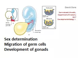
Development of gonads David Dora Semmelweis University Department of Anatomy 2017 Developmental biology I Determination of sex Formation and migration of PGCs Formation of gonad primordia colonisation by PGCs ID: 912302
Download Presentation The PPT/PDF document "Sex determination Migration of germ cell..." is the property of its rightful owner. Permission is granted to download and print the materials on this web site for personal, non-commercial use only, and to display it on your personal computer provided you do not modify the materials and that you retain all copyright notices contained in the materials. By downloading content from our website, you accept the terms of this agreement.


Slide1
Sex determinationMigration of germ cellsDevelopment of gonads
David Dora
Semmelweis University,
Department of Anatomy
2017
Developmental biology I.
Slide2Determination of sex
Formation and migration of PGCs
Formation of gonad primordia, colonisation by PGCs
Establisment of primary and secondary sexual characteristics
Slide3Genetical sex, chromosomal sex
Slide4In higher vertebrates the genetical sex determination dominates homotherm animals
In heterotherm animals it can be temperature-dependent (TSD), or environmental-dependent SD, ESD)
E.G.: turtles, crocodiles
Genetic sex is determined during fertilization
Slide5Slide6The male sex depends on the presence of Y chromosome
Gondadal sex
XY
– testes develop
XX
– ovaries develop
Slide7The genotype will decide whether male (testis) or female gonads (ovaries) will develop.
When the gonadal and phenotypical sex differs
pseudohermaphrodi
sm
.
The only „normal” man
Causes of male pseudohermaphroditism:
Development disorder of testes
5-alpha-reduktase deficiency
lack of testosteron
Androgenic insensitivity (receptor defect)
Testicular feminisation
Causes of female pseudohermaphroditism:
- Congenital adrogenital syndrome
Congenitális adrogenitalis hyperplasia (CAH)
-
Androgen producing tumor in the mother
Slide8Inactivation of X chromosome in females, „dosage compensation”
Many genes on the X chromosome have nothing to do with sexual traits
they have no homologous area on the Y chromosome
These genes would be present in „two doses” in females
The solution is the inactivation of one X chromosome in somatic cells
X-Inactive Specific Transcript (XIST) gene is essential for inactivation
The inactivated chromosome persists as a „Barr-body”
Activating factor: RNF12
Inhibitor: OCT4, SOX2, Nanog
Slide9Barr-body
Slide10Fluorescent X chromosomes in mouse- paternal:
red- maternal: green
Az X-inactivation is not uniformal in our somatic cells
mosaicism
Neuron,
January
8, 2014
During early development every cell choose, which X chromose it will inactivate (maternal or paternal)
undescribed random mechanism
After the „choosing” the cell line derived from the progenitor cell will all express the same X chromosome and the genes localised in it.
Slide11Genetical sex is decided at the moment of fertilization
The only morphological sign until the end of 7th gestational week:
The presence of Barr-body
Slide12On the 6th week the PGCs residing in the wall of the yolk sac start to migrate to the dorsal mesogastrium and the dorsal body wall and will colonize the gonad primordia medially from the mesonephros (plica genitalis, genital ridge) at the level of the 10. thoracal segment. From the coeloma epithelium of the body wall will the somatic supporting cells develop from, that will assist their long maturation process
Slide13Primordial germ cells, PGCs
Migration of PGCs in mouse embryo.
PGCs express alkalic phosphatase
TNAP (tissue nonspecific alkaline phosphatase) - histochemistry
Slide14Slide15Migrating
(A,B) and arrived (C) PGCs in mouse embryo (SSEA-1 IH)
http://embryology.med.unsw.edu.au/embryology/index.php?title=Primordial_Germ_Cell_Migration_Movie#Mouse_E9.0_Primordial_Germ_Cell_Migration
Slide16Vincent, S. D. et al. Development 2005;132:1315-1325
Expression of BLIMP1 in early mouse embryo
Specific marker for PGC precursors
Transcriptional repressor, product of PRDM1 gene
In adult, it is a repressor of TGF-beta
promotes immunological response in viral infections B-cell recruitement
Important regulator protein in hematopoiesis
Repressors of Hox genes, that is essential for PGC specification and differentiation
Slide17Vincent, S. D. et al. Development 2005;132:1315-1325
Morfogenesis and tissue patterning is intact in BLIMP-1 deficiency
Slide18Vincent, S. D. et al. Development 2005;132:1315-1325
A Blimp 1 mutant mice’s PGC development is impaired
Slide19ALK2
is essential for PGC formation
a
ctivin receptor-
l
ike
k
inase-2
Slide20BMP
signaling
and
PGC
formation
ExE
Extraembryonic ectoderm ALK2 BMP receptor type I
VE Visceral endoderm
EE Proximal epiblast
Slide21Human fetus 5,5 embryonic week
PGCs among hindgut cells
(Oct4-immunohistochemistry)
Slide22Molecular Human Reproduction, Vol.16, pp. 621–631, 2010
Recent studies suggest PGCs migrate along autonomic nerve fibers in dorsal mesentery
Schwann cells have important role in the direction of PGC migration by chemotactic factors and growth homones
PGCs migrate along autonomic nerve fibers
Slide23Slide24Schmoll 2002
Disorders of PGC migration
Sacrococcygeal (A) és oropharyngeal (B) teratomas in fetuses
Migrating PGCs arrest early in migration, but continue to proliferate
Slide25Dermoid cyst
Slide26Slide27II. Formation of phenotypeGonadal sex
Gonads are derivatives of intermedier mesoderm
The gonadal primordia (anlage) are localised in the posterior wall of the embryo, medially from the mesonephros található. At the 5th week of development the posterior body wall thickens, altogether with the coeloma epithelium
genital ridge
(plica genitalis).
The phoenomen happens simultaneusly in both sexes! The gonads here are still indifferent
no sex determination is possible
Slide28Formation of the male gonad needs the presence of the Y chromosome
(Sry gene) on the short arm of Y chromosome).
The male phenotype is determined by the testosteron production of the differentiationg male gonad
2. Formation of the female gonad requires the presence of 1 X chromosome and the
lack of Y chromosome
, but the formation of a functionally sound ovarium needs both X chromosomes.
Turner syndrome (X0) infertility
Slide29Molecular background
Short arm of Y chromose is essential for the formation of testes
http://physrev.physiology.org/content/78/1/1
The Sry gene codes for the 223 AA polypeptide SRY protein
Slide30Product of the SRY gene the SRY protein, or TDF (testis determining factor) is required for testis-formation
Slide31The supporting cells (Sertoli, Leydig) are the primary target of TDF
- Testis Determining Factor has no effect on PGCs
-THE TESTIS STILL DEVELOPS WITHOUT MALE PGCs
- a
TDF (SRY)
‘s main target is the
Dax1 gene
, on the X chromosome
-In the ovaries it acts as an anti-testis gene
- During the formation of testis DAX1 downregulates
- DAX1 activity persists in the ovaries
DAX1 (dosage-sensitive sex reversal, adrenal hypoplasia critical region, on chromosome X, gene 1)
Mechanism of action of Testis Determining Factor
Slide32A SOX9 transcription factor expression
is continous in Sertoli cells expression starts immediately after the production of
SRY protein
SOX-9 +/- human: growth impairment and
¾ of
XY-patients are females
Sex-determination cascade:
SRY
expression induces SOX9 expression
New gene is activated
SF-1
-et.
Az SF-1 (steroidogenetic factor-1) is important in the synthesis of sexual steroid hormones, it also binds to the promoter of AMH (Anti-müller) hormone.
Autosomal genes in sex-determination: SOX-9
Slide33Szex determináció, korai lépések
Slide34Slide35Slide361) Supporting cell line - Derivatives of primary sex chords of the coelomic epithelium:
Sertoli cells in testes – AMH – Regression of Müller duct
- Derivatives of secondary sex chords
of the coelomic epithelium:
Granulosa cells (follicular cells) in the ovaries
2) Steroid – secreting cell line:
- From mesenchyme: Leydig cells in testes (8th week from: testosteron, androstendion production
Peak of testosteron production is on the 17-18th week: Differentiation of male genital tract and external genitalia
After week 18th Leydig cells go through a relative regression
Theca cells appear in ovaries during puberty
Somatic cells of the gonads:
Slide37- Primitive sex chords persist in the medulla – testis chords
- In the chords pre-Sertoli cells differentiate first male specific cell type
- PGCs arrive and establish contact with pre-Sertoli cells
they not yet begin meiosis
-
They divide with mitosis slowly until puberty
- From sex chords: tubuli seminiferi, rete testis, tubuli recti develop
- From the ducts of mesonephros: ductuli efferentes testis, ductus epididymis
XY embryo
Slide38Primitive sex chords degenerate in the medulla (rete ovarii)
Coelomic epithelium forms new sex chords in the mesenchyme secondary sex chords
Definitive ovaries form from the cortical part
Oogonia proliferate with mitosis during fetal life
The first meiotic division begins in some oogonia during the 12-16th week → PRIMARY OOCYTES – sorrounded by follicular cells: primordial follicle
Meiosis of primary oocytes arrest in the diploten phase of prophase
Coelomic epithelial cells invade the mesenchyme and prefollicular cells differentiate from the
No more oogonia goes through mitosis after birth!
XX embryo
Slide39In the absence of Y chromose
female development
Slide40Slide41Slide42Slide43