
Degeneration Figure LegendFigure 1Skeletal muscleDegeneration in a male Harlan SpragueDawley rat from a subchronic study A central myofiber is swollen and hypereosinophilicarrows and a fragmented ID: 946102
Download Pdf The PPT/PDF document "Skeletal Muscle" is the property of its rightful owner. Permission is granted to download and print the materials on this web site for personal, non-commercial use only, and to display it on your personal computer provided you do not modify the materials and that you retain all copyright notices contained in the materials. By downloading content from our website, you accept the terms of this agreement.


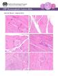
Skeletal Muscle Degeneration Figure Legend:Figure 1Skeletal muscleDegeneration in a male Harlan SpragueDawley rat from a subchronic study. A central myofiber is swollen and hypereosinophilic(arrows), and a fragmented segment of another fiber (arrowhead) demonstrates segmental degeneration. Figure 2Skeletal muscleDegeneration in a male Harlan SpragueDawley rat from a subchronic study. An enlarged, hypereosinophilic muscle fiber with subtle vacuolation is present in an otherwise normal muscle bundle. Skeletal muscleDegeneration in a male Harlan SpragueDawley rat from a subchronic study. A rounded fiber contains multiple peripheral nuclei, a single internal nucleus, and abundancytoplasmic vacuoles. Figure 4Skeletal muscleDegeneration in a male Harlan SpragueDawley rat from a subc
hronic study. Multiple macrophages and interstitial cells have phagocytized degenerative portions of an enlarged muscle fiber. Figure 5Skeletal muscleDegeneration in a male F344/N rat from a chronic study. In this cross section of muscle, several adjacent muscle fibers exhibit multiple features of degeneration;ncreased sarcolemmal nuclei are indicative of an early regenerative response. FigurSkeletal muscleDegeneration in a male Harlan SpragueDawley rat from a subchronic study. Degeneration is represented by a swollen, hyalinized, and partly fragmented muscle fiber. Figure 7Skeletal muscleDegeneration in a male Harlan SpragueDawley rat from a subchronic study. A fragmented and partly hyalinized muscle fiber has lost its striations and is accompanied by early infiltration of macropha
ges. CommentDegenerated muscle can grossly appear either pale or dark. Histologically, degenerating myofibers can exhibit a variety of microscopic changes, including cell swelling, hypereosinophilia, 2 Skeletal Muscle Degeneration inflammation and hemorrhage, should not be diagnosed separately unless warranted by severity but should be described in the narrative. References: Berridge BR, Van Vleet JF, Herman E. 2013. Cardiac, vascular, and skeletal muscle systems. In: Haschek and Rousseaux’s Handbook of Toxicologic Pathology, 3rd ed (Haschek WM, Rousseaux CG, Wallig MA, Bolon B, Ochoa R, Mahler MW, eds). Elsevier, Amsterdam, 1635-Greaves P. 2007. Musculoskeletal system. In: Histopathology of Preclinical Toxicity Studies, 3rd ed. Elsevier, Oxford, 160-214. Greaves P, Seely
JC. 1996. Non-proliferative lesions of soft tissues and skeletal muscle in rats, MST-1. In: Guides for Toxicologic Pathology. STP/ARP/AFIP, Washington, DC. Greaves P, Chouinard L, Ernst H, Mecklenburg L, Pruimboom-Brees IM, Rinke M, Rittinghausen S, Thibault S, von Erichsen J, Yoshida T. 2013. Proliferative and non-proliferative lesions of the rat and mouse soft tissue, skeletal muscle, and mesothelium. J Toxicol Pathol 26(3 suppl):1S-26S. Abstract: http://www.ncbi.nlm.nih.gov/pubmed/25035576 Haschek WM, Rousseaux CG, Wallig MA. 2010. Cardiovascular and skeletal muscle systems. In: Fundamentals of Toxicologic Pathology, 2nd ed. Academic Press, San Diego, . Leninger JR. 1999. Skeletal muscle. In: Pathology of the Mouse (Maronpot R, Boorman Gaul eds). Cache River Press, St Louis, .
McDonald MM, Hamilton BF. 1990. Bones, joints, and synovia. In: Pathology of the Fischer Rat: Reference and Atlas (Boorman G, Eustis SL, Elwell MR, Montgomery CA, MacKenzie WF, eds). Academic Press, San Diego, 193-Vahle JL, Leininger JR, Long PH, Hall DG, Ernst H. 2013. Bone, muscle, and tooth. In: Toxicologic Pathology Nonclinical Safety Assessment (Sahota PS, Popp JA, Hardisty JF, Gopinath C, eds). CRC Press, Boca Raton, FL, 561-Van Vleet JF, Valentine BA. 2007. Muscle and tendon. In: Jubb, Kennedy, and Palmer’s Pathology of Domestic Animals, 5, Vol 1 (Grant MG, . Elsevier, Edinburgh, . Weller AH, Magliato SA, Bell KP, Rodenberg NL. 1997. Spontaneous myopathy in the SJL/J mouse: Pathology and strength loss. Muscle Nerve 20:72-Abstract: http://www.ncbi.nlm.nih.gov/pubmed/8995586