
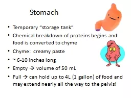
alnoubani MDMRCS asymmetrical pearshapedmost proximal abdominal organ of the digestive tract Cardia connected to esophagus Fundus bounded superiorly by diaphragm and laterally by the spleen ID: 928166
Download Presentation The PPT/PDF document "Stomach and duodenum Omar" is the property of its rightful owner. Permission is granted to download and print the materials on this web site for personal, non-commercial use only, and to display it on your personal computer provided you do not modify the materials and that you retain all copyright notices contained in the materials. By downloading content from our website, you accept the terms of this agreement.


Slide1
Stomach and duodenum
Omar alnoubani MD,MRCS
Slide2asymmetrical,
pearshaped,most proximal abdominal organ of the digestive tract.Cardia connected to esophagus.
Fundus
bounded superiorly by diaphragm and laterally by the spleen.
The angel of His is where the fundus meets the left side of the GE junction.The body contain most of parietal cells .Antrum the distal 25-30 percent of the stomach .
Slide3The organs that commonly abut the stomach are the liver,
colon, spleen, pancreas, and occasionally the kidneyThe lesser curvature is tethered to the liver by the
hepatogastric
ligament
Slide4Slide5Slide6the
left (anterior) and right (posterior)vagal trunks mnemonic LARP
The
antrior
to liver ,hepatoduodenal ligament as ant. N. of laterjetThe post. To celiac plexus .The Ns of laterjet terminate near the angularis
incisura
as crow’s foot
Slide7criminal nerve of
Grassi The branch that post
vagus
send to post.
Fundus arise above the esophageal hiatus Easley missed durning truncal vagotomy and HSV
The Ach is the most imp. Neurotransmitter in acid secretion .
The
sympathatic
supply T5-T10
Slide8Slide9Slide10Parietal
: • Location: Body • Function: secrete acid and intrinsic factor
Mucus:
Location: Body,
Antrum Function: mucus production Chief: Location: Body Function: produce Pepsin
Slide11Surface epithelium:
Location: Diffuse Function: produce mucus, bicarb, prostaglandins
ECL:
Location: Body
Function: Histamine production G cells: Location: Antrum Function: Gastrin production
Other cells ….
Slide12Acid Secretion
Two forms: Basal Acid Secretion Stimulated Acid Secretion
Slide13Stimulated Acid Secretion
Three Phases: Cephalic phase Gastric phase Intestinal Phase These phases occur concurrently NOT consecutively.
Slide14Cephalic Phase
Originates with the sight, smell, thought or taste of food. Stimulates the cortex and hypothalamus. Signals cause
Vagus
to release Ach, Ach causes increase in parietal cell acid production.
Accounts for 20-30% of acid production.
Slide15Gastric Phase
Begins when food enters the gastric lumen (gastric distention). Digestion products stimulate the G cells, they release gastrin, parietal cells release acid. Distention alone can increase acid production.
Accounts
for 60-70%
of acid production. Phase lasts until the stomach is empty.
Slide16Intestinal Phase
Poorly understood. initiated by chyme entering the small bowel.
Accounts for ~10% of acid production.
Slide17Gastric Hormones
Gastric Hormones: Chemical messengers that regulate intestinal and pancreatic function. The “gut” is the largest endocrine organ in the body. The messengers can act as:
Endocrine: distant target
Paracrine: close target Autocrine: self target Neurocrine: neurotransmitter or stimulator
Slide18Gastric Hormones
Gastrin Somatostatin Gastrin-releasing peptide (GRP)
Histamine
Leptin
Ghrelin
Slide19Gastrin
Synthesis: G-cells in the antrum Release: AA, protein, vagal tone,
antral
distention, GRP, pH > 3.0, ETOH, Histamine.
Inhibition: pH < 3.0, somatostatin, secretin, CCK, VIP, GIP, glucagon. Target cells: Parietal and Chief cells
Slide20Gastrin
Action: Stimulates acid secretion :
Direct action on parietal cells
Potentiating interaction with histamine
Possible: releasing of histamine Increases release of lytes & water from stomach, pancreas, liver and Brunner’s glands
Stimulates motility in stomach, intestine, and gall bladder
Inhibits contraction of pylorus and sphincter of
Oddi
.
Stimulates GI mucosal growth.
Slide21Somatostatin
Synthesis: CNS, antrum, fundus, sm. bowel, colon, and D cells in pancreas.
Release:
Antral acidification Fats, protein, acid in duodenum Pancreatic: glucose, amino acids, CCK Inhibition: Release of acetyl-
choline
from
vagal
nerve fibers
Slide22Action:
The “master off switch” Inhibits the release of most GI hormones •Inhibits pancreatic and GI secretion(s)
Inhibits intestinal motility.
Clinical:
Octreotide- decrease fistula output Treatment of esophageal variceal bleed
Can ameliorate symptoms of endocrine tumors
Slide23Gastrin-Releasing Peptide.
GRP is the mammalian equivalent of bombesin, a hormone discovered more than two decades ago in an extract of skin from a frog.
Synthesis: Gastric
antrum
, small bowel mucosa Release: vagal stimulation
Slide24GRP
Action: The “master on switch” Stimulates the release of all GI hormones (Secretin).
Stimulates GI secretion
Stimulates GI motility
most important: stimulates gastric acid secretion and release of antral gastrin Stimulates bowel and pancreatic mucosal growth
Slide25Histamine
Stimulates parietal cells Found in the acidic granules of ECL cells and resident Mast cells. Release is stimulated by: Gastrin
, acetyl-
choline
, epinephrine Inhibited by Somatostatin.
Slide26Leptin and
ghrelin Leptin is a protein primarily synthesized in adipocytes
.
It is also made by chief cells in the stomach, the main source of
leptin in the GI tract. Leptin works at least in part via vagally mediated pathways to decrease food intake in animals. Not surprisingly,
leptin
, a satiety signal hormone, and
ghrelin
, a hunger signal hormone
, are both primarily synthesized in the stomach.
Slide27PEPTIC ULCER DISEASE
Peptic ulcers are focal defects in the gastric or duodenal mucosa that extend into the submucosa or deeper. They may be acute or chronic and, ultimately, are caused by an imbalance between mucosal defenses and acid/peptic injury.
Slide28Slide29Epidemiology
PUD is one of the most common GI disorders in the United States with a prevalence of about 2%, and a lifetime cumulative prevalence of about 10%, peaking around age 70 years .Recent studies have shown an
increasein
the rates of hospitalization and mortality in elderly patients for the peptic ulcer complications of bleeding and perforation.
This may be due in part to the increasingly common use of NSAIDs and aspirin in this elderly cohort, many of whom also have H. pylori infection.
Slide30Pathophysiology and Etiology
No acid no ulcer
Slide31In general
H. pylori predisposes to ulceration, both by acid hypersecretion and by compromise of mucosal defense
mechanisms.
NSAID use causes ulcers predominantly by compromise of mucosal defenses.
Slide32Helicobacter pylori Infection
With specialized flagella and a rich supply of urease, H. pylori is uniquely equipped for survival in the hostile environment of the stomach.
About50% of the world’s population is infected with
H. pylori, a
major cause of chronic gastritisHave a role in gastric lymphoma
Slide33The organism possesses the enzyme
urease, which converts urea into ammonia and bicarbonate, thus creating an environment around the bacteria that buffers the acid secreted by the stomach. The ammonia is damaging to the surface epithelial cells.
Slide34Slide35Gastric ulcers
Slide36Slide37The most common, Johnson type I gastric ulcer, is typically located near the
angularis incisura on the lesser curvature, close to the border between antral and corpus mucosa.
Patients with type I gastric ulcer usually have normal or decreased acid secretion .
Slide38Nonsteroidal
Anti-Inflammatory Drugs in Peptic UlcerDiseaseChronic use of NSAIDs (including aspirin) increases the risk of peptic ulcer disease about 5-fold and upper GI bleeding about 4-fold.
Complications of PUD (specifically hemorrhage and perforation) are much more common in patients taking NSAIDS
Slide39Indications of acid suppression in patients taking NSAIDs
Slide40Clinical Manifestations
More than 90% of patients with PUD complain of abdominal pain. The pain is typically nonradiating, burning in quality, and located in the epigastrium
.
Slide41Slide42Medical management
Slide43Surgical Treatment of Peptic Ulcer Disease
Indications :BleedingPerforationObstructionIntractability or
nonhealing
Malignancy .
Slide44Mainly
oversewing of the bleeders and patch closure for perforating .The use of vagotomy is increasingly uncommon because of PPI.
Slide45Before PPI !!!
Slide46Bleeding
The most common complication Three-fourths of the patients who come to the hospital with bleeding peptic ulcer will stop bleeding if given acid suppression and nothing by mouth. However, one fourth will continue to bleed or will rebleed
after an initial quiescent period, and virtually all the mortalities (and all the operations for bleeding) occur in this group .
Slide47Slide48Slide49Patients with massive bleeding from
high-risk lesions (e.g., posterior duodenal ulcer with erosion of gastroduodenalartery, or lesser curvature gastric ulcer with erosion of left gastric artery or branch) should be considered for
early operation
.
Early operation should also be considered in patients more than 60 years of age, those presenting in shock, those requiring more
than four units of blood in 24
hours or eight units of blood in 48 hours, those with
rebleeding
, and those with
ulcers >2 cm
in diameter
The Mortality rate of surgery is around 20%.
Slide50Slide51Perforation
2nd M.C.C.Acute abdominal pain Initially chemical peritonitis then bacterial
80% of x-rays show free air .
Mx
: resuscitation Operative vs non operative Graham patch
Slide52Slide53G.O.O
occurs in no more than 5% of patients with PUD. Presented with nonbiliuos vomiting It is usually due to duodenal or
prepyloric
ulcer disease, and may be acute from inflammatory swelling or chronic from scarring .
Always role out malignant cause .The standard operation for obstructing PUD is vagotomy and antrectomy
Slide54Intractable or
Nonhealing Peptic UlcerShould raise red flags for the surgeon: Maybe the patient has a missed
cancer
maybe the patient is
noncompliant (not taking prescribed PPI, still taking NSAIDs, still smoking)maybe the patient has Helicobacter despite the presence of a negative test or previous treatment.
Slide55Slide56Slide57Slide58Zollinger-Ellison Syndrome
ZES is caused by the uncontrolled secretion of abnormal amounts of gastrin by a duodenal or pancreatic neuroendocrine
tumor (
i.e.,gastrinoma
). Most cases (80%) are sporadic, but 20% are inherited.Familial with MEN I usually the have multiple gastrinoma tumors, and surgical cure is less
common.
Slide59sporadic
gastrinomas are more often solitary and are more often amenable to surgical cure
Currently, about 50% to 60% of
gastrinomas are malignant, with lymph node, liver, or other distant metastases at operation. Five-year survival in patients presenting with metastatic disease is approximately 40%. The larger the primary gastrinoma, the
higher
the likelihood of
metastatic
disease.
More
than 90% of patients with sporadically, completely resected
gastrinoma
will be cured
Slide60The most common symptoms of ZES are epigastric pain, GERD, and
diarrhea. More than 90% of patients with gastrinoma have peptic ulcer.
Most
ulcers are in the typical location (proximal duodenum), but
atypical ulcer location (distal duodenum, jejunum, or multiple ulcers) should prompt an evaluation for gastrinoma.
Slide61Slide62Slide63About 80% of primary
tumors are found in the gastrinoma triangle , and many tumors
are small (<1 cm), making preoperative localization difficult.
Transabdominal
ultrasound is quite specific, but not very sensitive. CT will detect most lesions >2 cm in size and MRI is comparable. EUS is more sensitive than these other noninvasive
imaging tests, but it still misses many of the smaller lesions, and may confuse normal lymph nodes for
gastrinomas
Slide64Currently, the preoperative imaging study of choice for
gastrinoma is somatostatin receptor scintigraphy (the octreotide scan). When the
pretest
probability of
gastrinoma is high, the sensitivity and specificity of this modality approach 100%.
Slide65All patients with
sporadic (nonfamilial) gastrinoma should be considered for surgical resection and possible cure.
The
lesions should be located in 90% of patients, and the large majority is cured by extirpation of the
gastrinoma.The management of gastrinoma in patients with MEN I is controversial because the patients are often not cured by operation, and the
tumors
tend to be small and multiple.
If
the
tumor
can be imaged preoperatively, operation by an experienced
gastrinoma
surgeon is reasonable
Slide66GASTRITIS AND STRESS ULCER
Gastritis is mucosal inflammation. The most common cause of gastritis is H. pylori. Other causes of gastritis include alcohol, NSAIDs, Crohn’s disease, tuberculosis, and bile reflux
different mechanisms
,immune
cell infiltration , disruption the mucosal barrier.
Slide67Stress
gastritis and stress ulcer are probably due to inadequate gastric mucosal blood flow during periods of intense physiologic stress.The rationale for routine acid suppression in the ICU, supported by excellent data from clinical trials and the laboratory, is that less mucosal injury will be caused in the potentially weakened gastric mucosa if there is less luminal acid
Slide68There are some studies suggesting that routine acid suppression leads to overgrowth of gastric bacteria, which increases the incidence and/or severity of
aspiration pneumonia in the ICU.
Slide69MALIGNANT NEOPLASMS OF THE STOMACH
Slide70Adenocarcinoma
Epidemiology : Globally, gastric cancer is the fourth most common cancer type and the second leading cause of cancer death. Decreased in Western industrialized countries
This decrease has been largely in the so-called
intestinal
form rather than in the diffuse form of gastric cancer.
Slide71In general, gastric cancer is a disease of the elderly, and it is twice as common in blacks as in
whites.In younger patients, tumors are more often of the diffuse variety and tend to be large,
aggressive
, and more
poorly differentiated, sometimes infiltrating the entire stomach (linitis plastic).
Slide72Etiology
Gastric cancer is more common in patients with pernicious anemia, blood group A, or a family history of gastric cancer.Environmental factors appear to be more related etiologically to the intestinal form of gastric cancer than the more aggressive diffuse form.
Slide73Slide74Slide75Genetics
Slide76Gastric polyps
Benign gastric polyps are classified as neoplastic (adenoma and fundic gland polyps) or nonneoplastic (hyperplastic polyp
, inflammatory polyp,
hamartomatous
polyp). In general, inflammatory and hamartomatous polyps have little or no malignant potential. Fundic
gland polyps, commonly seen in patients on long term
ppi
therapy
,
are not premalignant
but in patients with
FAP,
dysplasia in these lesions is not
uncommon.
Slide77Hyperplastic
polyps usually occur in the setting of chronic inflammation. Large hyperplastic polyps (>2cm) may harbor dysplasia or carcinoma in
situ.
Gastric adenomas
are premalignant, similar to colon adenomas.Patients with FAP have a high prevalence of gastric adenomatous polyps (about 50%), and are
10
times more likely to develop adenocarcinoma of the stomach than the general
population.
Slide78Slide79Early Gastric Cancer
defined as adenocarcinoma limited to the mucosa and submucosa of the stomach,
regardless of lymph node status
.
The entity is common in the Orient, where gastric cancer is a common cause of cancer death, and where aggressive surveillance programs have therefore been established. Approximately 10% of patients with early gastric cancer will have lymph node metastases
Slide80Approximately 70% of early gastric cancers are
well differentiated, and 30% are poorly differentiated. The overall cure rate with adequate gastric resection and lymphadenectomy is 95%.
Slide81Gross Morphology and Histologic Subtypes
polypoid, fungating, ulcerative, and scirrhousIn the first two, the bulk of the
tumor
mass is
intraluminal.In the latter two gross subtypes, the bulk of the tumor mass is in the wall of the stomach
scirrhous
tumors
infiltrate the
entire thickness
of the stomach and cover a very large surface area.
Scirrhous
tumors
(
linitis
plastica
) have a particularly
poor prognosis
, and commonly involve the entire stomach.
Slide82Histology
The most important prognostic indicators in gastric cancer are both histologic: lymph node involvement and depth of tumor
invasion.
Tumor
grade (degree of differentiation: well, moderately, or poorly) is also important prognostically.
Slide83WHO and Ming classification
The Ming classification also is useful and easy to remember, with only two types—expanding (67%) and infiltrative (33%).
Slide84Lauren classification
commonly used separates gastric cancers into intestinal type (53%), diffuse type (33%), and unclassified (14
%).
The
intestinal type is associated with chronic atrophic gastritis, severe intestinal metaplasia, and dysplasia, and tends to be less aggressive than the diffuse type. The
diffuse
type of gastric cancer is more likely to be
poorly differentiated
and is associated with
younger
patients and
proximal
tumors
Slide85Pathologic Staging
Ultimately, prognosis is related to pathologic stage. The most widespread system for staging of gastric cancer is the tumor-node-metastasis (
TNM
) staging system based on
depth of tumor invasion, extent of lymph node metastases, and presence of distant metastases
.
This system was developed by the American Joint Committee on Cancer and the International Union Against Cancer, and has under gone several modifications since it was originally conceived
Slide86Slide87Clinical Manifestations
Most patients have advanced stage Weight loss , anorexia ,dysphagia,early satiety.Abdominal pain ,nausea ,vomiting .
Acute upper GI bleeding …
Paraneoplastic syndromes such as Trousseau’s syndrome , acanthosis nigricans .
Slide88Physical exam
Signs of wt loss and malnutrition Virchow’s node ,Irish node, Krukenberg’s tumor , Sister Joseph’s nodule ,Blumer’s shelf
ascites
.
Slide89Diagnostic Evaluation
Upper endoscopy and Bx.Virtual endoscopy Magnifying endoscopy with narrow-band imaging (NBI) has undergone technological improvements and can observe the microvascular
architecture of the mucosa and
microsurface
pattern of the lesion.
Slide90Staging
Abdominal/pelvic CT scanning with IV and oral contrast.EUS .. Depth of tumor Whole-body PET scan in evaluation of distant mets .
Staging Laparoscopy and Peritoneal Cytology
Slide91Treatment
Surgical resection is the only curative treatment for gastric cancer .The goal of curative surgical treatment is resection of all tumor (i.e., R0 resection).Grossly 5 cm margins , more in diffuse type Frozen section
At least 15 lymph node should be
resected
.
Slide92Slide93Slide94Extent of
Lymphadenectomy D1 vs D2D1 lymphadenectomy in total gastrectomy requires dissection of stations 1 through 7 and in D2, from 8a to 12a too.
Slide95Chemotherapy and Radiation for Gastric Cancer
Role of neoadjuvant chemotherapy .Different agents , 5-fluorouracil (5-FU), cisplatin, doxorubicin, methotrexate, taxanes, and
camptothecin
.
5-year survival for resected gastric adenocarcinoma stages I, II, and III is about 75%, 50%, and 25%, respectively .
Slide96Endoscopic Resection
differentiated tumors , less than 2 cm , not ulcerated , when there is a low risk for L.N mets .
Slide97Gastric Lymphoma
4 % of cases B-cell ,non-Hodgkin’s lymphoma.Stomach is the commonest site .MALT lymphoma .systemic symptoms in 50 % of pts.May cause obstruction or bleeding
Tx
mainly chemo-radiotherapy .
Slide98Gastrointestinal Stromal Tumor
Arise from (ICC) . Prognosis depends mostly on size , mitotic
count, and
metastasis.
hematogenous spreadExpress c-KIT (CD117) or the related PDGF receptor A, as well as CD34.2/3 of all GISTs occur in the stomach.
Epithelial
cell stromal GIST is the most common cell
and
cellular
spindle
type is the next most common.
The
glomus
tumor
type is seen only in the stomach
Slide99GIST
almost always solitary.Wedge resection with clear margins is adequate surgical treatment.
Imatinib
(
Gleevec), a chemotherapeutic agent that blocks the activity of the tyrosine kinase product of c-kit, yields excellent results in many patients with metastatic or unresectable GIST
Slide100Gastric Carcinoid Tumor
1% of all carcinoid tumors and less than 2% of gastric neoplasms.Arise from (ECL) . Type I is
the commonest ,75
%
associated chronic hypergastrinemia secondary to pernicious anemia or chronic atrophic gastritis.
Slide101Type
II gastric carcinoids are associated with MEN1 and ZES. Type III gastric carcinoids are sporadic tumors
the most aggressive .
Slide102BENIGN GASTRIC NEOPLASMS
Leiomyoma : umbilicated appearance , Asymptomatic Lipoma : small , asymptomatic .
Slide103Other stomach conditions
Ménétrier’s Disease : epithelial hyperplasia +PLE +hypochlorhydria .Watermelon Stomach (GAVE) :dilated mucosal blood vessels that often contain thrombi, in the lamina
propria
.
Dieulafoy’s Lesion a congenital arteriovenous malformation characterized by an unusually large tortuous submucosal artery , my erode and bleed .
Slide104Mallory-Weiss Syndrome : longitudinal tear in the mucosa of the GE junction, caused by forceful vomiting
, common in alcoholic.Bezoars: concretions of indigestible matter , Trichobezoars
Phytobezoars
Isolated Gastric Varices .
Slide105Volvulus
Associated with hiatus hernia Borchardt's triadOrgano-axial from a line connected the cardia to pylorus
Mesntro
-axial from a line connected the greater and lesser curvature .
Slide106Slide107POSTGASTRECTOMY PROBLEMS
Dumping Syndrome : phenomenon caused by the destruction or bypass of the pyloric sphincter.symptoms caused by abrupt
delivery of a hyperosmolar load into the
S.B .
Early occurs after 15-30 minutes ,crampy abdominal pain ,diaphoresis ,diarrhea .Late 2-3 hours “reactive hypoglycemia” .
Slide108Diarrhea
.Gastric Stasis.Bile Reflux Gastritis and Esophagitis .Roux Syndrome
.
Weight Loss
.Anemia .Bone Disease Gallstones .
Slide109Thank you