
Can we reduce the need for Colonoscopy Gangi A Ball C Beable R Higginson A Queen Alexandra Hospital Portsmouth Background 700000 adults are investigated in England per year for suspected colorectal cancer ID: 911274
Download Presentation The PPT/PDF document "The diagnostic accuracy of colonic ultra..." is the property of its rightful owner. Permission is granted to download and print the materials on this web site for personal, non-commercial use only, and to display it on your personal computer provided you do not modify the materials and that you retain all copyright notices contained in the materials. By downloading content from our website, you accept the terms of this agreement.


Slide1
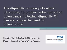
The diagnostic accuracy of colonic ultrasound, to problem solve suspected colon cancer following diagnostic CT. Can we reduce the need for Colonoscopy?
Gangi A, Ball C, Beable R, Higginson, A. Queen Alexandra Hospital, Portsmouth
Slide2Background
~ 700,000 adults are investigated in England per year for suspected colorectal cancer.In symptomatic patients:Colonoscopy is the gold standard.CT colonography (CTC) is the gold standard when radiological imaging of the whole colon is required.
Alternative investigations are not in routine clinical use.
Slide3Background
CTC+/- colonoscopy at times can be limited byResidual faeces and luminal fluidSuboptimal luminal distensionSegmental colitis
Complex diverticular disease
Patient discomfort
At our centre we carry out transabdominal ultrasound
to:
problem
solve cases where CTC is indeterminate
avoid colonoscopy and proceed directly to surgery
select
patients for endoscopic mucosal resection (EMR)
Slide4Aims
To determine the diagnostic accuracy of focused ultrasound in characterisation of colonic pathology in patients following abdominal CT or CT colonography.
Slide5Method
Consecutive patients from 2010- 2017 were extracted from a database by patient number and included in this preliminary retrospective study. Inclusion criteria:
Patients
with suspected colonic
cancer
A CT abdomen/CTC followed by a colonic US carried out within 3
months
Histopathology
, colonoscopy or 2 year clinical follow up
results
Slide6Method
Referral pathway
CT colon/CT abdomen
Colonoscopy
Colonic US
CT colon/CT
abdomen
Colonoscopy
Colonic US
+/-
Surgery
Slide7Method
Ultrasound technique:2 GI radiologists (25 years total experience; >10,000 bowel US)Toshiba Aplio 500, Canon
Aplio
i800
Low (3-5MHz) and high (5-17MHz) frequency probes
Overall assessment
Assessment from caecum to
sigmoid
Bowel wall layer assessment
Loss of submucosal layer
Hypoechoic echotexture
Superb micro-vascular ultrasound (
SMI) -Initial experience
Slide8Results
44 patients selected from the database met the criteria and were reviewed (22 female; mean age 74)Reference standard: 29/44 patients had histopathological results15/44 had 2+
years clinical/radiological
follow up
Slide9Results
Ultrasound results:Overall, US correctly evaluated for colonic cancer in 42 of 44 (95%)25/44
patients had a final diagnosis of colonic
cancer. US correctly identified 25/25 (100%).
3/44
had tubulovillous adenoma (TVA).
US correctly identified
1/3 (33.3%).
Technically inadequate in 1
case due to body habitus
Both
US and CT missed a
TVA
with adenocarcinoma transformation in a complex diverticular disease patient.
Slide10Results
Ultrasound results:
Sensitivity of 93%, specificity 100%, PPV 100%, NPV 89%,
Slide11Results
CT results:In 10/44 (23%) US had a greater diagnostic accuracy than CT7 indeterminate2 false positive
1 false negative
Indeterminate:
3 x technical factors (untagged faeces, sub-diagnostic)
4 x focal or eccentric thickening
2 of which had adenocarcinoma, 1 TVA
Slide12Transverse colon adenocarcinoma
Figure
1
(a
)–(b):
CTC: T2 circumferential mid distal transverse colon tumour.
US: T3b transverse colon tumour with anterior abdominal glide.
Histopathology confirmed a pT3 adenocarcinoma.
b)
a)
Slide13TVA
Figure
2
(a
)–(b): CTC
: 2cm
C-shaped lesion within the proximal sigmoid colon. US: a focal lesion with appearances in keeping with a TVA which was confirmed at histopathology.
a)
b)
Slide14Untagged faeces
Figure
3
(a
)–(b): US vs CTC. CTC: untagged faecal debris in caecum giving impression of a rolled edged mucosal lesion. US: normal bowel wall layers.
Clinical follow up and CTC 4 years later confirmed this.
a)
b)
Slide15Diverticulosis
Figure 4
(a
)–(b): CTC: Thickening of the sigmoid colon ? Diverticular or cancer. US: Resolving inflammatory change around the sigmoid colon.
Confirmed with colonoscopy and
biopsy to be sigmoid diverticulosis.
a)
b)
Slide16Diverticulitis
Figure 5
(a
)–(b): CTC:
Ascending colon
colitis. US: ulcerating tumour within the diverticular segment.
Confirmed with
histopathology to be adenocarcinoma.
a)
b)
Slide17Results
Colonoscopy results:17/44 (39%) patients had a colonoscopy as part of the investigation work up. In 8, colonoscopy pre USIn 9, colonoscopy post US
Of these 11/17 (65%) were non diagnostic :
Not
safe to proceed x1
Insufflation problems x1
Did not tolerate
x3
Residual
stool/poor prep x3 (
3x for 1 patient)
Unable to pass through inflammatory stricture x3
Slide18Discussion
Financial considerationsUS <20 mins = £30Colonoscopy = £440Only 39% of patients had colonoscopyCost if all had colonoscopy = £19,360Cost for 17 colonoscopy + 27 US = £8,260
Which is a saving of £11,100
From 2010- 2017 there are projected to be a total of 250 patients who meet the inclusion criteria
Projected saving of £62,525
Slide19Key findings
Colonic ultrasound has a high sensitivity of 93% and specificity 100%.
Higher diagnostic accuracy
than CTC
in complex patients.
There is a financial advantage to carrying out a problem solving
colonic US
rather than colonoscopy.
Slide20Conclusion
Colonic ultrasound in the hands of a GI specialist radiologist has high diagnostic accuracy.In suspected colon cancer, we have demonstrated that colonic US
has a role in
problem
solving
where colonoscopy has failed or
in complex/technically
inadequate
CTC examinations.
At our hospital,
CTC + coloni
c US
is used to confirm colon cancer thereby allowing patients to go
directly to surgery
.
A larger retrospective cohort or prospective study is needed to confirm these findings.
Slide21Thank you.
Anmol.Gangi@porthosp.nhs.uk