
ested Com p anion Products C atal og m r N o n e S 554656Stain Buffer FBS500 mLnone554657Stain Buffer BSA500 mLnone556454Annexin V Binding Buffer 10X concentrate50 mLnone556419FITC Annexin ID: 848225
Download Pdf The PPT/PDF document "Recommended Assay ProcedureStaining of L..." is the property of its rightful owner. Permission is granted to download and print the materials on this web site for personal, non-commercial use only, and to display it on your personal computer provided you do not modify the materials and that you retain all copyright notices contained in the materials. By downloading content from our website, you accept the terms of this agreement.


1 Recommended Assay Procedure:Staining of
Recommended Assay Procedure:Staining of Live Cells for Viability Analysis by Flow Cytometry1.Obtain a single cell suspension.2.Resuspend cells in BD PharmingenStain Buffer(FBS) (Cat..554656)or1×Dulbeccos Phosphate Buffered Saline(DPBS)containing0.05-0.2.The optimal concentration of DAPI for viability analysis may vary by cell type.We recommend titrating the reagent for yourcell type of interest in early experiments.b.Additionally, apoptotic cells may stain with variable amounts of DAPI.We recommend co-staining with BD PharmingenFITC Annexin V(Cat.No.556419)if further analysis of apoptotic cells is desired.3.Incubate5minutes at room temperature.No wash is necessary prior to analysis4.Proceed to analysis by flow cytometry.Staining of Fixed Cells for DNA Content Analysis by Flow Cytometry1.Obtain a single cell suspension.2.Treat cells on ice for30minutes with70-80%ice-cold ethanol..Ethanol fixation typically provides the most resolved histograms.However, this reagent has also been successfully used forDNA content analysis with the Transcription Factor Buffer Set(Cat.No.562574/562725)or BD CytofixFixation Buffer(Cat..554655)and BD PhosflowPerm Buffer III(Cat..558050)protocol.3.Wash cells once with BD PharmingenStain Buffer(FBS).4.Dilute DAPI solution to0.5-1 g/mL in Stain Buffer(FBS)or1×DPBS immediately prior to use.5.Stain cells for5-15minutes at a cell density of1-2x10^6cells/mL.No further wash is necessary prior to analysis..The optimal cell density and concentration of DAPI for DNA content analysis may vary by cell type.Assay conditions shouldbe optimized in early experiments for best results.6.Proceed to analysis by flow cytometry.Immunofluorescent Staining of Fixed Cells for Nuclear Visualization1.Fix and permeabilize cells as desired.2.Dilute DAPI solution to1µg/ml in1×DPBS immediately prior to use.3.Add DAPI solution to each sample at least15minutes before analysis.4.Proceed to imaging.Note:This reagent has been developed and certified for the Bioimaging application.However, routine Bioimaging testing is not performevery lot. ested Com p anion Products C atal og m r N o n e S 554656Stain Buffer (FBS)500 mL(none)554657Stain Buffer (BSA)500 mL(none)556454Annexin V Binding Buffer, 10X concentrate50 mL(none)556419FITC Annexin V200 Tests(none)562574Transcription Factor Buffer Set100 Tests(none)562725Transcription Factor Buffer Set2
2 5 Tests(none)550524Retrievagen A (pH 6.0550524Retrievagen A (pH 6.0")
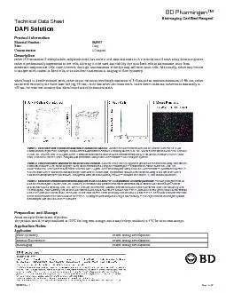
5 Tests(none)550524Retrievagen A (pH 6.0)1000 mL(none)564731Purified Mouse Anti-Nkx2.20.1 mg74.5A5564750Alexa Fluor® 647 Mouse Anti-PTF1A50 µgS25-763558050Perm Buffer III125 mL(none)Product NoticesFlowJo is a trademark of Tree Star Inc. 1. Alexa Fluor® is a registered trademark of Molecular Probes, Inc., Eugene, OR. 2. For fluorochrome spectra and suitable instrument settings, please refer to our Multicolor Flow Cytometry web page at www.bdbiosciences.com/colors. 3. Please refer to www.bdbiosciences.com/pharmingen/protocols for technical protocols. 4. Darzynkiewicz Z, Bruno S, Del Bino G, et al. Features of easured by flow cytometry. Cytometry. 1992; 13(8):795-808. (Methodology: Flow cytometry)rzynkiewicz Z. Flow cytometricosis: Comparison of the assays of in situ DNA degradation and chromati n changes. Cytometry. 1994; 15(3):237-244. (Methodology: Flow cytometry)Otto F. DAPI staining of fixed cells for high-resolution flow cytometry of nuclear DNA. Methods Cell Biol. 1990; 33:105-110. (Methodology: Flow cytometry)Shapiro HM. Practical Flow Cytometry, 3rd Edition. New York: Wiley-Liss, Inc; 1995:280-282. (Methodology: Flow cytometry)Tanious FA, Veal JM, Buczak H, Ratmeyer LS, Wilson WD. DAPI (4',6-diamidino-2-phenylindole) binds differently to DNA and RNA: minor-groove binding at AT sites and intercalation at AU sites. Biochemistry. 1992; 31(12):3103-3112. (Biology) 564907 Rev. 1Page 2 of 2 Bioima Certified Rea g Product InformationMaterial Number:564907Size: Concentration:Description Panel 1. Two-color flow cytometric analysis of Jurkat cell viability. Jurkat cells were treated with DMSO vehicle (Left Plot) or 5 Camptothecin (Right Plot) overnight. Cells were resuspended in Annexin V Binding Buffer (Cat. No. 556454) and stained with FITC with DAPI (pseudo-colored blue). The nuclei of islet cells (DAPI+) stain positively for both NKX2.2 and PTF1A. The nuclei of acinar cells and some islet precursor cells stain positive for PTF1A. All images were analyzed using a BD Pathway 435 High-Content Bioimager System and merged with BD Attovision Software.Preparation and StorageAvoid multiple freeze-thaws of product.The product should be kept undiluted at -20°C for long term storage, A pplication NotesFlow cytometryTested During DevelopmentImmunofluorescenceTested During DevelopmentBioimagingTested During Development 564907 Rev. 1Page 1 of