
Cardiac region near the heart Fundus lateral to the cardia The body mid and largest portion Pylorus funnelshaped terminal part of the stomach Most digestive activity occurs in this region ID: 1032452
Download Presentation The PPT/PDF document "Stomach The stomach is on the left side ..." is the property of its rightful owner. Permission is granted to download and print the materials on this web site for personal, non-commercial use only, and to display it on your personal computer provided you do not modify the materials and that you retain all copyright notices contained in the materials. By downloading content from our website, you accept the terms of this agreement.


1.
2. StomachThe stomach is on the left side of the abdominal cavityCardiac region (near the heart)Fundus (lateral to the cardia)The body (mid and largest portion)Pylorus (funnel-shaped terminal part of the stomach). Most digestive activity occurs in this region.Two sphincters:Cardioesophageal sphincter, through which food enters the stomach from esophagusPyloric sphincter, which is continuous with small intestineGreater curvature and lesser curvatureThe lesser omentum (double layer of peritoneum extends from liver to lesser curvature)The greater omentum (another extension of the peritoneum riddled with fat and covers the abdominal organs (cushioning and insulation)
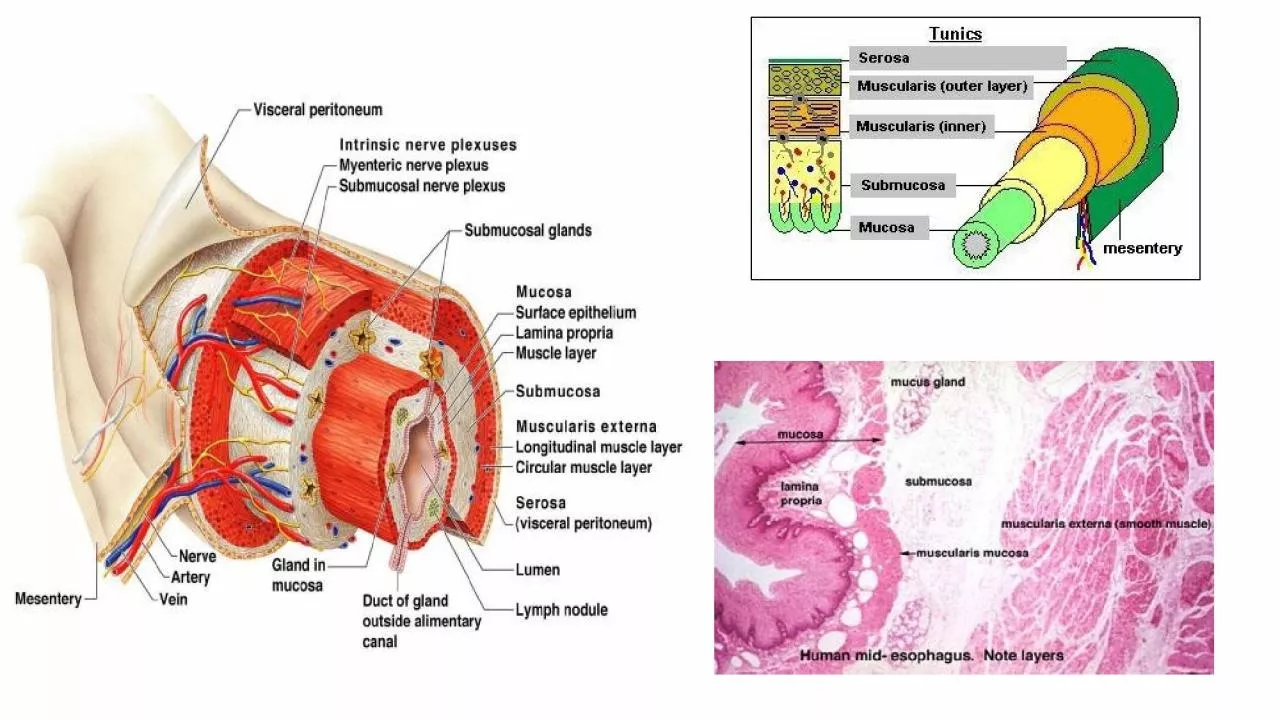
3. StomachHistology of the stomach is similar to the esophagus except that the mucosa is lined with simple columnar epithelium which produces large amounts of mucusDigestion:Special arrangement of muscular cells in muscularis externa helps to move, churn and mix the food to break it down to smaller fragments (physical)Chemical breakdown of proteins beginsStomach lining contains deep gastric pits that secrete gastric juiceChief cells produce protein digesting enzymes pepsinogensParietal cell produce HCL makes stomach acidic and activates enzymesLow PH of stomach stimulates stomach cells to produce gastrin hormone stimulates stomach glands to produce more protein digesting enzymes, mucus and hydrochloric acidHow is the stomach wall protected from HCL’s corrosive nature? mucous neck cells produce sticky alkaline mucusAfter food has been processed in the stomach, it resembles heavy cream and is called chime. The chime then enters the small intestine through the pyloric sphincter
4. Small IntestineThe body’s major digestive organLongest section of the alimentary tube (increase surface area for absorption)Extends from pyloric sphincter to ileocecal valve (valve that separates small intestine and large intestine)The large intestine encircles it and frames it in the abdominal cavityThree subdivisions:DuodenumJejunum (longest part) Ileum (joins large intestine at ileocecal valve)
5. Small Intestine cont.Small intestine can digest only a small amount of food at a time (pyloric sphincter controls food movement from the stomach so that the small intestine is not overwhelmed)Digestion:Most of the digestion occurs in small intestineEnzymes are produced my intestinal cells help in digestionEnzymes produced by pancreas and ducted into the duodenum through the pancreatic ductsBile (formed by liver) enters duodenum through bile ductAbsorptionMost of the food absorption occurs in the small intestine
6. Small Intestine cont.Structures help increase the absorptive surface:Microvilli: tiny projections of the plasma membrane of the mucosal cells, which give the cell surface a fuzzy appearance (brush border)Villi: fingerlike projections of the mucosa. Within each villus is a rich in capillaries and contains a lymphatic capillary called a lactealDigested food is absorbed through the mucosa cells into the capillaries and lacteal Circular folds, called plicae circulares: deep folds of mucosa and sub-mucosaPeyer’s Patches:Collection of lymphatic tissue (high in lymphocytes+ other immune cells) found towards the end of the small intestine (remaining undigested food residue is high in bacteria that must be prevented from entering the blood stream)
7.
8.
9. Large IntestineFunctionsPrimary task is absorption of waterAbsorption of other substances (e.g :Bacteria in large intestine that produce vitamin K that is absorbed in the LI)Excretion of wastesParts of LI:Cecum (where small intestine contents enter the large intestine)Appendix (wormlike structure that hangs from the cecum)ColonAscending colon (follows the cecum)Transverse colonDescending colon Sigmoid colon (has an S-shaped curve)RectumAnal canal (opens to the exterior)Has an external voluntary sphincter and internal involuntary sphincter act together to open and close (usually closed except during defecation)
10. Accessory digestive organsPancreas:Considered both an Endocrine gland (insulin, glucagon) Exocrine gland (digestive enzymes): trypsinogen that becomes activated into trypsin in the small intestine to digest proteins into amino acids.*Endocrine are ductless glands that secret hormones that travel through the blood to regulate a target organ*Exocrine glands produce secretions that reach their target through a ductTogether insulin and glucagon maintain our blood glucose levels insulin lowers blood glucose levels, glucagon increases blood glucose levels
11. Accessory digestive organsLiver and Gall Bladder:The liver is the largest gland in the bodyLocated under the diaphragmLiver functions include:Helps in digestion by producing bile (yellow to green watery solution containing bile salts,bile pigments, cholesterol and phospholipids)Bile salts helps to break down large fat globules into smaller ones to aid the action of fat-digesting enzymes (lipases)Liver also produces blood clotting factorsThe gall bladder stores and secretes bile
12. Overview of digestion