
what ductal carcinoma in situ DCIS is how it is diagnosed and how it is treated DCIS is an early form of breast cancer sometimes called precancerous noninvasive or intraductal cancer T ID: 961410
Download Pdf The PPT/PDF document "This information sheet explains" is the property of its rightful owner. Permission is granted to download and print the materials on this web site for personal, non-commercial use only, and to display it on your personal computer provided you do not modify the materials and that you retain all copyright notices contained in the materials. By downloading content from our website, you accept the terms of this agreement.


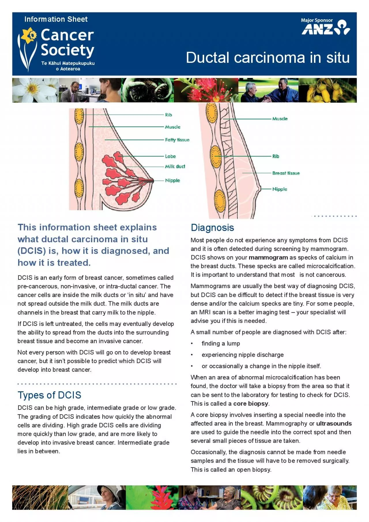
This information sheet explains what ductal carcinoma in situ (DCIS) is, how it is diagnosed, and how it is treated. DCIS is an early form of breast cancer, sometimes called pre-cancerous, non-invasive, or intra-ductal cancer. The cancer cells are inside the milk ducts or ‘in situ’ and have not spread outside the milk duct. The milk ducts are channels in the breast that carry milk to the nipple. If DCIS is left untreated, the cells may eventually develop the ability to spread from the ducts into the surrounding breast tissue and become an invasive cancer. Not every person with DCIS will go on to develop breast cancer, but it isn’t possible to predict which DCIS will develop into breast cancer. Types of DCIS DCIS can be high grade, intermediate grade or low grade. The grading of DCIS indicates how quickly the abnormal cells are dividing. High grade DCIS cells are dividing more quickly than low grade, and are more likely to develop into invasive breast cancer. Intermediate grade lies in between. Diagnosis Most people do not experience any symptoms from DCIS and it is often detected during screening by mammogram. DCIS shows on your mammogram as specks of calcium in the breast ducts. These specks are called microcalcification. It is important to understand that most is not cancerous. Mammograms are usually the best way of diagnosing DCIS, but DCIS can be difficult to detect if the breast tissue is very dense and/or the calcium specks are tiny. For some people, an MRI scan is a better imaging test – your specialist will advise you if this is needed. A small number of people are diagnosed with DCIS after: • finding a lump • experiencing nipple discharge • or occasionally a change in the nipple itself. When an area of abnormal microcalcification has been found, the doctor will take a biopsy from the area so that it can be sent to the laboratory for testing to check for DCIS. This is called a core biopsy . A core biopsy involves inserting a special needle into the affected area in the breast. Mammography or ultrasounds are used to guide the needle into the correct spot and then several small pieces of tissue are taken. Occasionally, the diagnosis cannot be made from needle samples and the tissue will have to be removed surgically. This is called an open biopsy. Information Sheet Ductal carcinoma in situ Treatment The aim of treatment is to prevent DCIS from developing into an invasive breast cancer. The type of treatment you receive depends on how much of the breast is affected and the grading of the cells. Some people with DCIS are concerned about having treatment for a condition that isn’t life-threatening. However, most accept that it’s important to treat DCIS to prevent it from developing into an invasive cancer. Surgery Treatment with surgery varies from removal of the area affected by DCIS with some surrounding normal tissue, to complete removal of the breast (mastectomy). Wide local excision is the removal of the area of DCIS. It is more commonly used for small areas of low grade DCIS. Mastectomy is recommended for women who have a large area of DCIS or several separate areas of DCIS within the breast. DCIS rarely spreads to the lymph nodes in the armpit so it is not usual to remove them. It may be possible to have breast reconstruction surgery if you have a mastectomy. This can be discussed with your surgeon. F
or more information, see Cancer Council Australia’s booklet:Breast Prostheses and Reconstruction – A guide for women affected by breast cancer. Other treatments Radiation treatment If you have wide local excision for high grade DCIS it is likely that it will be followed by radiation treatment. Hormone treatment Some forms of DCIS have receptors for the hormone oestrogen on their cells. This means that the DCIS needs oestrogen to grow. Hormone treatments may be recommended to block the effect of oestrogen on the cancer cells, or to lower the levels of oestrogen in your body, and stop the DCIS growing. Hormone treatments include tablets such as tamoxifen, anastrozole or letrozole. Making decisions about treatment Some people find it very hard to make decisions about treatment, especially when there may be different options offered. DCIS doesn’t need to be treated urgently, so you can often take a little time to think things over. If you’re asked to choose between treatments, make sure that you have enough information so you can make an informed decision.You should ask questions or discuss anything you don’t understand with your surgeon or breast care nurse. Your specialist can explain what’s involved and any possible side effects of the treatment so you can decide what’s right for you. For more information, read our information sheet ‘Making decisions about your cancer treatment’ on our website (www.cancernz.org. nz). You can also contact your local Cancer Society or telephone 0800 CANCER (226237) to speak confidentially with a cancer information nurse. Suggested websites BreastScreen Aotearoa Cancer Australia Breast Cancer Care (UK) Macmillan Cancer Support (UK) Glossary Biopsy— the removal of a small amount of cells or tissue from your body, so that it can then be examined under a microscope. Cells— the ‘building blocks’ of the body. A human is made of millions of cells, which are adapted for different functions. Cells are able to reproduce themselves exactly, unless they are abnormal or damaged, as are cancer cells. Core biopsy— a larger needle than that used for fine needle aspiration is used to remove a small piece of tissue from the abnormal area. This is done with a local anaesthetic. Invasive cancer— cancer cells that have spread beyond their site of origin into the surrounding tissue. Lobule— part of the breast capable of producing milk. Local excision or breast conserving surgery— surgical removal of the affected area of the breast with some surrounding tissue, but not total removal of the breast. Grade/Grading— the grade indicates how quickly the abnormal cells are dividing. High grade DCIS is when the cells are dividing more quickly than low grade DCIS. Lymph nodes— glands that are found throughout the body that remove bacteria and other harmful agents from the body. Mammography/Mammogram— an X-ray of the breast that can be used to examine a breast lump. Mammograms are also used for people without any breast changes because they may detect areas of microcalcification (possible sites of DCIS). Radiation treatment— the use of high-energy X-ray beams to kill cancer cells or prevent them from reproducing. Cancer Society of New Zealand. Te Kāhui Matepukupuku o Aotearoa 2018 0800 CANCER (226 237) or www.cancernz.org.nz Ductal carcinoma in sit