
Agrawal Additional Professor Department of Ophthalmology AIIMS Rishikesh Acknowledgement Kanskis Clinical Ophthalmology 8 th Edition Becker Schaffers Diagnosis and therapy of The ID: 917555
Download Presentation The PPT/PDF document "Secondary Glaucoma Dr.Ajai" is the property of its rightful owner. Permission is granted to download and print the materials on this web site for personal, non-commercial use only, and to display it on your personal computer provided you do not modify the materials and that you retain all copyright notices contained in the materials. By downloading content from our website, you accept the terms of this agreement.


Slide1
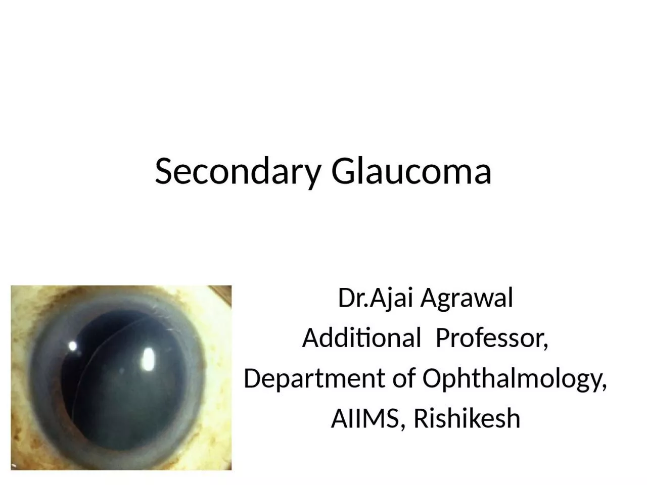
Secondary Glaucoma
Dr.Ajai AgrawalAdditional Professor,Department of Ophthalmology,AIIMS, Rishikesh
Slide2Acknowledgement
Kanski’s Clinical Ophthalmology (8th Edition).Becker- Schaffer’s Diagnosis and therapy of The Glaucomas (8th Edition).Comprehensive Ophthalmology (A.K.Khurana
) (7
th
Edition).Glaucoma - Medical Diagnosis & Therapy (Tarek M Shaarawy )(2nd Edition).
2
Slide3Learning Objectives
At the end of this class the students shall be able to :Define secondary glaucoma.Classify secondary glaucoma.Understand the aetiopathogenesis and clinical features of secondary glaucoma’s.
Understand
the fundamentals of managing secondary
glaucoma’s.3
Slide4Question
A 12 year old boy is diagnosed as having an angle recession glaucoma. It is a type ofprimary open angle glaucomasecondary open angle glaucomaprimary angle closure glaucomasecondary angle closure glaucoma
4
Slide5Definition
Secondary Glaucoma A group of disorders in which rise in intraocular pressure(leading to glaucoma) is
associated with some primary
ocular or
systemic disease
.
5
Slide6Classification of secondary glaucoma's
Based on mechanism of IOP rise Secondary open angle glaucoma Secondary angle closure glaucoma
6
Slide7Classification of secondary glaucoma's
Depending on causative primary diseasePhacogenic (Lens induced) glaucomaPigmentary glaucomaNeovascular glaucomaInflammatory glaucoma (
Uveitic
)
Traumatic glaucomaSteroid induced glaucomaPseudoexfoliative glaucomaGlaucomas associated with intraocular tumours (Malignant
melanoma,
retinoblastoma)
7
Slide88
Slide99
Slide10Lens induced glaucoma
Raised IOP secondary to a disorder of crystalline lensSecondary angle closure Secondary open angle
Phacomorphic
glaucoma Phacolytic glaucoma Phacotopic glaucoma Lens particle glaucoma
Phacoanaphylactic
glaucoma
10
Slide11Phacomorphic glaucoma
Causes -Intumescent lensAnterior subluxation or dislocation of the lens and spherophakia (Phacotopic variant)
Pathogenesis
– Swollen lens pushes iris forwards
, obliterating the anglePresentation – Acute congestive glaucoma 11
Slide12Phacomorphic glaucoma
Treatment –Medical treatment – Control of IOP by iv mannitol, systemic acetazolamide
and
topical
beta blockersSurgical Cataract extraction with implantation of PCIOL
12
Slide13Phacolytic glaucoma
Trabecular meshwork is clogged by lens proteins and macrophages which
phagocytose
the
lens proteins and inflammatory debrisTreatment
M
edical
therapy
to lower
IOP followed by extraction
of
cataractous
lens with PCIOL
implantation.
13
Slide14Lens particle glaucoma
Trabecular meshwork is blocked by lens particles floating in aqueous humour.ManagementMedical therapy to lower IOP and
irrigation – aspiration
of lens particles from anterior chamber
14
Slide15Phacoanaphylactic glaucoma
Fulminating acute inflammatory reaction due to antigen – antibody reactionGranulomatous inflammation in involved eyePreceding disruption of lens capsule and leakage of proteins from capsuleIOP
is raised due to inflammatory reaction of
uveal
tissue excited by lens matter.
15
Slide16Phacoanaphylactic glaucoma
Management includes medical therapy to lower IOP. Treatment of iridocyclitis
with steroids and cycloplegics . Irrigation – aspiration of
lens matter
from
anterior
chamber ( if
required
).
16
Slide17Pigmentary glaucoma
Clogging up of trabecular meshwork by pigment particles in patients with Pigment Dispersion Syndrome(PDS)Pigment released by mechanical rubbing of posterior pigment layer of iris with zonular fibrilsClinical
features
–
Young myopic malesFeatures similar to POAGDeposition of pigment granules in anterior segment
Pigment deposition on lens
zonules
and
equatorial
region. The deposits are clearly
visible
in full
mydriasis
.
17
Slide18REVERSE PUPILLARY BLOCK IN PIGMENTARY GLAUCOMA
18
Slide19CLASSIC DIAGNOSTIC TRIAD
Krukenberg spindle (Pigment deposition on the endothelium, in a vertical spindle-shaped distribution).Midperipheral iris transillumination defectsDense trabecular meshwork pigmentation
19
Slide20Pigmentary glaucoma
Gonioscopy – pigment accumulation along the Schwalbe’s line especially inferiorly (Sampaolesi’s line
)
Iris
transillumination – radial slit – like defects in the peripheryTreatment is similar to that of POAG
20
Slide21Neovascular glaucoma
Intractable glaucoma due to neovascularisation of iris and angle of anterior chamber.Due to retinal ischaemia Diabetic retinopathy CRVO
Sickle cell retinopathy
Eales’ disease Chronic intraocular inflammation21
Slide22PATHOGENESIS
CHRONIC RETINAL ISCHAEMIAANGIOGENIC FACTORS RELEASED NEOVASCULARISATION ON IRIS AND ANGLENEOVASCULAR GLAUCOMA
22
Slide23Stages of neovascular glaucoma
Pre-glaucomatous stageOpen angle glaucoma stageSecondary angle closure glaucoma
23
Slide24TREATMENT
Panretinal photocoagulationIntra- vitreal Anti -VEGFMydriatics and CorticosteroidsFiltering surgeries
Glaucoma drainage devices
Cyclodestructive
procedures
24
Slide25INFLAMMATORY GLAUCOMA
Non specific inflammatory glaucomaOpen angle Angle closureSpecific hypertensive uveitis syndromesFuchs’ uveitis syndromeGlaucomatocyclitic
crisis (Posner
Schlossman syndrome)
25
Slide2626
Slide27Open angle inflammatory glaucoma
Acute open – angle
inflammatory glaucoma
Chronic open – angle
inflammatory glaucoma
Mechanism of rise in
IOP
Trabecular clogging ,
trabecular oedema and
prostaglandin – induced
rise in IOP
Chronic
trabeculitis
and
trabecular scarring
Clinical features
Features of acute
iridocyclitis
associated with
raised IOP with open-angle
of anterior chamber
Raised IOP, open angle, no
active inflammation but
signs of previous episode
of uveitis present
Management
Treatment of
iridocyclitis
Medical therapy to
lower IOP by use of
hyperosmotic agents,
acetazolamide and beta –
blockers eye drops
Medical therapy
Trabeculectomy
Cyclodestructive
procedures
27
Slide28Angle closure inflammatory glaucoma
Mechanism of rise in IOP –Secondary angle – closure with pupillary blockSecondary angle – closure without
pupillary
block
Clinical features – Raised IOP, seclusio papillae, shallow anterior chamberManagement –Prophylaxis
– Local steroids and atropine to
prevent
formation of
synechiae
Curative
treatment – Medical therapy, surgical or
laser
iridotomy
and filtration surgery
28
Slide29Specific
hypertensive uveitis syndromesGlaucomatocyclitic crisis (Posner Schlossman syndrome)Recurrent attacks of unilateral, acute mild uveitis with secondary open angle glaucoma.
Glaucoma
is
out of proportion to inflammation. Due to accompanying acute
trabeculitis
.
Fuchs’ uveitis syndrome
Chronic low grade anterior uveitis.
Occurs unilaterally in middle aged persons
Heterochromia
of iris
No posterior
synechia
.
Associated with cataract and secondary glaucoma
29
Slide30Blunt Trauma
30
Slide3131
Slide32Causes of glaucoma after trauma
Inflammatory glaucomaGlaucoma due to hyphemaLens induced glaucomaAngle recession glaucomaEpithelial or fibrous ingrowthAngle closure due to PAS
32
Slide33Angle recession glaucoma
Rupture in ciliary body face Bimodal onset at 1 year and 10 year post trauma270 degree recession- risk of glaucoma- 5%360 degree recession- risk of glaucoma- 24%
Gonioscopic
view of angle recession, demonstrated by a widened ciliary body band.
There is a disruption in the ciliary body between the external longitudinal muscle
fibers
and the internal oblique and circular muscle
fibers
.
33
Slide34Traumatic glaucoma
Management Medical therapy with topical 0.5% timolol and oral acetazolamide Surgical intervention needs to be individualized according to nature and site of trauma
34
Slide35Steroid induced glaucoma
Secondary open angle glaucoma following steroid therapyIn the general population:High steroid responders – 5%Moderate steroid responders – 35%Non steroid responders – 60% (IOP rise after six weeks of steroid therapy) Precise mechanism of IOP rise not knownPrevented by judicious use of steroids and regular IOP monitoring
Treated by stopping steroids gradually and anti glaucoma medications
35
Slide36Pseudoexfoliative glaucoma
Pseudo exfoliation syndrome(PES)/Glaucoma capsulare is associated with Secondary OAG in 50% of the cases.Deposition of an amorphous grey dandruff – like material on the pupillary border, posterior surface of iris and ciliary processes
Trabecular blockage by
exfoliative
materialManaged on the same lines as POAG
36
Slide37Causes of elevated IOP post cataract surgery
Early phaseInflammationHaemorrhageRetained viscoelastic/lens matterLate phase
Tight suture
Excessive cautery
Pupillary block(IOL/Vitreous)Aqueous misdirectionEpithelial/Fibrous down growth37
Slide38Glaucoma associated with iridocorneal
endothelial syndromesThree clinical entities:Progressive iris atrophyChandler’s syndromeCogan-Reese syndrome/Iris nevus syndrome
Pathogenesis
: Abnormal corneal endothelial cells proliferate to form a membrane in angle of AC. Contraction of membrane leads to
secondary angle closureTreatment: Difficult and usually surgical
38
Slide39Other causes of secondary glaucoma
Glaucoma in aphakia/pseudophakiaCiliary block glaucomaGlaucoma associated with intraocular haemorrhage Red cell glaucoma Haemolytic glaucoma Ghost cell glaucoma Hemosiderotic glaucoma
39
Slide4040
Slide4141
Slide4242
Slide43Question
A 50 year old lady with uncontrolled diabetes presented with painful red eye and decreased visual acuity in her right eye. On examination there was raised Intraocular Pressure and new blood vessels on the iris. The treatment includes all except?atropinebeta blockerssteroidspilocarpine
43
Slide44Question
The laser procedure, most often used for treating neovascular glaucoma:a) Goniophotocoagulationb) Laser trabeculoplastyc) Panretinal photocoagulation (PRP)d) Laser iridoplasty
44
Slide45Question
What is the most likely type of glaucoma in this patient ?Phacolytic glaucomaPhacoanaphylactic glaucomaPhacotopic glaucomaLens particle glaucoma
45
Slide4646