
pulmonary NUTmidline carcinoma with Epithelioid morphology and prominent eosinophilic nucleoli 400X magnification A D C B Supplemental Figure 2 A NUTmidline carcinoma with a florid ID: 1010035
Download Presentation The PPT/PDF document "Supplemental figure 1. Primary" is the property of its rightful owner. Permission is granted to download and print the materials on this web site for personal, non-commercial use only, and to display it on your personal computer provided you do not modify the materials and that you retain all copyright notices contained in the materials. By downloading content from our website, you accept the terms of this agreement.

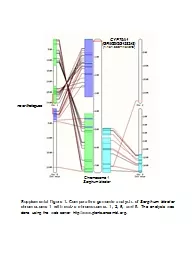


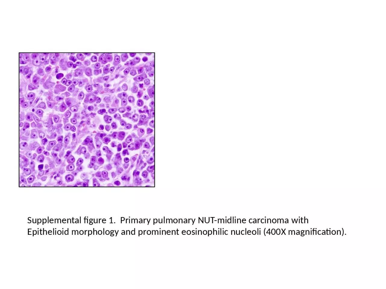
1. Supplemental figure 1. Primary pulmonary NUT-midline carcinoma withEpithelioid morphology and prominent eosinophilic nucleoli (400X magnification).
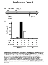
2. ADCBSupplemental Figure 2:A) NUT-midline carcinoma with a florid pneumocyte proliferation.B) p63 highlights tumor cells. C) TTF-1 is negative in tumor cells and highlights the adjacent pneumocytes.D) NUT protein is expressed in the tumor cells, negative in adjacent pneumocytes.