/
Surgical anatomy of the esophagus Surgical anatomy of the esophagus

Surgical anatomy of the esophagus - PowerPoint Presentation

jaena
jaena . @jaena
Follow
75 views
Uploaded On 2023-07-09

Surgical anatomy of the esophagus - PPT Presentation

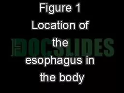
Asses Prof Dr Rafid majeed Esophagus The esophagus is the connecting tube between the pharynx and stomach that functions to transport food fluids and saliva The 3 portion of the esophagus ID: 1007369

esophageal esophagus cervical portion esophagus esophageal portion cervical thoracic left dorsal cranial drain form vagal trachea caudal branches veins

Share:

Link:

Embed:

Download Presentation from below link

Download Presentation The PPT/PDF document "Surgical anatomy of the esophagus" is the property of its rightful owner. Permission is granted to download and print the materials on this web site for personal, non-commercial use only, and to display it on your personal computer provided you do not modify the materials and that you retain all copyright notices contained in the materials. By downloading content from our website, you accept the terms of this agreement.


Presentation Transcript

1. Surgical anatomy of the esophagus Asses. Prof. Dr. Rafid majeed

2. Esophagus The esophagus is the connecting tube between the pharynx and stomach that functions to transport food , fluids and saliva.The 3 portion of the esophagus cervical portionthoracic portion abdominal portion

3. The cervical portion of the esophagusThe cervical portion of the esophagus begins dorsal to the caudal border of the cricoid cartilage, inclines to the left of the trachea as it runs caudally, and ends at the thoracic inlet.

4. The thoracic portion of the esophagus The thoracic portion extends from the thoracic inlet, where it is located to the left of the trachea, crosses the trachea to regain its dorsal position at the tracheal bifurcation, and extends caudally to the esophageal hiatus of the diaphragm

5. The aorta obliquely crosses the left side of the midthoracic esophagus. In the caudal thorax, the dorsal branches of the left and right vagal nerves run across the side of the esophagus and unite dorsally to form the dorsal vagal trunk, and the left and right ventral branches similarly unite to form the ventral vagal trunk. Both vagal trunks enter the abdomen through the esophageal hiatus of the diaphragm.

6. The abdominal portion of the esophagusThe abdominal portion of the esophagus is short and wedge shaped, extending from the diaphragmatic hiatus to the stomach.

7. Layers of the Esophageal WallThe outer layer of the esophagus is the adventitia. In the neck, the esophageal adventitia blends with the deep cervical fascia. In the thorax and abdomen, the adventitia is largely covered with pleura and peritoneum, respectively. The esophagus is loosely connected to the diaphragm by a phrenicoabdominal membrane.

8. The muscularis is composed of striated muscle for the entire length of the esophagus in dogs, but it changes to smooth muscle in the terminal esophagus in cats.

9. functional and physiological esophageal sphinctersAt its cranial and caudal ends, the esophagus has functional and physiological sphincters that are difficult to delineate anatomically. There is no obvious thickening of the esophageal wall at the pharyngoesophageal junction to form a true cranial (or upper) esophageal sphincter. However, the thyropharyngeus and cricopharyngeus muscles and associated elastic tissue serve the function of a cranial esophageal sphincter.وضيفتها تمنع رجوع الطعام للمجاري التنفسية وتمنع دخول الهواء للمريء اثناء عدم الاكل  There is an increase in thickening of the circumferential striated muscle layer at the gastroesophageal junction in dogs, which correlates with the accepted location of the highpressure zone at the gastroesophageal junction and may represent the caudal (or lower) esophageal sphincter

10. The submucosaThe submucosa loosely connects the mucosa and muscularis, allowing mucosa to move independently and form mucosal folds in the undistended esophagus. The submucosa contains blood vessels, nerves, and simple mucous glands that secrete mucus, which lubricates the mucosal surface.

11.

12. The esophageal mucosaThe esophageal mucosa is composed of a stratified squamous epithelium. In the nondistended esophagus, the mucosa forms numerous large longitudinal folds that can be seen with positive-contrast esophagography. In cats, the terminal esophagus is also folded transversely, producing a herringbone appearance on esophagrams. 

13. Esophageal Blood SupplyThe main arterial blood supply to مهمthe cervical esophagus is from branches of the cranial and caudal thyroid arteries. A. The bronchoesophageal artery is the main source of blood for the cranial two thirds of the thoracic esophagus; B. the remaining thoracic esophagus is supplied by esophageal branches of the aorta or dorsal intercostal arteries. The terminal portion is supplied by a branch of the left gastric artery.  

14. Vein drain The veins that drain the esophagus are largely satellites of the arteries that supply it. Veins leaving the cervical esophagus drain into the external jugular veins, and those from the thoracic esophagus drain mostly into the azygous vein. The vein that accompanies the left gastric vein drains into the portal venous system.  The esophageal arteries and veins form a rich, intramural plexus of anastomosing vessels in the submucosal layer.

15. Esophageal lymphatic vesselsEsophageal lymphatic vessels drain into 1. the medial retropharyngeal, 2. deep cervical, cranial mediastinal, 3. portal, splenic, and gastric lymph nodes.

16. The esophagus innervationThe esophagus is innervated by nerve fibers arising from various branches of the vagus, beginning with 1. the paired pharyngoesophageal nerves followed by 2. the recurrent laryngeal and paralaryngeal nerves and concluding with 3. the dorsal and ventral vagal trunks

17.  Approach to the cervical esophagus. A, Position the patient in dorsal recumbency with the neck resting on a rolled towel. B, The skin is incised from the larynx to the manubrium, and the sternohyoid muscles are separated to expose the trachea. C, The trachea is retracted to the right to expose the esophagus, thyroid, carotid sheath, and recurrent laryngeal nerve