
The length of the small bowel varies from 300 to 850 cm B etween the duodenojejunal DJ flexure to the ileocaecal valve The proximal 40 of the small intestine is referred to ID: 913670
Download Presentation The PPT/PDF document "The small intestine ANATOMY" is the property of its rightful owner. Permission is granted to download and print the materials on this web site for personal, non-commercial use only, and to display it on your personal computer provided you do not modify the materials and that you retain all copyright notices contained in the materials. By downloading content from our website, you accept the terms of this agreement.


Slide1
The small
intestine
ANATOMY
The
length of the small bowel varies
from 300
to 850
cm.
B
etween
the duodenojejunal (DJ) flexure
to the
ileocaecal
valve.
The
proximal 40% of the small intestine is referred to
as the
jejunum; the remainder is the ileum.
There
is no
clear demarcation
between jejunum and
ileum.
The
jejunum tends to have a wider diameter and
a thicker
wall, with more prominent mucosal folds (
valvulae conniventes).
T
he
ileum has a thicker, more fatty
mesentery with
more complex arterial arcades.
I
t also contains
larger aggregates of lymph nodes (Peyer’s patches
).
The blood
supply,
derived from
the superior mesenteric
artery.
T
he
venous drainage
is via
the portal venous system, into which the superior
mesenteric vein
drains blood rich in nutrients after a meal.
Slide2This
arrangement facilitates processing of the nutrients by the liver, into which the portal vein drains in turn.
The lymphatic drainage of the small intestine follows the arterial supply.
The small intestine has a rich autonomic innervation arising from the splanchnic nerves, which contribute a dense network of sympathetic fibres around the superior mesenteric artery and its branches.
Referred pain from the small intestine is usually felt in the periumbilical region (T10).
The blood and nerve supply to the small intestine runs in the attached mesentery, which originates on the posterior abdominal wall and runs obliquely downwards to the right between the duodenojejunal flexure to the left of the second lumbar vertebra and the right sacroiliac joint.
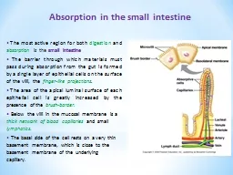
Slide3PHYSIOLOGY OF THE
SMALL INTESTINE
The principal function of the small intestine is the
digestion of
food and the absorption of nutrients, water and electrolytes.
Carbohydrates
and
proteins
are broken down in
the intestinal
lumen by pancreatic
enzymes,
after
which they
are absorbed.
Fats
are digested
by the actions
of pancreatic
lipase and bile salts.
The
products of fat
digestion, (fatty
acids and
monoglycerides),
separate from bile salts in
the jejunum
and are absorbed for further processing.
The jejunum is
the principal site for digestion and absorption of fluid,
electrolytes, iron
, folate, fat, protein and
carbohydrate
.
The absorption
of bile salts and vitamin B12 only occurs in
the terminal ileum
.
Slide4If the jejunum is resected, the ileum can assume all the required absorptive functions.
Resection of the terminal ileum will result in a diminished bile salt pool, B12 deficiency and may lead to deficiency of the fat-soluble vitamins A, D, E and K.
The small intestine plays an important role in the metabolism of plasma lipoproteins, as it is the main site of synthesis of high-density, low-density and very low-density
lipoproteins (HDL
, LDL, VLDL).
These particles transport most of the absorbed dietary fat to the systemic circulation via the lymph.
The small bowel also synthesises intestinal hormones such as glucagon-like peptides GLP-1 and 2, peptide YY and motilin, which interact with the enteric nervous system to modulate intestinal function, growth and differentiation.
Slide5INFLAMMATORY
BOWEL DISEASE
It is a
conditions characterised by the presence
of idiopathic
intestinal
inflammation.
Crohn’s
disease (regional enteritis)
CD
is characterised by
a chronic
full-thickness inflammatory process that can
affect any
part of the gastrointestinal tract from the lips to the
anal margin
.
It
is most common in North America and
Northern Europe
with an annual incidence of 8 per 100 000.
Over
the last four decades, the incidence appears to
have increased
three-fold, thought to possibly be a
consequence of;
E
nvironmental factors
I
mproved
diagnostic
modalities
B
oth.
Slide6It is slightly more common in women than in men, and is most commonly diagnosed between the ages of 25 and 40 years.
There is a second peak of incidence around the age of 70 years.
In those countries with high prevalence of CD, the groups with the highest prevalence seem to be Caucasian, notably American Whites and Northern Europeans, whereas it is less common, even in high prevalence countries, in those originating from Central Europe and less prevalent still in those originating from South America, Asia and Africa.
CD seems to be especially prevalent (three- to five-fold higher) in the Ashkenazi Jewish population, although interestingly, the prevalence of CD in the Jewish population in Israel is lower than that in Europe or the United States, suggesting that environmental factors are also important.
Slide7Aetiology
I
s
incompletely understood
T
hought to
involve a complex interplay of genetic and
environmental factors.
N
o
causative organism has ever been
demonstrated.
Smoking
increases the relative risk of
CD three-fold
.
Smoking
cessation has a beneficial effect on
disease activity.
Genetic
factors are also clearly extremely important.
10% of patients have a first-degree
relative with
the disease,
and
50% in monozygotic twins.
Inheritance
is
thought to
involve multiple genes with low penetrance.
The NOD2/ CARD15
gene
have
strong associations with CD.
The
vast majority of individuals with CD have no abnormalities of these genes.
Since these genes are involved in intracellular recognition of bacteria, their discovery provides potentially valuable insight into the pathogenesis of CD, as a disease in which the
relationship between
the gut mucosa and the normal gut bacteria becomes deranged, resulting in uncontrolled intestinal inflammation
.
Slide8Pathogenesis
I
ncreased
gut mucosal permeability appears
to develop
at
a
early stage of the disease.
This lead
to increased passage of luminal antigens, which
then induce
a cell-mediated inflammatory response.
This results in
the release of proinflammatory cytokines, such as
interleukin- 2
and tumour necrosis factor, which coordinate local
and systemic
inflammatory responses.
It
has been suggested
that CD
is associated with a defect in suppressor T-cells,
which usually
act to prevent escalation of the inflammatory process.
I
ncrease
in
gut permeability
,
combined with
an abnormal immune-mediated
response to
colonisation of the gut with some subspecies of the
normal enteric
microflora, may initiate the disease.
Slide9Pathology
The terminal ileum is most commonly involved (65%),
either in
isolation or in combination with colonic disease.
Colitis alone
occurs in up to a one-third of cases and the
remainder are
patients with more proximal small bowel
involvement.
The
stomach and duodenum are affected in around 5
%, but
perianal lesions are common, affecting up to 50–75%
of patients.
Macroscopically,
resection specimens show fibrotic
thickening of
the intestinal wall with narrowing of the lumen
and fat
wrapping (encroachment of mesenteric fat around
the bowel).
There
is usually dilated bowel just proximal to the stricture and deep mucosal ulcerations with linear or snake-like patterns in the strictured area itself.
Oedema in between the ulcers gives rise to a cobblestone appearance of the mucosa.
The transmural inflammation (which is a characteristic feature of CD) may lead to segments of bowel becoming adherent to each other and to surrounding structures, inflammatory masses with mesenteric abscesses and fistulae into adjacent organs
.
.
Slide10The
serosa is usually opaque, with thickening of the mesentery and enlarged mesenteric lymph nodes.
CD is characteristically discontinuous, with inflamed areas separated by normal intestine, so-called ‘skip’ lesions.
Microscopically,
there are focal areas of chronic inflammation involving all layers of the intestinal wall with lymphoid aggregates.
Non-caseating giant cell granulomas are found in 60% of patients and when present clearly allow a confident diagnosis of CD.
They are most common in anorectal
disease Multifocal
arterial occlusions are found in the muscularis propria, which is thickened.
There may be nerve cell hyperplasia and there is deep, fissuring ulceration within affected areas.
Characteristically, and unlike in UC, there may be completely normal areas immediately next to areas of severe inflammation
Slide11Clinical
features
Occasionally, CD
presents acutely with ileal inflammation and
symptoms and
signs resembling those of acute
appendicitis or
even
with free
perforation of the small intestine, resulting in a local
or diffuse
peritonitis.
M
ay
present with fulminant
colitis.
M
ore
commonly presents with features of chronicity.
Small bowel CD is often characterised by abdominal
colicky pain
, which may be postprandial, and mild diarrhoea
extending over
many months occurring in bouts.
A
tender mass
may be
palpable in the right iliac fossa.
Intermittent
fevers,
secondary anaemia
and weight loss are common.
After months of
repeated attacks
of acute
inflammation,
the affected
area of intestine begins to narrow with fibrosis,
causing more
chronic obstructive symptoms
.
Slide12Children developing the illness before puberty may have retarded growth and
sexual development
.
With progression of the disease, adhesions and transmural fissuring, intra-abdominal abscesses and fistulae may develop.
Fistulation may occur into adjacent loops of bowel (enteroenteric or interloop fistulae).
Occasionally
, the (healthy) sigmoid loop may become adherent to the affected terminal ileum, resulting in ileosigmoid fistulation.
The fistula tracks in such cases are usually small and the profuse diarrhoea that results from ileosigmoid fistulation is
due
to bacterial overgrowth (attributable to colonisation of
the small
bowel with colonic flora) rather than passage of small bowel content into the colon.
Fistulation
may also occur into the bladder (ileovesical) or the female genital tract and,
less commonly
, the duodenum.
Slide13Fistulation
into the
abdominal wall
(enterocutaneous fistulation) may also develop
spontaneously, or more
commonly occurs as a complication
of abdominal surgery.
Colonic
CD presents with symptoms of colitis and
proctitis as
described for
UC,
although toxic
megacolon is
much less common.
Many patients with CD present with perianal
problems.
In
the presence of active disease, the perianal skin
appears bluish
.
Superficial
ulcers with undermined edges are
relatively painless
and can heal with bridging of epithelium.
Deep cavitating ulcers
are usually found in the upper anal canal;
they can
be painful and cause perianal abscesses and fistulae,
discharging around
the anus and sometimes forwards into
the genitalia
.
Fistulation
through the posterior wall of the
vagina may
lead to rectovaginal fistula and continuous leakage of
gas and/or
faeces per vaginam.
Slide14The rectal mucosa is often spared in CD and may feel normal on rectal examination.
If it is involved, however, it will feel thickened, nodular and irregular.
Perianal
disease is frequently associated with dense, fibrous stricturing at the anorectal junction.
Incontinence may develop as a result of destruction of the anal sphincter musculature because of inflammation, abscess formation, fibrotic change and repeated episodes of surgical drainage.
In
severe cases, the perineum may become densely fibrotic, rigid and covered with multiple discharging
openings.
Each
patient with CD should have their disease phenotype (manifestations) classified according to the Montreal classification.
This is important as it allows an overview of disease progression in the individual patient over time, and it enables group comparisons and evaluations.
The Montreal classification specifies age of onset, location and behaviour.
T
he
behaviour of CD can be dominated by inflammation without stricturing or penetration, stricturing or penetration (causing phlegmons, abscesses and fistulae).
Slide15Montreal classification for Crohn's disease
Montreal
Age at diagnosis
A1
below 16 y
A2
between 17 and 40
y
A3
above 40 y
Location
L1
ileal
L2
colonic
L3
ileocolonic
L4
isolated upper disease*
Behaviour
B1
non‐stricturing, non‐penetrating
B2
stricturing
B3
penetrating
p
perianal disease modifier
†
----------------------------------------------------------------------------------------------------------
*L4 is a modifier that can be added to L1–L3 when concomitant upper gastrointestinal disease is present.
†“p” is added to B1–B3 when concomitant perianal disease is present.
Slide16Investigations
LABORATORY
A full blood count should be performed, as
anaemia
is
common and
usually multifactorial.
It
may result from the
anaemia of
chronic
disease
.
From
iron deficiency as a result of
blood loss
or malabsorption.
Vitamin
B12 deficiency may occur
as a
consequence of terminal ileal disease
or resection
.
Folate deficiency
may also result from diffuse small bowel disease
or resection
.
Active
inflammatory disease is usually
associated with
a fall in serum albumin, magnesium, zinc and
selenium.
Acute
phase protein measurements (C-reactive
protein)
and the erythrocyte sedimentation rate
may correlate
with disease activity.
Finding an elevated concentration in the stools of
calprotectin
, a
specific marker of
inflammation.
M
ay
support
a diagnosis
of CD in patients with new onset of persistent
gastrointestinal symptoms
.
It
can also be used to monitor
disease activity
in the long-term management of established CD.
Slide17ENDOSCOPY
M
ay
be normal or show
patchy inflammation
.
Characteristically
, there are areas of
normal mucosa
in between areas of inflammation that are
irregular and
ulcerated, with a mucopurulent exudate.
The earliest findings
are of aphthous ulcers surrounded by a rim of
erythematous mucosa
.
These
become larger and deeper
with increasing
severity of disease.
There
may be stricturing,
and it
is important to exclude malignancy at these sites by
multiple and
often repeated mucosal biopsies.
An
irregular
Crohn’s stricture
with polypoid mucosa may be almost
macroscopically indistinguishable
from malignancy.
The
terminal
ileum may
be ulcerated and strictured
.
Slide18In patients who have had previous ileocaecal resection and anastomosis, recurrent disease usually presents first with aphthous ulceration just proximal to the anastomosis.
Interval colonoscopy is therefore important in the follow-up after surgery for CD.
Upper gastrointestinal symptoms may
require
upper
gastrointestinalendoscopy
, which may reveal deep longitudinal ulcers and cobblestoning of mucosa in the duodenum,
stomach or
, rarely, in the oesophagus.
Enteroscopy
may reveal jejunal ulceration and stricturing.
Capsule endoscopy
should not be undertaken where there is a suspicion of stricture, because of the possibility of the capsule becoming stuck in the narrow segment.
Slide19IMAGING
High-resolution ultrasound
can demonstrate inflamed
and thickened bowel loops, as well as fluid
collections and
abscesses.
The
small intestine is traditionally
imaged by
a
small bowel
enema
.
This
is performed
by instilling
contrast into the small bowel via a
nasoduodenal tube
, and will show up areas of stricturing and
prestenotic dilatation
.
The
involved areas tend to be narrowed,
irregular and
, sometimes, when a length of terminal ileum is
involved, there
may be the ‘string sign’ of
Kantor.
Computed tomography (CT) scans
with oral
contrast are
widely used in the investigation of abdominal
symptoms and
can demonstrate fistulae, intra-abdominal abscesses
and bowel
thickening or dilatation.
Slide20Magnetic resonance imaging (MRI)
is useful in assessing complex perianal disease and, more recently, has been shown to be an excellent method for investigating the small bowel.
MR enterography (oral contrast) or enteroclysis (contrast administered via nasoduodenal tube) is particularly effective at demonstrating small bowel stricturing and avoids the need for repeated exposure to large doses of ionising radiation in young patients
.
A labelled white cell scan
is occasionally of value to determine whether or not a segment of bowel is actively inflamed and guide decisions on medical treatment.
In patients with enterocutaneous fistulae,
fistulography
will be required to demonstrate the anatomy and complexity of the fistulae and allow adequate planning for future surgery.
Slide21Treatment
MEDICAL TREATMENT
Steroids
are the traditional method for
inducing remission
in CD, and remain important when rapid
remission is
required.
They
induce remission in 70–80% of
cases with
moderate to severe disease.
They
should be used
in short
courses only and tapered when a response has
been achieved
.
They
reduce inflammation and are therefore
ineffective in
fibrostenotic disease, where the symptoms
relate mainly
to obstruction.
Steroids
can also be used as
topical agents
in the rectum where the benefits include reduced
systemic bioavailability
, but long-term use can still cause
adrenal suppression.
More recently, oral steroid formulations
such as
budesonide have been devised, to ensure that the
steroid moiety
is removed in the portal circulation, reducing
systemic side
effects.
Steroids
should not be used for maintenance
therapy for
CD and are usually replaced with
immunomodulatory agents
in order to minimise the risk of side
effects associated
with long-term steroid use.
Slide22Aminosalicylates
Colonic symptoms can be treated by 5-ASA agents in a similar manner to that in UC.
These agents
have limited efficacy in small bowel CD.
Antibiotics
Metronidazole
and ciprofloxacin may be used, particularly for periods of a few weeks at a time, especially in perianal disease.
Long-term
use of metronidazole is to
be especially
avoided, as there is a risk of peripheral neuropathy.
Ciprofloxacin
also has significant side effects when used in the long term, such as Achilles tendinitis and tendon rupture
.
Antibiotics may also be used to decrease systemic symptoms resulting from an inflammatory mass or an abscess.
In
general
,
abscess should be treated by percutaneous drainage and/or surgery as antibiotics alone will not treat a Crohn’s mass effectively.
Slide23Immunomodulatory
agents
Azathioprine is used for
its additive
and steroid-sparing effects and currently
represents standard
maintenance therapy.
It
is a purine analogue,
and
works
by inhibiting
cell-mediated immune responses.
6-MP (6-Mercaptopurine) may be given
directly for the same
effects.
Cyclosporin
also acts by inhibiting cell-mediated
immunity.
Short-course
intravenous cyclosporin treatment is
associated with
80% remission; however, there is relapse
after completion
of treatment in many cases.
Monoclonal
antibody therapy
monoclonal antibodies
targeting tumour necrosis factor alpha and
other key
pro-inflammatory mediators.
Infliximab, a murine chimeric monoclonal antibody,
was the
first available monoclonal antibody for the treatment
of CD.
This needs to be administered as an intravenous
infusion and
is typically given every 8 weeks for maintenance of remission.
Adalimumab
, an entirely human monoclonal
antibody, is
an alternative to
infliximab.
This
is administered
subcutaneously every
1–2 weeks, depending on response, and
most patients
can self-administer this agent.
Slide24Third-generation monoclonal antibody therapies
include integrin
antibodies vedulizumab and etrolizumab.
Both prevent leucocyte
migration
in
the
gastrointestinal tract
and may therefore have fewer side effects than the
earlier monoclonal antibodies.
The
roles of monoclonal antibodies have expanded
from initially
being used exclusively in the most severe cases of
CD when
other therapies failed, to having a more central role
in the
management of moderate to severe CD.
They
are
currently widely
used for induction and maintenance of remission.
Slide25There is evidence that early and aggressive use of these agents in patients at high risk for early recrudescent disease after surgery (for example, penetrating phenotype, early mucosal inflammation or aphthous ulceration at follow-up colonoscopy) may reduce the need for subsequent surgery.
These agents also appear to be effective treatments for perianal disease.
Recent studies have suggested, however, that while they may reduce the inflammation associated with the process of fistulation and can achieve healing of fistula openings, the fistula tracks may remain patent and cessation of therapy is usually associated with a high risk of
reactivation of
the fistulae.
Monoclonal antibodies are expensive forms of treatment that are associated with a small but definable risk of overwhelming bacterial infection and specific malignancies over the long term.
Active
infection, tuberculosis and a past history of malignancy are specific contraindications
.
Slide26Nutritional
support
Patients
with moderate nutritional
impairment will
require nutritional supplementation and
severely malnourished
patients may require enteral tube or even
intravenous feeding
.
Anaemia
, hypoproteinaemia and
electrolyte, vitamin
and metabolic bone problems must all be
addressed.
Elemental
diet or parenteral nutrition can induce
remission in
up to 80% of patients, an effect comparable to steroids.
However, almost all patients relapse rapidly after cessation of therapy
.
Principles
of management of Crohn’s disease (CD)
●
Close liaison between physician and surgeon is crucial
●
Medical therapy should always be considered as
an alternative
to surgery, although surgery should not be
delayed when
a clear indication for surgery exists
●
Patients must be optimised as far as possible prior to
surgery, and
this may require preoperative total parenteral nutrition
●
CD is a chronic relapsing disease with a high likelihood
of reoperation
; the surgeon must take every reasonable effort
to
preserve
bowel length and sphincter function
Slide27ENDOSCOPIC DILATATION IN CROHN’S DISEASE
S
tricturing
may be
amenable to
endoscopic treatment, provided the strictures can
be reached
with an
endoscope.
This may be accomplished by enteroscopy or
colonoscopy, depending
on the site of the stricture.
Dilatation
of
an inflamed
or ulcerated stricture is contraindicated because
of the
risks of perforation, but balloon dilatation of
fibrostenotic disease
may result in substantial symptomatic
improvement and
obviate the need for surgery in selected cases.
Slide28INDICATIONS FOR SURGERY
Surgical resection will not cure CD.
Surgery therefore focuses on managing the complications of the disease.
As many of these indications for surgery may be relative, joint
management by
an aggressive physician and a conservative surgeon is ideal.
CD is a complicated condition and decisions regarding management are best made jointly
by members
of a
team
.
Complications or manifestations of CD for which surgery is usually appropriate include the following
:
Recurrent
intestinal obstruction;
Persistent
or, less commonly, massive acute bleeding;
Free
perforation of the bowel;
Failure
of medical therapy;
Steroid
dependent disease;
Intestinal
fistula;
Perianal
disease (abscess, fistula, stenosis);
Malignant
change (notably in the colon and less commonly as a complication of small bowel disease).
Slide29TOP-DOWN APPROACH TO MANAGEMENT
OF CROHN’S
DISEASE
A
ctive
ileocolic CD may be treated
initially with
a thiopurine, adding steroids and then a
monoclonal antibody
only if and when required.
Some
centres
instead advocate
a top-down approach, where rapid remission
is obtained
by initiating therapy with a monoclonal
antibody agent
(unless contraindicated), often in combination with
a thiopurine
.
Studies
suggest advantages mainly in the form
of rapid
remission, steroid sparing and increased rates of
mucosal healing
.
Slide30Intra-abdominal septic
complications are
more common if one or more of the following risk factors are present:
● Current
high-dose steroid therapy (≥10 mg prednisolone for ≥4 weeks before surgery);
● Current
preoperative monoclonal antibody therapy;
● Preoperative
significant weight loss (>10% premorbid weight);
● Pre-existing
abdominal sepsis (notably abscess or fistula);
● Serum
albumin <32 g/L.
If any these risk factors are present (and particularly if more than one risk factor is present as the risks appear to be additive), one should consider exteriorising the bowel and planning a delayed anastomosis when the risk factor has been corrected
.
Slide31A range of operations is performed for CD, depending on the pattern of disease – the most common are outlined below
:
● Ileocaecal resection is the usual procedure for terminal ileal disease, with a primary anastomosis between the ileum and the ascending or transverse colon, depending on the extent of the disease.
● Segmental resection of short segments of small or large bowel strictures can be performed.
● Colectomy and ileorectal anastomosis may be undertaken for colonic CD with rectal sparing and a normal anus.
● Subtotal colectomy and ileostomy for Crohn’s colitis accounts for 8% of such procedures for acute colonic disease.
● Temporary loop ileostomy.
This
can be used either in patients with acute distal CD, allowing remission and later restoration of continuity, or in patients with severe perianal or rectal disease.
Slide32●
Proctectomy and proctocolectomy. Many patients
with severe
anal disease failing to respond to medical
treatment will
eventually require a permanent colostomy. When
this occurs
in a setting of severe colonic disease,
proctocolectomy and
permanent ileostomy may be required.
●
Strictureplasty.
Strictured
areas of CD
can be
treated by strictureplasty, a local widening
procedure, to
avoid small bowel resection and is thus an
important bowel
sparing technique
Strictureplasty is
particularly useful for the treatment of
fibrostenotic disease
, when there is little or no active
inflammation in
the involved segment. Multiple strictureplasties
can be
performed and strictureplasty can be combined
with resection
.
.
Slide33Anal disease should be treated conservatively by simple drainage of abscesses and the use of setons through fistulae to avoid sphincter injury.
Infliximab or adalimumab therapy may be combined with seton insertion in the early phase of management of perianal fistulae.
Once the fistula has dried up, typically after 2–3 doses, the seton can be removed.
Laying open of fistulae (fistulotomy), commonly performed for fistulae resulting from the common cryptoglandular perianal abscess, should be avoided in CD as the wound edges heal very slowly or not at
all.
Slide34Extraintestinal manifestations of Crohn’s disease
●
Related to disease activity
Erythema nodosum
Pyoderma gangrenosum
Arthropathy
Eye complications (iritis/uveitis)
Aphthous ulceration
Amyloidosis
●
Unrelated to disease activity
Gallstones
Renal calculi
Primary sclerosing cholangitis
Chronic active hepatitis
Sacroiliitis
Slide35Differences between ulcerative colitis (UC)
and Crohn’s
disease (CD)
●
UC affects the colon; CD can affect any part of
the gastrointestinal
tract, but particularly the small and
large bowel
●
UC is a mucosal disease, whereas CD affects the
full thickness
of the bowel wall
●
UC produces confluent disease in the colon and
rectum, whereas
CD is characterised by skip lesions
●
CD more commonly causes stricturing and fistulation
●
Granulomas may be found on histology in CD, but not in UC
●
CD is often associated with perianal disease, whereas this
is unusual
in UC
●
CD affecting the terminal ileum may produce
symptoms mimicking
appendicitis, but this does not occur in UC
●
Resection of the colon and rectum cures the patient with
UC, whereas
recurrence is common after resection in CD
Slide36INFECTIVE ENTERITIS
Campylobacteriosis
Infection with Campylobacter jejuni (a gram-negative rod
with a distinctive spiral shape) is the most common form of bacterial gastroenteritis
in the UK, typically acquired from
eating infected
poultry.
It
causes diarrhoea and abdominal pain
and may
mimic an acute abdomen.
Severe
cases may
resemble UC
, with rectal bleeding and colorectal ulceration,
causing diagnostic
difficulty.
The
organism is very sensitive and
may take
several days to isolate on stool culture.
Toxic dilatation and
even disintegrative colitis have rarely been reported
to occur
.
Treatment
is generally supportive as the condition
usually resolves
without antibiotics.
It
is a notifiable disease
.
Slide37Yersiniosis
Yersinia enterocolitica is a gram-negative rod that can infect the terminal ileum, appendix, ascending colon and mesenteric lymph nodes, and can cause a granulomatous
inflammatory process
that mimics CD.
Yersinia
typically causes a fever and gastroenteritis, but may persist and cause a terminal ileitis, which, on occasion, may perforate.
The diagnosis may
be made on stool culture, but is more often confirmed serologically
.
If discovered at laparotomy, the terminal ileum and mesenteric nodes will look thickened and inflamed and
a lymph
node biopsy can be taken for diagnostic purposes.
The
disease is normally self-limiting, but responds to treatment with cotrimoxazole or chloramphenicol.
Slide38Salmonellosis, typhoid
and paratyphoid
Salmonella are a family of gram-negative rods that can
cause a
range of enteric infections.
Salmonella
gastroenteritis is
typically caused
by S. enteritidis from poultry, and is most
often a
self-limiting illness comprising headache, fever and
watery diarrhoea
.
When
severe,
antibiotics, hospitalisation and intravenous
fluids may be needed.
The
diagnosis is based
on stool
culture.
Shigella
and enteropathogenic strains of E.
coli may
cause similar diarrhoeal illnesses.
Typhoid fever is caused by S. typhi and presents with
fever and
abdominal pain after an incubation period of 10–20
days.
Over
the next week, the patient can develop distension,
diarrhoea, splenomegaly
and characteristic ‘rose spots’ on
the abdomen
caused by a vasculitis.
Typhoid
is a systemic
infection and
diagnosis of typhoid is confirmed by culture of
blood or
stool.
Treatment
is by antibiotics, usually chloramphenicol.
A number of surgical complications can result, including
paralytic ileus
, intestinal haemorrhage, free ileal perforation
and cholecystitis
.
Slide39I
nvasion
of the systemic circulation,
which is
a characteristic feature of salmonellosis, may cause
severe gram-negative
sepsis, resulting in septic shock.
Some patients develop
metastatic sepsis, including septic arthritis and
osteomyelitis, meningitis
, encephalitis, disseminated
intravascular coagulation
and pancreatitis.
Perforation of a typhoid ulcer characteristically
occurs during
the third week of the illness, although it is
sometimes the
first clinical sign of the disease.
The
ulcer is parallel
to the
long axis of the gut and is usually situated in the
distal ileum
.
Perforation
requires surgery to wash out and close
the ulcer
and intestinal resection is usually avoided.
In unstable patients
, notably with evidence of septic shock, the
bowel should
be exteriorised and the perforation closed after
recovery.
Paratyphoid
infection (with S. paratyphi)
resembles typhoid
fever and is treated in a similar manner.
Slide40Tuberculosis of the intestine
Tuberculosis,
can
affect any part of the gastrointestinal tract from the mouth to the anus.
The
sites affected most often are the ileum, proximal colon and peritoneum.
The disease presented as
;
Ulcerative tuberculosis
Ulcerative tuberculosis develops secondary to pulmonary tuberculosis and arises as a result of swallowing tubercle bacilli.
Multiple
ulcers, lying transversely,
in
the
terminal ileum
and the overlying serosa is thickened, reddened and covered in tubercles.
Patients
typically present with diarrhoea and weight loss, although subacute obstruction
and even
local perforation and fistula formation can occur.
A
barium follow-through or CT examination fails to show filling of the lower ileum, caecum and the ascending colon as a
result of
narrowing of the ulcerated
segment.
A
course of antituberculous chemotherapy usually leads to cure, provided the pulmonary tuberculosis is adequately treated.
Surgery
is usually undertaken only in the rare event of a perforation or complete intestinal obstruction.
Slide41Hyperplastic
tuberculosis
This is caused by the ingestion of Mycobacterium
tuberculosis by
patients with a high resistance to the organism.
The infection usually
occurs in the ileocaecal region, although
solitary and
multiple lesions in the lower ileum are also
sometimes seen
.
The
infection establishes itself in lymphoid
follicles, and
the resulting chronic inflammation causes thickening
of the
intestinal wall and narrowing of the lumen.
There
is
early involvement
of the regional lymph nodes, which may
caseate.
A
bscess
and fistula formation are rare.
Patients usually present with attacks of abdominal
pain and
intermittent diarrhoea.
There
is incomplete ileal
obstruction, leading
to stasis and bacterial overgrowth.
This
in
turn causes
steatorrhoea, anaemia and loss of weight.
Patients may present
with a mass in the right iliac fossa and vague ill health.
The differential diagnosis is that of an appendix mass,
lymphoma, carcinoma
of the caecum, CD, tuberculosis or actinomycosis
.
Slide42A barium follow-through or small bowel
enema will
show a long narrow filling defect in the terminal
ileum.
CT will also
demonstrate the narrowed segment with proximal
distension and
the associated lymphadenopathy.
When
the
diagnosis is
clear and the patient has not yet developed
obstructive symptoms
, treatment with chemotherapy is advised and
may be
curative.
Where
obstruction is present, or the possibility
of CD
or lymphoma require clarification, ileocaecal resection
is often
required.
Slide43Actinomycosis
Abdominal actinomycosis is rare.
It is caused by infection with Actinomyces israelii and infection usually develops several weeks after an apparently straightforward perforated appendicitis.
An
abscess develops and spreads to the retroperitoneal tissues and the adjacent abdominal wall, eventually becoming the seat of multiple indurated discharging sinuses.
At
first,
the discharge
from the sinuses is thin, watery and inoffensive, but it may later become thicker and malodorous.
Secondary
fistulation may occur and the tissues may become
extensively indurated
and woody.
M
esenteric
lymph nodes are not involved and the lumen of the intestine is not narrowed.
Haematogenous
spread via
the portal
vein may lead to multiple liver abscesses.
Pus should be sent for bacteriological examination, which will reveal the characteristic sulphur granules.
Penicillin
or cotrimoxazole treatment is required and should be
prolonged and
in high dosage.
Slide44Human immunodeficiency virus
Human immunodeficiency virus (HIV) infection is
associated with
a number of proctological
problems.
Intestinal
complications are common
after the
development of AIDS when opportunistic organisms
can cause gastroenteritis.
HIV
may
also cause
a specific enteropathy.
Treatment
is directed towards
the relevant
organism and surgery should be avoided if possible.
Opportunistic
intestinal infections in patients with AIDS
●
Bacteria
Salmonella
Shigella
Yersinia
Campylobacter
Mycobacterium avium intracellulare (MAI)
●
Viral
Cytomegalovirus
●
Protozoa
Cryptosporidium
Giardia
●
Fungal
Candida albicans
Slide45TUMOURS OF THE
SMALL INTESTINE
Small bowel tumours are rare and in total account for
less than
10% of gastrointestinal neoplasia
.
Benign
The majority of small bowel neoplasms are benign,
comprising adenomas
, lipomas, haemangiomas and neurogenic
tumours.
They
are frequently asymptomatic and identified
incidentally, but
can present with intussusception, small bowel
obstruction and
bleeding that may cause anaemia or may even be
overt.
Where
these lesions do cause anaemia, the cause can be
difficult to
diagnose, as CT or small bowel contrast studies do
not show
them easily.
Capsule
endoscopy or small bowel
endoscopy has
been used successfully where the facilities exist.
Symptomatic lesions can be treated by small bowel
resection and
anastomosis
.
Slide46Peutz–Jeghers syndrome
This is an autosomal dominant disease characterised by melanosis of the mouth and lips, with multiple hamartomatous
polyps
in the small bowel and
colon.
Melanin
spots can also occur on the digits and perianal skin.
The
gene STK11 on chromosome 19 has been found in a proportion of patients with this condition.
Long-term
follow-up of the original family described by Peutz has shown reduced survival as a consequence of complications of bowel obstruction and the development of a range of cancers
.
Slide47It is logical to perform regular colonic surveillance and encourage female patients to attend breast and cervical screening.
Despite the increased risk of malignancy in general, malignant change in the polyps themselves rarely occurs and the polyps can be left alone unless they are the cause of symptoms.
Resection may be indicated, however, for heavy and persistent or recurrent bleeding or intussusception.
Polyps may be removed by enterotomy or, at laparotomy, snared via a colonoscope introduced via an enterotomy.
Heavily involved segments of small intestine may occasionally be resected.
Slide48Malignant
Small bowel malignancy is rare and classically presents
late, most
often diagnosed after surgery for small bowel
obstruction.
Four
types will be considered, which account for
over 99
% of small bowel malignancies:
Adenocarcinoma
C
arcinoid tumours
L
ymphomas
M
esenchymal
tumours (
gastrointestinal stromal
tumours [
GIST]
Adenocarcinoma
Small
bowel adenocarcinoma is more often found in the
jejunum than
the ileum and although the aetiology is
unknown, it
is more common in patients with CD, coeliac disease,
familial adenomatous
polyposis, hereditary non-polyposis
colon cancer
and Peutz–Jeghers syndrome.
Slide49The tumours
present
with anaemia, overt gastrointestinal bleeding, intussusception or obstruction.
Prognosis
is poor, particularly in patients with CD, in whom these tumours often present late, because the symptoms are commonly mistaken for those of CD and treated conservatively.
When suspected, the advised surgical
treatment
is a resection of 5 cm of non-involved bowel either side of the lesion and the affected mesentery.
A right hemicolectomy is likely to be required for tumours of the distal ileum.
Slide50Carcinoid tumours
These
neuroendocrine tumours
occur throughout the gastrointestinal tract, most commonly in the appendix, ileum and rectum in decreasing order of frequency.
Appendicular carcinoid tumours
are commonly noted as an incidental finding at appendicectomy, and are said to occur in approximately 2% of appendices removed surgically.
The
tumour arises from Kulchitsky cells at the base of intestinal crypts (of Lieberkuhn).
The
primary is usually small, although
significant lymph
node metastases can occur.
In
up to one-third of cases of small bowel carcinoids, the tumours are multiple.
They may
produce dense fibrosis in the surrounding tissues, resulting in distortion and scarring of the bowel and associated mesentery, giving them a characteristic radiological appearance.
Slide51Carcinoid
tumours can produce a number of
vasoactive peptides
, most commonly 5-hydroxytryptamine (serotonin
), but
also histamine, prostaglandins and kallikrein.
When they metastasise
to the liver, the ‘carcinoid
syndrome’ can become evident
, because the vasoactive substances escape the
filtering actions
of the liver.
The
clinical syndrome itself
consists of
reddish-blue cyanosis, flushing attacks, diarrhoea,
borborygmi, asthmatic
attacks and, eventually, pulmonary
and tricuspid stenosis.
Slide52Classically, the flushing attacks are induced by alcohol.
Surgical resection is usually sufficient for patients with primary disease, but the incidence of recurrence is significant.
The extent of disease can be assessed preoperatively using
octreotide scanning
, which may detect otherwise clinically unapparent primary and secondary tumours.
Plasma markers of tumour bulk, such as
chromogranin A
concentrations, may be useful markers of disease recurrence, as well as of prognostic value.
Slide53Hepatic resection can be carried out in patients with metastatic disease.
The
treatment
has been transformed by the use of octreotide (a somatostatin analogue), which
reduces both
flushing and diarrhoea, and octreotide cover is usually used in patients with a carcinoid syndrome who have surgery to prevent a carcinoid crisis resulting from liberation
of vasoactive
substances following handling of the tumour.
Carcinoid
tumours generally grow more slowly than most metastatic malignancies and patients may live with the syndrome of metastatic disease for many years.
They
are not usually sensitive to chemo- or radiotherapy
Slide54Lymphoma
primary or, secondary
The
incidence
is
increased in patients with CD and
immunodeficiency syndromes
.
It
is rare for Hodgkin’s lymphoma
to affect
the small bowel and most western-type lymphomas
are
non-Hodgkin’s
B-cell lymphomas.
They
usually present
with anaemia
, bleeding, perforation, anorexia and weight loss
.
T-cell lymphoma
develops in patients with coeliac
disease.
It
usually presents with worsening of the patient’s
diarrhoea, pyrexia
of unknown origin and local obstructive
symptoms.
Mediterranean
lymphoma
is found mostly in North
Africa and
the Middle East and is often widespread at
diagnosis.
Burkitt’s
lymphoma
can aggressively affect the
ileocaecal region
, particularly in children
.
The
treatment
for
these conditions is chemotherapy; however, surgery
may be
required for obstruction, perforation or bleeding.
Slide55Gastrointestinal stromal
tumours (
GISTs)
A
re mesenchymal tumours
and the distinction between benign or
malignant types
is difficult even on histological examination
.
Increased size
and high levels of c-kit (CD117) staining are
associated with
malignant potential.
GISTs
are found most
commonly in
the stomach, but can be found in other parts of the gut.
They occur most commonly in the 50- to 70-year age
group.
Although
the cause is unknown, patients with
neurofibromatosis have
an increased risk of developing these
types of
tumour.
Patients
may be asymptomatic and the
tumour may
present as an incidental mass on a CT scan.
Symptoms include
lethargy, pain, nausea, haematemesis or melaena.
Surgery
is
the most effective way of treating GISTs, as the
tumour is
radioresistant and is not sensitive to conventional
chemotherapy.
Glivec
® (imatinib) is a tyrosine kinase inhibitor
that has
been shown to be effective in advanced cases and
may also
have a role in adjuvant treatment
.
Slide56CONNECTIVE TISSUE DISORDERS
Intestinal diverticulae
Diverticulae (hollow out-pouchings) are a common structural abnormality that can occur from the oesophagus to the rectosigmoid junction (but not usually in the rectum).
Small
bowel diverticulae may be congenital or acquired.
Congenital diverticulae
A
ll
three coats of the bowel are present in the wall of
the diverticulum
(e.g. Meckel’s diverticulum
).
Acquired diverticulae
These
develop
in the jejunum and arise from the mesenteric side of the bowel as a result of mucosal herniation at the point of entry of the blood vessels.
There
is thus no muscularis layer present in the wall.
Jejunal
diverticulae can vary in size and are frequently multiple.
They
are commonly asymptomatic and present as an incidental finding at surgery or on radiological
imaging.
However
, they can result in malabsorption, as a result of bacterial stasis, or present as an acute abdominal emergency if they become inflamed or perforate.
Bleeding
from a jejunal diverticulum is a rare complication (compared with sigmoid diverticular disease).
Slide57Elective
resection of an affected small bowel
segment that
is causing malabsorption can be effective, provided
there is
only a limited amount of jejunum affected by the
condition.
If
perforated jejunal diverticulitis is found at
emergency laparotomy
, a small bowel resection should be performed
and a
decision made between anastomosis and stoma
formation.
This
will depend on the degree of contamination,
physiological stability
and local resources for managing a patient with
a high
output
jejunostomy.
Complications
resulting from extensive jejunal
diverticulosis can
be extremely difficult to treat.
In
severe cases,
much of
the proximal small intestine may be involved,
effectively precluding
resection.
Prolonged
antibiotic therapy for
bacterial overgrowth
may be preferable, and antibiotics (
metronidazole, ciprofloxacin
, rifaximin) are frequently rotated in
an attempt
to avoid the development of antibiotic
resistance.
.
Slide58Meckel’s diverticulum
A Meckel’s diverticulum is a persistent remnant of the
vitellointestinal duct
and is present in about 2% of the
population.
It
is found on the antimesenteric side of the ileum,
commonly approximately
60 cm from the ileocaecal valve and is
classically 5
cm
long.
A
Meckel’s diverticulum
is a
congenital diverticulum.
It
contains all three coats of
the bowel
wall and has its own blood supply.
It
may be
vulnerable to
obstruction and inflammation in the same way as
the appendix
; indeed, when a normal appendix is found at
surgery for
suspected appendicitis, a Meckel’s diverticulum
should be
looked for by examining the small bowel, particularly
if free
fluid or pus is found
.
In approximately 20% of cases,
the mucosa
of a Meckel’s diverticulum contains heterotopic
epithelium of
gastric, colonic or pancreatic type.
The presence of
heterotopic mucosa may predispose to the development
of complications
.
The
vast majority of Meckel’s diverticulae
are asymptomatic
and Meckel’s diverticulum is notoriously
difficult to
see with contrast radiology
.
Slide59Meckel’s diverticulum may, however, present clinically in the following ways:
●
Haemorrhage
. If gastric mucosa is present, peptic ulceration can occur and present as painless dark rectal bleeding or melaena. If the stomach, duodenum and colon are excluded as a source of bleeding by endoscopy, radioisotope scanning with technetium-99m may demonstrate aMeckel’s diverticulum.
●
Diverticulitis
. Meckel’s diverticulitis presents like appendicitis, although if perforation occurs the presentation may resemble a perforated duodenal ulcer.
●
Intussusception.
A Meckel’s diverticulum can be the lead point for ileoileal or ileocolic intussusception.
●
Chronic ulceration
. Pain is felt around the umbilicus, as the site of the diverticulum is midgut in origin.
●
Perforation
.
When
found in the course of abdominal surgery, a Meckel’s diverticulum can safely be left alone, provided it has
a wide
mouth and is not thickened. When there is doubt, it can be resected. The finding of a Meckel’s diverticulum in an inguinal or femoral hernia has been described as ‘Littre’s hernia’.
Slide60Meckel’s
diverticulectomy
A broad-based Meckel’s diverticulum should not
be amputated at
its base and invaginated (as for an appendix
), as
there is the risk of stricture and of leaving heterotopic
epithelium behind
.
It
is safer simply to excise the
diverticulum, either
by resecting it and suturing the defect at its base,
or with
a linear stapler-cutter.
If
the base of the diverticulum
is indurated
, it is on balance more logical to perform a
limited small
bowel resection of the involved segment followed
by an
anastomosis.
Slide61VASCULAR ANOMALIES OF
THE INTESTINE
Mesenteric ischaemia
Classified to;
A
cute intestinal ischaemia
– with or without
venous occlusion
Chronic arterial ischaemia, central
or peripheral.
The
superior mesenteric
vessels
A
re
the visceral vessels most likely to be affected by
embolisation or
thrombosis, with the former being most common.
Occlusion at the origin of the superior mesenteric
artery (SMA
) is almost invariably the result of thrombosis,
whereas emboli
tend to lodge at the origin of the middle colic
artery.
Inferior
mesenteric
vessels
I
nvolvement
is usually clinically
silent because
the collateral circulation is better
.
Slide62Possible sources for embolisation of the SMA
include;
T
he
left atrium in atrial fibrillation,
T
he
left ventricle after mural myocardial infarction,
V
egetations
on mitral and aortic valves associated with endocarditis
A
theromatous
plaque from an aortic aneurysm.
Primary thrombosis is associated with atherosclerosis and vasculitides, including conditions such as thromboangitis obliterans and polyarteritis nodosa.
Primary
thrombosis of the superior mesenteric veins may occur in association
with;
F
actor V Leiden
P
ortal hypertension
P
ortal pyaemia
S
ickle cell disease
W
omen taking the oral contraceptive pill.
A specific form of ‘non-occlusive mesenteric ischaemia’ may complicate critical illness, possibly due to alterations in splanchnic blood flow
.
Slide63Irrespective of whether the occlusion is arterial or
venous, haemorrhagic
infarction occurs.
The
mucosa is
especially sensitive
to ischaemic injury because of its high
metabolic activity
.
The
intestine and its mesentery become
swollen and
oedematous, especially with venous occlusion.
Bloodstained fluid
exudes into the peritoneal cavity and
bowel lumen
.
The
changes develop rapidly and irreversible
injury, ranging
in severity from mucosal necrosis and sloughing,
to full-thickness infarction
usually occurs within a few hours
at most.
If the main trunk of the SMA is involved, the
infarction usually
covers an area from just distal to the DJ
flexure to
the splenic flexure.
Usually
, a branch of the main trunk
is implicated
and the area of infarction is less.
Slide64Clinical
features
S
udden
onset of severe
abdominal
pain in a patient with atrial fibrillation or atherosclerosis.
The pain is
typically in the central
abdomen.
Persistent
vomiting and defaecation occur early, with the subsequent passage of altered blood.
Abdominal tenderness may be mild initially, with rigidity being a late feature.
Shock
, with features of both hypovolaemia and sepsis, rapidly ensues.
Investigation
A
profound neutrophil leucocytosis
A
bsence
of gas in the lumen of the thickened small intestine on abdominal radiographs
and
CT scans.
Gas
may be present within the intestinal wall and occasionally in the mesenteric and portal veins.
Slide65Treatment
F
ull resuscitation
L
aparotomy
with embolectomy
via the
ileocolic artery or revascularisation of the SMA by
vascular bypass
may be considered in early cases.
Anticoagulation should
be implemented early in the postoperative
period.
However
, the condition is usually diagnosed late in the
disease process
and the mortality rate is extremely high.
In
the
young, all
affected bowel should be resected, whereas in the elderly
situation
may
be incurable
.
After
extensive enterectomy, it is usual for patients
to require
intravenous alimentation.
The
young, however,
may sometimes
develop sufficient intestinal digestive and
absorptive function
to lead relatively normal lives.
In
selected
cases, consideration
may be given to small bowel transplantation
.
Slide66Chronic small intestinal ischaemia
A
lmost
invariably results from atherosclerosis and affects the proximal superior mesenteric and coeliac vessels.
Patients
classically present with symptoms of severe central abdominal pain that comes on within 30–60 minutes of eating (mesenteric angina).
Weight
loss and diarrhoea due to malabsorption may also occur.
The
condition may be difficult to diagnose and is often overlooked initially, the symptoms being mistaken for those of peptic ulcer disease or irritable bowel syndrome.
The
presence of significant vascular disease on visceral angiography is common in elderly patients and in those with severe vascular
disease, and
should not necessarily be assumed to indicate that abdominal symptoms are attributable to chronic ischaemia.
Treatment
By
selective visceral angiography, with
stenting/angioplasty
If not
possible
, so
bypass surgery.
Smoking
cessation in
imperative.
P
atients
are usually anticoagulated.
Slide67STOMAS
A colostomy (or ileostomy)
stoma;
I
s
an artificial
opening made
in the colon (or small intestine) to divert faeces
and flatus
out-side the abdomen where they can be collected
in an
external appliance.
A
stoma may
be;
Temporary
Permanent
Loop ileostomy
;
I
s often used
for defunctioning a low
rectal anastomosis
or an ileal pouch.
A
knuckle of ileum is exterio-rised through a skin trephine in the right iliac fossa.
An incision is
made in the distal part of the knuckle, and this is
then pulled
over the top of the more proximal part to create a
spout on
the proximal side of the loop with a flush distal side
still in
continuity
.
Slide68This allows near perfect defunctioning, but also the possibility of restoration of continuity, by taking down the spout and reanastomosing the partially divided ileum.
The advantages of a loop ileostomy over a loop colostomy are the ease with which the bowel can be brought to the surface and the relative absence of odour.
Care
is needed when the ileostomy is closed, so that suture line obstruction does not occur.
Closure
of a loop ileostomy can be a technically challenging procedure, particularly if there are dense adhesions resulting from previous surgery.
Slide69End ileostomy
Is
formed after a subtotal colectomy without anastomosis, when it may later be reversed, or may be permanent after a panproctocolectomy.
The
ileum is
normally brought
through the rectus abdominis muscle.
Careful
attention to the terminal ileal mesentery should be taken to ensure that it is not too bulky.
The
use of a spout was
should project some 2–4 cm from the skin
surface.
A
disposable appliance is placed over the ileostomy so that it is a snug fit at skin level.
Slide70There may be an ‘ileostomy flux’ while the ileum adapts to the loss of the colon.
A
consistent ileostomy output in excess of 1.5 litres is usually associated with dehydration and sodium depletion in the absence of intravenous therapy.
The
stools thicken in a few weeks and are semisolid in a few months.
The help and
advice of the stoma care nurse specialist are essential.
Modern
appliances have transformed stoma care, and skin problems are
unusual.
Stoma
bags and appliances
Stoma output is collected in disposable adhesive bags.
Ileostomy appliances tend to be drainable bags, which are leftin place for 48 hours, while colostomy appliances are simply changed two or three times each day.
A wide range of such bags is currently available.
Many now incorporate an adhesive backing, which can be left in place for several days.
In most hospitals, a stoma care service is available to offer advice to patients
Slide71Stoma complications
● Skin
irritation ●
Prolapse
●
Retraction ●
Ischaemia
●
Stenosis ●
Parastomal hernia
●
Bleeding ●
Fistulation
Complications
of
stomas are common.
On
occasion,
require
surgical
revision.
Sometimes
, this can be achieved with an incision
immediately around
the stoma, but on occasion reopening the
abdomen and
freeing up the stoma may be necessary.
Repair of parastomal
hernias is particularly technically challenging
and the
recurrence rate is high.
Simple
suture of the
parastomal hernia
is associated with an almost 100% risk of
recurrence and it may need to either;
T
ransfer
to the opposite side of the
abdomen
.
I
nsertion of
a piece of prosthetic material within the abdominal
wall around
the stoma may be necessary
.
Slide72CONDITIONS
CAUSING MALABSORPTION
Coeliac
disease
I
s
the most common cause of
malabsorption in
the UK with a stated prevalence of
1:1800.
It is characterised by a
hypertrophic small
bowel mucosa with atrophic villi and deep crypts.
It is thought
that the loss of surface area and brush border
enzymes results
in malabsorption.
I
s
caused by an abnormal immune
response to gluten.
There
is a genetic component, as the
disease is
more common in first-degree relatives and has an
association with
HLA B8.
In
children, coeliac disease presents
with steatorrhoea
and growth retardation.
In
adults, it may
result in
diarrhoea and weight loss but many patients simply
present with
an iron deficiency anaemia
.
Slide73Some patients develop a characteristic skin rash (dermatitis herpetiformis)
The
diagnosis is usually made after an endoscopic duodenal biopsy allows pathological examination of mucosa.
The
antiendomysial antibody tests have a very high sensitivity and specificity for coeliac disease, but a duodenal biopsy is usually indicated to confirm the diagnosis.
The
biopsy usually shows flattening of the mucosa, marked inflammatory changes and characteristic findings of intraepithelial lymphocytes.
Patients
with coeliac disease may develop an acute inflammatory condition of the small intestine (ulcerative jejunoileitis) and have an increased risk of small bowel
lymphoma and
adenocarcinoma.
The
main treatment for coeliac disease is the
withdrawal of
gluten from the diet by avoiding wheat, rye and barley.
Surgery
does not usually play a role in the management of coeliac disease and is primarily reserved for resection of malignancy
.
Slide74Bacterial overgrowth
The
small intestine can become colonised with bacteria
normally confined
to the colon if there is stasis resulting in
delayed bacterial
clearance (blind loop
syndrome).
Similar complications may result from chronic small
bowel obstruction
, jejunal diverticulosis and ileosigmoid
fistulation.
If
overgrowth occurs in the upper small
intestine, the
defect is chiefly of fat absorption; if in the lower
intestine, there
is vitamin B12 deficiency.
There
is usually
relatively little
effect on carbohydrate or protein metabolism.
Stasis results
in an abnormal bacterial flora, which prevents
adequate breakdown
of
fat.
Sometimes
, the only manifestation
is anaemia
, resulting from vitamin B12 deficiency but, if
steatorrhoea occurs
, other serious malabsorption features may
follow, including
glossitis, osteomalacia, paraesthesia and
peripheral neuropathy
.
Improvement normally follows after intermittent
therapy with
oral antibiotics; metronidazole, ciprofloxacin,
tetracycline and
rifaximin are commonly used.
Definitive treatment is
surgical when the anatomical abnormality can be
corrected, but
this is not always possible.
Slide75ENTEROCUTANEOUS FISTULA
An abnormal connection between the small intestine and
the skin
can occur as a result of fistulating CD, radiotherapy
or abdominal
trauma, but the condition most commonly
follows a
surgical complication – either a leak from an anastomosis
or an
inadvertent enterotomy during dissection.
At
least 50%
of small
bowel enterocutaneous fistulae develop after surgery
in which
no small bowel has been resected, as a result of injury
to the
intestine following division of adhesions.
The
frequency
of this
complication has been shown to increase with the number
of
previous laparotomies.
Management
of patients
with enterocutaneous
fistulae can be very challenging,
especially when
the fistula output is high (usually defined as >500
mL of
effluent/day).
The
majority of fistulae can be expected
to heal
spontaneously, provided there is no distal obstruction
or disease
at the fistula site.
Reasons
for failure of
spontaneous healing
also include epithelial continuity between the
gut and
the skin and an associated complex abscess.
Slide76The management of fistulas
An
early return to theatre to try and treat the problem definitively in a septic, malnourished patient is doomed to failure.
Infected
collections are best identified at
CT
and can be drained percutaneously.
Skin
protection is important, as small bowel effluent is caustic.
Nutritional
support must include fluid and electrolytes, which can be
lost in
high quantities from a proximal fistula, as well as carbohydrates, protein, fat
and vitamins
.
Judgements
have to be made between enteral and parenteral feeding – enteral feeding
has advantages
but if the fistula is proximal or high output,
total parenteral
nutrition will be
required.
Defining
anatomy is best done after
a
sequence of contrast studies (follow-through, fistulogram and enema) may well be required to define bowel length and plan a surgical strategy.
Surgery
can be extremely technically demanding, and an anastomosis should not be fashioned in the presence of continuing intra-abdominal sepsis or when the patient is hypoalbuminaemic (<32 g/dL).
Slide77SHORT BOWEL SYNDROME
Intractable diarrhoea with impaired absorption of
nutrients following
resection or bypass of the small intestine,
ultimately leading
to progressive malnutrition, is referred to
as ‘short
bowel syndrome’.
The
most common causes of
short bowel
syndrome
are;
R
esection
resulting from the
management of
CD and its complications (which accounts for almost
half of
cases
)
M
esenteric
vascular
thrombosis
R
adiation enteritis
Tumours
The
features
are usually appear
when there is less than 200 cm of small
bowel length
and nature of the remaining
intestine.
In general, diseases which result in short bowel
syndrome tend
to preferentially affect the distal small
intestine, and
there is some evidence that the ileum, with its
tighter intercellular
junctions and consequently better fluid
absorptive capacity
, can assume the functions of a missing
jejunum, but
not vice versa
.
Slide78T
he
ileocaecal valve used to be considered important with regard to preservation of absorptive
function, of the distal ileum and right colon.
Patients
with an intact colon are relatively protected
from the
effects of massive small bowel resection because of the ability of the colon to absorb not only fluid and electrolytes, but a modest amount of nutrient energy.
Patients
with as
little as
100–200 cm of jejunum anastomosed to an intact colon may therefore be able to maintain satisfactory macronutrient, fluid and electrolyte status, although they will, of course, be
at risk
of fat-soluble and B12 vitamin deficiencies and will also generally need oral nutritional supplements of trace elements, vitamins and minerals.
Some
(but not all) patients with50–100 cm of small intestine and an intact colon will need long-term parenteral nutrition, as will almost all patients with 50 cm or less of jejunum anastomosed to an intact colon.
In
contrast, most patients with less than 200 cm of small intestine ending in a jejunostomy will require regular infusions of parenteral fluid and electrolytes, and almost all of those with less than 100 cm of small bowel ending in a jejunostomy will require long-term parenteral nutrition
.
Slide79Medical
management
The
use of antidiarrhoeal agents (
loperamide and
codeine phosphate), drugs to reduce diarrhoea
related to
bile-salt malabsorption
and enteral, parenteral vitamin
and trace element supplements.
Although there
has also been interest in the use of drugs to
promote intestinal
adaptation, such as growth hormone,
glutamine and
, most recently, glucagon-like peptide 2 agonists, the
mainstay of
treatment for short bowel
syndrome.
H
ome parenteral nutrition
(HPN
)
is, however, expensive and
demanding.
The patients
with short bowel syndrome receiving HPN are at
risk from
catheter-related complications
(sepsis
and occlusion), as well as
metabolic
complications
(fibrotic
liver disease, gallstones, metabolic bone disease
and kidney
stones).
Slide80Surgical
treatment
procedures
designed to improve the surface area or reduce the speed of transit of the remaining small intestine (and thus improve absorptive capacity) have shown some promise in children, but their place in managing adults with established short bowel syndrome is currently unclear.
In
some patients, the loss of venous access resulting from the complications of long-term intravenous feeding or the development of progressive liver dysfunction may represent indications for
small bowel transplantation
.
The
results of small bowel transplantation have progressively improved and 5-year patient survival now exceeds 80% in some centres.
Slide81Quiz 1
In
patients with short bowel syndrome
receiving Home
parenteral nutrition
(HPN)
are at risk
of metabolic and catheter-related complications. Enumerate these complications.
Slide82The large
intestine
ANATOMY
OF THE
LARGE INTESTINE
The large intestine begins at the ileocaecal valve and
extends to
the anus.
It
is divided into the caecum, ascending
colon, hepatic
flexure, transverse colon with attached greater
omentum, splenic
flexure, descending colon, sigmoid and
rectum.
The
large intestine is approximately 1.5 m long, but can
be straightened
at endoscopy so the caecum can be reached
with 70
cm of colonoscope.
The
colon is distinguished from the small bowel by
having fat-filled
peritoneal tags known as appendices
epiploicaem and
the taenia coli.
These
are three flat bands of
longitudinal muscle
that run from the appendix base to the
rectosigmoid junction
.
Slide83They act to pull the colon into its sacculated state,producing a series of haustrations.
Distended
small and large intestine can be distinguished on an abdominal radiograph as the small bowel has complete transverse markings caused by the valvulae conniventes, while the colon has incomplete lines from the sacculation caused by the taeniae.
The
important posterior relations of the caecum and ascending colon are the right ureter, right gonadal vessels and
duodenum and
these must be protected at surgery.
The
left ureter, left gonadal vessels and tail of the pancreas must be protected when operating on the left colon.
The
blood supply of the large intestine is derived from branches of the superior mesenteric artery from the
caecum to
the distal transverse colon and the inferior mesenteric artery and its branches more distally.
Adjacent
branches anastomose so there is usually a complete vascular supply along the colon, named the marginal
artery.
Slide84This vessel is often the key blood supply to the
vascular arcades
, ensuring adequate perfusion of a colonic
anastomosis, but
blood flow in the ‘watershed’ area of the
splenic flexure
representing the junction between the superior
and inferior
mesenteric supply may be tenuous.
Sudden occlusion of
the inferior mesenteric artery may leave the area
of the
splenic flexure poorly perfused, leading to an
ischaemic colitis
.
Venous
and lymphatic drainage of the colon
follows the
arterial supply and venous drainage is into the
portal system
.
High
ligation of the artery supplying a segment
of colon
will therefore also remove the draining lymph
nodes, a
key technical point in cancer surgery.
The
nerve
supply to
the large intestine is derived from the splanchnic
nerves via
a dense sympathetic plexus surrounding the superior
and inferior
mesenteric arteries.
Visceral
pain from the part
of the
colon supplied by the superior mesenteric artery is
thus felt
, like that of the small intestine, in the
periumbilical region
, while pain from the colon distal to that point is
felt suprapubically
.
Slide85PHYSIOLOGY OF THE LARGE INTESTINE
The principle function of the colon is absorption of water; 1000 mL of ileal content enters the caecum every 24 hours, of which only approximately 200 mL is excreted as faeces.
Sodium
absorption is
by
an active transport system, while chloride and water are absorbed passively.
Fermentation
of dietary fibre in the colon by the normal colonic microflora leads to the generation of short chain fatty acids, which are an important metabolic fuel for the colonic mucosa.
Diversion
of the faecal stream may lead to inflammatory changes in the colon downstream (diversion colitis).
Absorption
of nutrients including glucose, fatty
acids, amino
acids and
vitamins.
Colonic
motility is variable.
In
general, faecal residue reaches the caecum 4 hours after a meal and the rectum after 24 hours.
Passage
of stool is not orderly because of mixing within the colon, so it is not uncommon for residue from a single meal to still be passed 4 days later
.
Slide86TUMOURS OF THE
LARGE INTESTINE
Benign
The
‘
polyp’
is
any protrusion
of the
mucosa
.
Polyps
can
occur;
S
ingly
Small
numbers
A polyposis syndrome
Adenomatous
polyps
A
tubular
adenoma
T
he
villous
adenoma
A
flat spreading
lesion
Villous
tumours can cause
diarrhoea, mucus
discharge and, occasionally hypokalaemia and
hypoalbuminaemia.
The
risk of malignancy developing in an
adenoma increases
with size; there is a 10% risk of cancer in
a 1-cm
diameter tubular adenoma, whereas almost one-third
of large
(>3 cm) colonic adenomas will have an area of
invasive malignancy
within them.
Slide87Adenomas larger than 5 mm in diameter are usually excised because of their malignant potential.
Snare
polypectomy is usually possible for
colonic polyps
but larger sessile polyps may require endoscopic mucosal resection after infiltration of a solution containing dilute adrenaline
.
Larger rectal adenomas may require transanal resection or, where the adenoma is too high, transanal endoscopic microsurgery or resection via transanal placement of laparoscopic instruments.
Extensive
villous lesions of the rectum may require argon beam ablation when the patient is frail but symptomatic.
Slide88Classification of intestinal polyps.
Inflammatory
Inflammatory polyps (pseudopolyps in UC)
Metaplastic
Metaplastic
or hyperplastic polyps
Hamartomatous
Peutz–Jeghers
polyp
Juvenile
polyp
Neoplastic
Adenoma Tubular
Tubulovillous
Villous
Adenocarcinoma
Carcinoid
tumour
UC,
ulcerative colitis
.
Slide89Familial adenomatous
polyposis (
FAP)
D
efined clinically by
the presence of more than 100 colorectal adenomas, but is also characterised by duodenal adenomas and
multiple extraintestinal manifestations.
Over
80% of cases come from patients with a positive family history.
The
remainder arise as a result of new mutations in the adenomatous polyposis coli (APC) gene on the short arm of chromosome 5.
FAP
is inherited as an autosomal dominant condition and is consequently equally likely in men and women.
The
lifetime risk of colorectal cancer is 100% in patients with FAP.
FAP
can also be associated with
benign mesodermal
tumours such as desmoid tumours and osteomas.
Epidermoid
cysts can also occur (Gardner’s syndrome); desmoid tumours in the abdomen spread locally to involve the intestinal mesentery and, although non-metastasising, they may become
unresectable.
Up
to 50% of patients with FAP have congenital hypertrophy of the retinal pigment epithelium (CHRPE), which can be used to screen affected families if genetic testing is unavailable.
Slide90CLINICAL FEATURES
Polyps are usually visible on sigmoidoscopy by the age of
15 years
and will almost always be visible by the age of 30
years.
Carcinoma
of the large bowel develops 10–20 years after
the onset
of the polyposis
.
If there are no adenomas by the
age of
30 years, FAP is unlikely.
If
the diagnosis is made
during adolescence
, surgery is usually deferred to the age of 17
or 18
years unless symptoms develop.
Malignant
change is
rare before
the age of 20 years
.
Examination of blood
relatives, including
cousins, nephews and nieces, is essential; a
family tree
should be constructed and a register of affected
families maintained.
Referral to a medical geneticist is essential.
If over
100 adenomas are present at colonoscopy, the
diagnosis can
be made
confidently.
Slide91TREATMENT
The aim of surgery in FAP is to prevent the development of colorectal cancer.
The
surgical options are:
Colectomy
with ileorectal anastomosis (IRA);
Restorative
proctocolectomy with an ileal pouch–anal anastomosis (RPC);
Total
proctectomy and end ileostomy.
The patient is almost always young and likely to prefer to avoid a permanent stoma and so the choice is normally between the first two options.
The
advantage of an IRA is that it avoids the temporary stoma frequently required for an RPC and avoids the potential compromise to sexual function accompanying proctectomy.
It
is also has a lower morbidity and mortality.
However
, the rectum requires regular surveillance
.
Even with optimal surveillance of the rectal remnant, up to 10% of patients will develop invasive malignancy within a 30-year follow up period.
Restorative
proctocolectomy has the advantage of removing the whole colon and rectum
.
Slide92T
here
is a pouch failure rate of approximately10
%.
In addition, and particularly where a
stapled anastomosis
has been created, there remains a small but
definite incidence
of cancer developing in the small strip of rectal mucosa between the pouch and the dentate line.
Some
advocate
complete mucosectomy of the residual cuff
and a
transanal anastomosis, although this may result in
worse function
.
In
experienced hands a laparoscopic approach
to these
operations can be
successful.
Slide93Extracolonic manifestations of FAP
●
Endodermal derivatives
Adenomas and carcinomas of the duodenum, stomach, small intestine, thyroid and biliary tree
Fundic gland polyps
Hepatoblastoma
●
Ectodermal derivatives
Epidermoid cysts
Pilomatrixoma
Congenital hypertrophy of the retinal pigment epithelium (CHRPE)
Brain tumours
●
Mesodermal derivatives
Desmoid tumours
Osteomas
Dental problems
Slide94POSTOPERATIVE SURVEILLANCE
F
ollow-up is important
and takes the form of rectal/pouch
surveillance, with
biopsy of the cuff recommended yearly.
Gastroscopies
to
detect upper gastrointestinal
tumours (notably
duodenal adenomas).
L
ifespan
is
reduced because
of the development of duodenal and
ampullary Cancers and
complications of desmoid tumours
.
Hereditary
non-polyposis colorectal
cancer (Lynch
syndrome)
Hereditary non-polyposis colorectal cancer (HNPCC)
is characterised
by an increased risk of colorectal cancer
and also
cancers of the endometrium, ovary, stomach and
small intestine
.
It
is an autosomal dominant condition caused
by a
mutation in one of the DNA mismatch repair genes.
The most
commonly affected genes are MLH1 and MSH2.
The lifetime
risk of developing colorectal cancer is 80%, and
the mean
age of diagnosis is 45 years.
Most
cancers develop
in the
proximal colon.
Females
have a 30–50% lifetime risk
of developing
endometrial cancer
.
Slide95DIAGNOSIS
HNPCC can be diagnosed by genetic testing or by
the Amsterdam
II criteria:
●
T
hree
or more family members with an
HNPCC-related cancer (colorectal, endometrial, small bowel, ureter, renalpelvis), one of whom is a first-degree relative of the other two;
●
T
wo
successive affected generations;
●
A
t
least one colorectal cancer diagnosed before the age of 50 years;
●
FAP excluded;
●
T
umours
verified by pathological examination.
Patients with HNPCC are offered regular endoscopic surveillance.
Slide96Malignant – colorectal cancer
Epidemiology
In the UK, colorectal cancer is the second most common cause of cancer death.
In
the
UK, approximately
35 000 patients are diagnosed with colorectal cancer every
year.
Approximately
one-third of these tumours are in the rectum and two-thirds in the colon.
The
burden of disease is similar in men and women.
Colorectal
cancer occurs less frequently in the resource-poor world than in resource-rich countries.
Slide97Aetiology
I
t
arises from adenomatous polyps after a sequence of genetic mutations influenced by environmental factors.
The
adenoma–carcinoma sequence is not a simple stepwise progression of
mutations but
a complicated array of multiple genetic alterations, ultimately resulting in an invasive tumour.
Mutations
of the adenomatous polyposis coli (APC) gene occur in two-thirds of colonic adenomas and are thought to develop early in the carcinogenesis pathway.
K-ras
mutations result in activation of cell signalling pathways and are more common in larger lesions,
suggesting
that they are later events in mutagenesis
.
Slide98The
p53 gene is frequently mutated in
carcinomas but
not in adenomas and therefore thought to be a marker of invasion.
There is association between diet
and colon cancer.
Worldwide
, the prevalence of
colorectal cancer
is closely associated with intake of red meat
and particularly
processed meat products (haem and
N-nitroso compounds
).
These
adversely affect DNA in the
colorectal mucosa
.
A
protective effect of dietary fibre is also
suggested by
epidemiological studies.
The
hypothesis is that
increased roughage
is associated with reduced colonic transit times,
and this
in turn reduces the exposure of the mucosa to dietary
carcinogens.
Increased
risk for colorectal cancer has also been associated with dietary animal fat, smoking and alcohol.
Cholecystectomy
may marginally increase the risk of rightsided colon cancer and inflammatory bowel disease is a well-recognized risk
factor.
Slide99Pathology
Macroscopically, the tumour may take one of four
forms.
Annular
Tubular
Ulcer
Cauliflower
The
annular variety tends to give rise to obstructive symptoms, whereas the others present more commonly with bleeding.
Most
large bowel cancers arise from the left colon, notably the rectum (38%), sigmoid (21%), and descending colon (4%).
Cancer
of the caecum (12%) and ascending colon (5%) are less common, but may be gradually increasing in incidence.
Cancer
of the transverse
colon (5.5
%), flexures (2–3%) and appendix (0.5%) are relatively uncommon.
Microscopically
, the neoplasm is a columnar cell adenocarcinoma.
Origin
from a benign polyp may be
evident in
early cases, before the benign architecture is destroyed by malignant infiltration.
Slide100Spread
Colonic cancer can
spread:
locally
the lymphatics
bloodstream
across
the peritoneal
cavity
Direct
spread may be longitudinal or radial.
Radial spread may
be retroperitoneal into the ureter, duodenum and
posterior abdominal
wall muscles or intraperitoneal into
adjacent organs
or the anterior abdominal
wall.
In
general, involvement of the lymph nodes by the
tumour progresses
from those closest to the bowel along the
course of
lymphatics to central nodes.
Haematogenous
spread is most
commonly to
the liver via the portal vein.
One-third
of
patients will
have liver metastases at the time of diagnosis and
50% will
develop them at some point, accounting for the
majority of
deaths.
The
lung is the next most common
site
M
etastasis to
ovary, brain, kidney and bone is less common.
Colorectal cancer
can spread from the serosa of the bowel or via
subperitoneal lymphatics
to other structures within the
peritoneal cavity
, including peritoneum, ovary and omentum.
Slide101Staging colon
cancer
Dukes
’
classification was
originally described
for rectal tumours but has been adopted for histopathological reporting of colon cancer.
Although
it is simple and widely
recognised the
more detailed
TNM
system is regarded as the international
standard.
Slide102Dukes
’ staging for colorectal cancer
●
A: invasion of but not breaching the muscularis propria
●
B: breaching the muscularis propria but not involving
lymph nodes
●
C: lymph nodes
involved.
Dukes
himself never described a stage D, but this is often
used to
describe metastatic
disease
TNM
classification for colonic cancer
●
T Tumour stage
T1 Into submucosa
T2 Into muscularis propria
T3 Into pericolic fat or subserosa but not breaching serosa
T4 Breaches serosa or directly involving another organ
●
N Nodal stage
N0 No nodes involved
N1 1-3 nodes involved
N2 Four or more nodes involved
●
M Metastases
M0 No metastases
M1 Metastases
Slide103Clinical features
Carcinoma of the colon typically occurs in patients over 50 years of age and is most common in the 8th decade of life.
Emergency
presentation occurs in 20% of cases and is associated with a considerably worse prognosis, even when matched for disease stage
.
A careful family history should be taken.
Those
with first-degree relatives who have developed colorectal cancer before the age of 45 years may be part of one of the colorectal cancer familial syndromes.
Tumours of the
left side of the colon usually present with a change in bowel habit or rectal bleeding, while proximal lesions typically present later, with iron deficiency anaemia or a
mass.
Patients
commonly present with metastatic disease.
Lesions
of the flexures may present with vague upper abdominal symptoms for many months before symptoms
suggestive of
colonic disease appear
.
Slide104Investigation of colon cancer
SCREENING
Colon cancer is suited to screening as prognosis is better
for early
stage disease and polypectomy allows the
prevention of
cancer development.
In
the UK, a screening
programme has
been introduced based on
faecal occult blood testing
of people
aged 60–69 years, followed by colonoscopy in
those who
test positive.
A
guaiac-based test
is used, which
detects peroxidase-like
activity of faecal haematin
.
Studies
have suggested
a 15–20% reduction
in
colorectal cancer
specific mortality in
the screened population.
Flexible sigmoidoscopy
can
also be used as the initial screening tool, with a
similar reduction
in colorectal cancer
specific mortality
.
Slide105ENDOSCOPY
The 60-cm, fibreoptic, flexible sigmoidoscope is increasingly being used in ‘one-stop’ rectal bleeding clinics.
The
patient is prepared with an enema and sedation is not usually necessary.
It is usually possible to assess the bowel up to the splenic flexure, which will detect up to 70% of cancers and almost all that cause fresh rectal
bleeding.
Colonoscopy
is the investigation of choice if colorectal cancer is
suspected.
It
has the advantage of not only picking up a primary cancer but also having the ability to detect
polyps
or other carcinomas, which occur in 3–5% of cases.
There
is a small risk of perforation (1:1000).
Slide106RADIOLOGY
Double-contrast
barium enema
has traditionally been
used and
shows a cancer of the colon as a constant irregular
filling defect
, often described as looking like an
apple-core.
False
positives occur in 1–2% of cases and
false negatives
in 7–9% of cases.
It
has now been largely
replaced by
computed tomography (CT)
virtual colonoscopy,
which is
extremely sensitive in picking up polyps down to a
size of
6
mm.
It
has the advantage of being
less invasive than
colonoscopy but if a biopsy is required,
an endoscopy
will still be needed.
CT
is used as a diagnostic
tool in
patients with palpable abdominal masses.
Spiral
CT of
the chest
, abdomen and pelvis now represents the standard
means of
staging colorectal cancer, although
chest x-ray and
liver ultrasound
are alternatives if CT is not readily available.
Rectal
cancer
usually requires additional staging for local
spread, using
magnetic resonance
imaging (MRI)
.
Slide107Surgical treatment
PREOPERATIVE PREPARATION
Mechanical bowel preparation
has fallen out of favour in surgery for colon cancer, with little evidence of benefit and some of an increased rate of wound infection
.
It currently
remains in
use largely for low rectal resection, where unprepared bowel may be associated with a higher infection rate.
Antiembolism
stockings
should be fitted and the patient started on prophylactic subcutaneous
low molecular weight heparin
.
If
available,
manual compression boots
are used perioperatively.
Intravenous
prophylactic antibiotics
are given immediately before the start of surgery, to reduce the risk of surgical site infection.
A
single dose of antibiotics covering bowel organisms is as effective as multiple doses.
In
all cases where a stoma seems likely, careful preoperative counselling and marking of an appropriate site by an enterostomal therapist is
essential.
Slide108OPERATIONS
The operations described are designed to remove the
primary tumour
and its draining locoregional lymph nodes.
It is
unusual to find unsuspected metastases at laparotomy (
or laparoscopy
) after CT staging, but the presence of
peritoneal metastases
may predicate a palliative strategy with a
segmental resection
and less aggressive lymphadenectomy.
The
use
of stapling
and hand-suturing techniques for colonic
anastomoses have
been compared, and there is probably little
difference in
leak rate.
It
is more important that healthy bowel, free
of tension
or distal obstruction, is used to construct an
anastomosis and
that patients are adequately nourished and
free from
active infection if anastomotic leakage is to be avoided.
Slide109Right hemicolectomy
Carcinoma
of the caecum or ascending colon
is treated by right
hemicolectomy.
At open surgery the peritoneum lateral to the ascending colon is incised, and the incision is carried around the hepatic flexure.
The mesentery of the distal 20 cm of ileum and the mesocolon as far as the proximal third of the transverse colon is divided.
When
it is clear that there is an adequate blood supply at the resection margins, the right colon is resected, and an anastomosis is fashioned between the ileum and the transverse colon.
If the tumour is at the hepatic flexure the resection must be extended further along the transverse colon and will involve dividing the right branch of the middle colic artery.
Slide110Extended right hemicolectomy
Carcinomas of the transverse colon and splenic flexure are most commonly treated by an extended right hemicolectomy.
The extent of the resection is from the right colon to the descending colon.
The mobilisation is as for a right hemicolectomy but dissection continues to take down the splenic flexure and the whole transverse mesocolon is ligated.
Some surgeons prefer to perform a left hemicolectomy for a splenic flexure cancer.
Slide111Left hemicolectomy
This is the operation of choice for descending colon and sigmoid cancers.
The
left half of the colon is mobilised completely along the ‘white line’ that marks the lateral attachment of the mesocolon.
As the sigmoid mesentery is mobilised, the left ureter and gonadal vessels must be identified and protected.
The splenic flexure may be mobilised by extending the lateral
dissection from
below and completed by entering the lesser sac
.
The bowel and mesentery can then be resected to allow a tension-free anastomosis.
A temporary diverting stoma may be fashioned upstream, usually by formation of a loop ileostomy.
This is usually undertaken if the anastomosis is below the peritoneal reflection of the rectum, because healing is more likely to be impaired distally.
Slide112Laparoscopic surgery
Laparoscopic surgery for colon cancer has been shown to have equivalent overall and cancer related outcomes to open surgery.
In the UK, the National Institute for
Health and
Care Excellence has stated that laparoscopic
colorectal surgery
should be offered to suitable patients.
Operation times are
longer but wound infection rates, blood loss and
postoperative pain
scores are lower than for open surgery.
The costs of
laparoscopic surgery are, however, generally higher and
this may
particularly relevant where funds are limited
.
Specimen retrieval and bowel anastomosis can then be performed via small incisions.
Training in laparoscopic colorectal surgery is important, as there is a relatively long learning curve.
Slide113Emergency surgery
In the UK, 20% of patients with colonic cancer will present as an emergency, the majority with obstruction, but occasionally with haemorrhage or perforation.
If
the lesion is right sided, it is usually possible to perform a right hemicolectomy and anastomosis in the usual manner.
If
there has been perforation with substantial contamination or if the patient is unstable, it may be advisable to bring out an ileocolostomy rather than forming an anastomosis.
For
a left-sided lesion the decision lies between a Hartmann’s procedure or resection and anastomosis.
Where
endoscopic and radiological facilities are present an obstructing left-sided lesion can be treated with an expanding metal
stent.
This
has the advantage of converting an emergency operation with a high chance of a stoma to a situation that can be
managed semi-electively
by resection and anastomosis.
Although
early studies cast doubt on the benefits of colorectal stenting, more recently evidence has emerged that stenting leads to a
reduction in
stoma rates.
Slide114Postoperative
care
After colonic surgery patients should be closely
monitored, as
there is a small incidence of postoperative bleeding.
Antithrombosis measures
should be continued
for 28
days postoperatively.
Wound infections are
relatively common after colonic surgery and
may well
be more frequent than the 10% usually quoted.
Anastomotic leaks
occur in 4–8% of ileocolic or colocolic anastomoses.
The possibility should be borne in mind in
any patient
not progressing as expected or with unexplained
cardiac abnormalities
, fever or worsening abdominal pain.
Early investigation
with contrast enhanced CT scan is
appropriate.
In
the presence of sepsis or peritonitis, early return to
theatre and
taking down the leaking anastomosis with the
formation of
stomas is usually advised.
Slide115Prolonged nasogastric drainage, intravenous fluid therapy and cautious introduction of oral fluid and diet represented traditional postoperative practice.
Enhanced
recovery
programmes have
been shown to reduce length of hospital stay from 10–14 days to as little as 2–3 days, by modulating the surgical stress response and reducing post-operative ileus
.
It
is important to appreciate that these programmes require multiple interventions to be instituted and considerable time, effort and education from the surgical, anaesthetic and ward teams.
Adjuvant
therapy
In most patients with colon cancer there is little clear benefit of preoperative chemotherapy, although trials in selected patient groups with locally advanced disease are ongoing.
There is evidence that adjuvant chemotherapy improves outcome after surgery in patients with node-positive disease (Dukes C).
Slide116Metastatic
disease
Hepatic metastases can be resected and series have
demonstrated 5-year
survival of over 30% in resectable disease.
Liver surgeons
are increasingly aggressive in treatment and the
only absolute
limitation on what can be resected relates to
leaving behind
sufficient functioning liver, although this
clearly has
to be moderated by patient factors.
It
is important not
to biopsy
potentially resectable hepatic metastases as this
may cause
tumour dissemination.
Imaging
will usually
correctly identify
colorectal metastases and assess patients suitable
for liver resection.
The
role of
chemotherapy and
the timing of colonic and hepatic surgery in
synchronous metastases
is still a matter of debate and such cases should
be carefully
discussed by a multidisciplinary team.
Isolated lung metastases
may occasionally be suitable for resection but
they are
more commonly accompanied by metastases
elsewhere.
In
patients with widespread disease, palliative
chemotherapy is
offered alongside symptomatic treatment and support by
a palliative
care team.
Slide117Prognosis
Overall 5-year survival for colorectal cancer is approximately 50%.
The
most important determinant of prognosis is tumour stage and, in particular, lymph node status.
Patients
with disease confined to the bowel wall (Dukes stage A) will usually have cure by surgical resection alone and over 90% will have disease-free survival at 5 years.
Spread
beyond the bowel
wall (Dukes
B) reduces 5-year survival to approximately 60–70%. Patients with lymph node metastases (Dukes C) have a 5-yearsurvival of 30%, while fewer than 10% of patients
presenting with
metastatic disease at the outset will be alive 5 years later.
Slide118Colorectal
cancer follow-up
Since the advent of safe liver resection for metastases
the outcome
benefit of follow-up has been clearly demonstrated.
Follow-up aims to identify synchronous bowel tumours (
present in
3%) that were not picked up at the time of
original diagnosis
due to emergency presentation or incomplete assessment.
Similarly, 3% of patients will develop a
metachronous (at
a different time) colonic cancer and surveillance
colonoscopy is
designed to diagnose these.
Up
to a half of all
patients with
colorectal cancer will develop liver metastases at
some point
and regular imaging of the liver (by ultrasound and
CT scan
) and measurement of carcinoembryonic antigen (
CEA) is
designed to diagnose this early, in order to allow
curative metastectomy
.
Trials
of the optimum follow-up pathway
have suggested
that CEA measurement alone can be as effective
as regular
imaging.
Slide119Quiz 2:
What are the advantages
of
the ileorectal
anastomosis (
IRA)
over
the Restorative
proctocolectomy with an ileal pouch–anal anastomosis (RPC
)?
Slide120INFLAMMATORY
BOWEL DISEASE
The term ‘inflammatory bowel disease’ is reserved for conditions characterised by the presence of idiopathic intestinal inflammation (i.e. ulcerative colitis [UC] and Crohn’s
disease [CD
].
Ulcerative
colitis
UC is a disease of the rectum and colon with extraintestinal manifestations.
The
incidence is 10 per 100 000 per year in the UK with a prevalence of 160 per 100 000 population.
UC
affects men and women equally in early life, although it is said to be more common in males in later life.
It
is most commonly diagnosed between the ages of 20 and 40.
UC
is far more common in the USA and Western Europe but relatively rare in the Far East and tropics.
Asians
who spent their childhood before the age of 14 in Asia have a much lower incidence of UC than Asians born and raised in the UK, suggesting an important effect of environmental exposure in childhood.
Slide121Aetiology
The cause of UC is unknown.
There
is clearly a genetic
contribution, as
10–20% of patients have a first-degree
relative with
inflammatory bowel disease.
Patients
with severe
colitis have
a reduction in the number of anaerobic bacteria and
in the
variability of bacterial strains in the colon, but no
causative link
with any specific organism has been
identified.
Unlike
CD, smoking seems to have a protective effect in
UC and
has even been the basis of therapeutic trials of
nicotine.
Relapses
are occasionally said to be associated with periods
of stress
, but personality and psychiatric profiles in patients
with UC
are the same as those of the normal population
.
Slide122Pathology
A
ll
cases the disease starts in the rectum and extends proximally in continuity.
Colonic
inflammation is diffuse, confluent and superficial, primarily affecting the mucosa and superficial submucosa
.
‘Pseudopolyposis’ occurs in almost one-quarter of cases.
Stricturing
in UC is very unusual (unlike CD) and should prompt urgent assessment because of the possibility of coexisting carcinoma.
A
small proportion of patients develop irregular mucosal swellings (
dysplasia associated
lesions or mass [DALMs]), which are highly predictive of coexisting carcinoma.
Histological examination reveals an increase in inflammatory cells in the lamina propria and the crypts of Lieberkuhn and there are ‘crypt abscesses’.
There
is depletion of goblet cell mucin.
With
time, precancerous changes can develop (dysplasia).
Slide123High-grade
dysplasia is regarded as an
indication for
surgery as 40% of colectomy specimens in which
highgrade dysplasia
was detected will have evidence of a
colorectal cancer
.
In
contrast, optimum management of
low-grade dysplasia
is currently controversial.
Ten
to twenty per cent
of patients
with low-grade dysplasia will have a cancer at
colectomy.
The
progression rate of low-grade dysplasia to
invasive cancer
is unclear and many cancers in patients with
low-grade dysplasia
probably develop without high-grade dysplasia
.
.
Slide124Symptoms
Clinical presentation depends in large part on the extent of disease.
If confined to the rectum (proctitis), there is
usually no
systemic upset and extra-alimentary manifestations are rare.
The main symptoms are rectal bleeding, tenesmus and mucous discharge.
The disease remains confined to the rectum in 90% of cases but proctitis may extend proximally.
Colitis
is almost always associated with bloody diarrhoea and urgency.
Severe and/or extensive colitis may result in anaemia, hypoproteinaemia and electrolyte disturbances.
Pain
is
unusual
Children with poorly controlled colitis may have impaired growth.
The
more extensive the disease the more likely extraintestinal manifestations are to occur.
Extensive
colitis is also associated with systemic illness, characterised by malaise, loss of appetite, and fever
.
Slide125Classification
of colitis severity
The assessment of severity of UC is determined by
frequency of
bowel action and the presence of systemic signs of illness:
●
Mild disease
is characterized by fewer than four
stools daily
, with or without bleeding. There are no
systemic signs
of toxicity.
●
Moderate
disease
corresponds
to more than four
stools daily
, but with few signs of systemic illness. There may
be mild
anaemia. Abdominal pain may occur.
Inflammatory markers
, including erythrocyte sedimentation rate
and C-reactive
protein, are often raised.
●
Severe disease
corresponds to more than six bloody
stools a
day and evidence of systemic illness, with fever,
tachycardia, anaemia
and raised inflammatory markers.
Hypoalbuminaemia is common.
●
Fulminant disease
is associated with more than 10
bowel movements
daily, fever, tachycardia, continuous
bleeding, anaemia
, hypoalbuminaemia, abdominal tenderness
and distension
, the need for blood transfusion and, in the
most severe
cases, progressive colonic dilation (‘toxic megacolon’). This is a very significant finding, suggestive of
disintegrative colitis
, and an indication for emergency
surgery if
colonic perforation is to be avoided.
Slide126Extraintestinal manifestations
Arthritis
occurs in around 15% of patients and is typically a large joint polyarthropathy, affecting knees, ankles, elbows and wrists.
Sacroiliitis
and ankylosing spondylitis are 20 times more common in patients with UC than the general population and are associated with the HLA-B27 genotype
.
Sclerosing cholangitis
is associated with UC and can progress to cirrhosis and hepatocellular failure.
Patients
with UC and sclerosing cholangitis are also at a significantly greater risk of development of large bowel cancer.
Cholangiocarcinoma
is an extremely rare association and its frequency is not influenced by colectomy.
The
skin lesions
erythema nodosum and pyoderma gangrenosum
are associated with UC and both normally resolve with good colitis control.
The
eyes can also be affected by
uveitis
and
episcleritis
.
Slide127Acute
colitis
Approximately 5% of patients present with severe acute (
fulminant) colitis
.
Intensive
medical treatment leads to
remission in
70% but the remainder require urgent surgery.
Toxic dilatation
should be suspected in patients who develop
severe abdominal
pain and confirmed by the presence on a
plain abdominal
radiograph of a colon with a diameter of more
than 6 cm.
A
reduction in stool frequency is
not always
a sign of improvement in patients with severe UC,
and a
falling stool frequency, abdominal distension and
abdominal pain
(resulting from progression of the inflammatory
process through
the colonic wall) are strongly suggestive of
disintegrative colitis
and impending perforation.
Plain abdominal radiographs
should be obtained daily in patients with
severe colitis
, and a progressive increase in colon diameter
despite medical
therapy is an indication for urgent surgery.
Colonic perforation
is a grave complication with a mortality rate
of 40
%. Steroids may mask the physical signs.
Severe haemorrhage is
uncommon (1–2%) but may occasionally
require urgent
surgical intervention.
Slide128Cancer risk in colitis
The risk of cancer in ulcerative colitis increases with duration of disease.
At
10 years from diagnosis it is approximately 1%, increasing to 10–15% at 20 years and 20% at 30 years.
Patients with pancolitis (defined as the presence of inflammation proximal to the splenic flexure) of more than ten years duration should be entered into screening programmes
in order
to detect clinically silent dysplasia, which is
predictive of
increased cancer risk.
The
value of screening programmes remains somewhat controversial, however, with most UC patients who develop cancer (approximately 3.5% of all patients) presenting with their tumours in-between attendances for screening colonoscopy.
Carcinoma
is more likely to occur if the whole colon is involved
or if the disease started early in life.
Malignant
change, often
atypical and
high grade, may occur at many sites at
once.
Colonoscopic
surveillance with dye-spray (chromo-endoscopy) or multiple biopsies every 10 cm is undertaken to look for
subtle mucosal abnormalities.
Slide129Investigations
ENDOSCOPY
AND BIOPSY
Rigid/flexible sigmoidoscopy can detect proctitis in the
clinic; the
mucosa is hyperaemic and bleeds on touch, and
there may
be a purulent exudate.
Where
there has been
remission and
relapse, there may be regenerative mucosal nodules
or pseudopolyps
.
Later
, tiny ulcers may be
seen.
Colonoscopy
and biopsy has a key role in
diagnosis and
management:
T
o
establish the extent of inflammation, although
colonoscopy is
contraindicated in severe acute colitis because
of the
risk of colonic perforation;
T
o
distinguish between UC and Crohn’s
colitis;
To
monitor the response to treatment;
T
o
assess longstanding cases for malignant change.
Slide130RADIOLOGY
A plain abdominal film
may indicate the severity of disease in the acute setting and is particularly valuable in demonstrating the development of toxic megacolon.
Barium
enema
has largely
been replaced by CT, although a contrast study will show a featureless colon.
CT
findings in pancolitis may show significant thickening of the colonic wall, as well as
inflammatory stranding
in the colonic
mesentery.
BACTERIOLOGY
A stool specimen should be sent for microbiological analysis when UC is suspected, in order to exclude infective colitides, notably Campylobacter, which may be very difficult to
distinguish from
acute severe UC.
Clostridium
difficile colitis may need to be considered in populations at risk of this
disease.
Slide131Treatment
MEDICAL
TREATMENT
Medical therapy is based on anti-inflammatory agents.
The 5-aminosalicylic
acid (5-ASA) derivatives can be given
topically (per
rectum) or systemically.
They
act as inhibitors of
the cyclo-oxygenase
enzyme system and are formulated to
protect the
aspirin-related drug from degradation before reaching
the colon
.
They
can be used long term as maintenance therapy.
Corticosteroids
are the mainstay of treatment for ‘flareups
’, either
topically or systemically, and have a
widespread anti-inflammatory
action.
The
immunosuppressive drugs
azathioprine and
cyclosporin can be used to maintain
remission and
as ‘steroid-sparing’ agents.
The
monoclonal
antibodies infliximab
and adalimumab both act against
antitumour necrosis
factor alpha, which has a central role in
inflammatory cascades
.
Most recently, vedolizumab, which blocks integrins, has been used as ‘rescue therapy’ for severe colitis, to try and avoid emergency colectomy.
Slide132ACUTE COLITIS
Patients with a mild attack usually respond to a course of oral prednisolone.
A
moderate attack often responds to oral prednisolone, twice-daily steroid enemas and 5-ASA. Failure to achieve remission as an outpatient is an indication for admission.
Severe
attacks of UC occur in up to 10% of patients and are emergencies, requiring hospital admission.
Regular assessment of
vital signs, weight and the abdomen is required.
A
stool chart should be kept and a plain abdominal radiograph is taken daily and inspected for dilatation of the transverse colon.
The
presence of mucosal islands or intramural gas on plain radiographs, increasing colonic diameter or a sudden increase in pulse and temperature may indicate a colonic perforation.
Fluid
and electrolyte balance is maintained, anaemia corrected and adequate nutrition is provided, sometimes intravenously in severe cases
.
Slide133The
patient is treated
with intravenous
hydrocortisone four times daily, as well as
rectal steroids
.
If
there is failure to gain an improvement within
48 hours
of commencing high-dose intravenous steroids,
then surgery
should be considered and it is certainly advisable
if there
has been no improvement within 3–5 days.
Regular and joint
review by gastroenterologist and surgeon is essential
to identify
patients who are failing to make anticipated
progress and
to ensure that surgery is neither inappropriately
delayed nor
undertaken.
Gastroenterologists
will use
azathioprine, cyclosporin
or infliximab in severe acute attacks to
attempt to
induce remission.
Slide134INDICATIONS FOR
SURGERY
●
S
evere
or fulminating disease failing to respond to
medical therapy
;
●
C
hronic
disease with anaemia, frequent stools,
urgency and
tenesmus;
●
S
teroid-dependent
disease – here, the disease is not
severe but
remission cannot be maintained without
substantial doses
of steroids;
●
I
nability
of the patient to tolerate medical
therapy required
to control the
disease;
●
N
eoplastic
change: patients who have severe dysplasia
or carcinoma
on review colonoscopy;
●
E
xtraintestinal
manifestations;
●
R
arely
, severe haemorrhage or stenosis causing obstruction.
Slide135OPERATIVE TREATMENT FOR UC
Emergency
In
the emergency situation, (or for a patient who is malnourished or on steroids), the ‘first aid’ procedure is a subtotal colectomy and end ileostomy.
The
rectosigmoid stump is left long and can either be brought out as a mucous fistula or closed just beneath the skin.
This
operation has the advantages that the patient avoids a pelvic operation while unwell, that colonic histology can be assessed and restorative surgery can be contemplated at a later date when the patient is no longer on steroids and has fully recovered.
The
mesentery is divided close to the bowel and the omentum should be preserved if possible.
Dissection
of the left colon is continued to divide the sigmoid at a level that will comfortably reach the skin as a mucous fistula.
The
temptation to close the rectal stump and leave it stapled off in the pelvis should be avoided if at all possible
.
Slide136The
diseased rectum may disintegrate,
causing a
pelvic abscess and severe sepsis, with potentially fatal
consequences.
Allowing
the rectal remnant to discharge
through the
mucous fistula not only minimises the risk of this
serious complication
but may also allow the delivery of a
high-dose topical
steroid or 5-ASA compound, via the mucous
fistula, into
the isolated rectum.
An
emergency subtotal
colectomy can
be performed laparoscopically, provided the surgeon
and theatre
team has adequate experience
.
Elective
surgery
The
indications for elective
surgery include
:
F
ailure
of medical therapy/steroid dependence;
G
rowth
retardation in the young;
E
xtraintestinal
disease (polyarthropathy and
pyoderma gangrenosum
respond to colectomy);
M
alignant
change
.
Slide137In the elective setting four operations are available – all
of these
can be successfully performed laparoscopically in
experienced hands
:
S
ubtotal
colectomy and ileostomy (as in an emergency);
P
roctocolectomy
and permanent end ileostomy;
R
estorative
proctocolectomy with ileoanal pouch;
S
ubtotal
colectomy and ileorectal anastomosis
.
Segmental resections are not recommended as even
when the
right side is not obviously involved there is a high
recurrence rate
in the remaining colon.
Subtotal
colectomy
with ileostomy
is performed electively for a frail patient, a
patient who
cannot be weaned from steroids and when there is
doubt as
to whether the colitis may represent CD.
A
pouch, a
completion proctectomy
and even an ileorectal anastomosis
can be
considered at a future date
.
Slide138Crohn’s
disease of the colon
Colonic
involvement is found in 30% of patients with
CD, frequently
in association with perianal disease and it
may coexist
with small bowel pathology.
Colonic
CD
presents with
symptoms of colitis and
proctitis.
Colonic
strictures may form just as are seen in
small bowel
CD.
Endoscopic
dilatation may be performed in
expert hands
as an alternative to surgical resection.
Distinguishing between
CD and UC is often difficult and requires
clinical and
pathological patterns to be combined.
The
presence
of skip
lesions, rectal sparing, non-caseating granulomas or
perianal disease
will point to CD.
Slide139Colonoscopic examination may be normal or show patchy inflammation.
There will be areas of normal colon or rectum in between areas of inflamed mucosa that are irregular and ulcerated, with a mucopurulent exudate.
The earliest appearances are aphthous ulcers surrounded by a rim of erythematous mucosa.
These become larger and deeper with increasing severity of disease.
There may be stricturing, and it is important to exclude malignancy in these sites.
An irregular Crohn’s stricture with polypoid mucosa may be almost indistinguishable from malignancy.
Slide140Treatment
Disease
activity can be controlled with 5-ASA compounds and flare-ups treated with steroids.
Rectal
agents
can be
particularly effective if the disease activity is localised to the rectum.
Immunomodulatory
agents are frequently used, particularly if there is evidence of CD activity in large
and small
bowel.
Although
CD is usually regarded as a contraindication to pouch surgery, the other options (panproctocolectomy or total colectomy with ileorectal anastomosis) are
frequently appropriate
and there may be considerable rectal sparing in CD, justifying the latter.
Where
the diagnosis of CD is firmly established, segmental rather than total colectomy may be appropriate.
Slide141INFECTIONS
OF THE
LARGE INTESTINE
Campylobacter
Infection with Campylobacter jejuni (a gram-negative rod
with a
distinctive spiral shape) is the commonest form of
gastroenteritis in
the UK, typically acquired from eating
infected poultry.
It causes diarrhoea and abdominal pain.
Severe cases may
resemble UC.
The
organism may take several days
to isolate
on stool culture
.
Treatment
I
s
supportive as it
usually resolves
without antibiotics, but severe colitis and even
perforation may
occur
.
It is a notifiable disease
.
Slide142Intestinal amoebiasis
Entamoeba histolytica has a worldwide distribution and is transmitted mainly in contaminated drinking water.
It can cause colonic ulcers, which are described as ‘bottlenecked’ because they have considerably undermined edges.
The ulcers typically also have a yellow necrotic floor, from which blood and pus exude.
In
the majority of cases they are confined to the distal sigmoid colon and the rectum.
Clinically amoebiasis can mimic UC, most commonly causing bloody diarrhoea, but more severe colonic complications can occur, including severe haemorrhage,
Slide143A pericolitis
is not uncommon and results in adhesions and may cause intestinal obstruction.
Amoebiasis
may cause liver abscesses or an amoebic mass (‘amoeboma’) of the
caecum or
sigmoid which is difficult to distinguish from a carcinoma.
Surgery
is fraught with danger as the bowel is extremely friable.
Endoscopic
biopsies or fresh hot stools are examined to look for the presence of amoebae
.
It
is important to emphasise, however, that the presence of the parasite does not indicate that it is pathogenic.
It
is especially important to exclude amoebic infection in patients suspected of having UC.
Treatment
Is
by metronidazole in the acute setting, three times daily for 7–10 days.
Diloxanide
furoate is
effective against
chronic infections associated with the passage of
cysts in
stools.
Slide144Salmonellosis
, typhoid
and paratyphoid
Salmonella are a family of gram-negative rods that can
cause a
range of enteric infections.
Salmonella
gastroenteritis is
typically caused
by S. enteritidis from poultry, and is most
often a
self-limiting illness comprising headache, fever and
watery diarrhoea
.
When
severe, antibiotics and indeed
hospitalisation and
intravenous fluids may be needed.
The
diagnosis
is based
on stool culture.
Shigella
and enteropathogenic
strains of
E. coli may cause similar diarrhoeal
illnesses.
Typhoid fever is caused by S. typhi and presents with fever and abdominal pain after a 10–20-day incubation period.
Over
the next week, the patient can develop
distension, diarrhoea
, splenomegaly and characteristic ‘rose spots’ on
the abdomen
caused by a vasculitis.
Slide145In addition, invasion of the systemic circulation,
which is
a characteristic feature of salmonellosis, may cause
severe gram-negative
sepsis and septic shock may develop.
Some patients
may develop metastatic sepsis, including
septic arthritis
and osteomyelitis, meningitis, encephalitis and pancreatitis.
A
number of surgical complications can result:
● Paralytic ileus
;
● Intestinal haemorrhage;
●
Perforation;
● Cholecystitis.
Human immunodeficiency
virus (HIV
)
Intestinal complications are common after the
development of
AIDS when opportunistic organisms can cause
gastroenteritis.
HIV1
may also cause a
specific enteropathy
.
Treatment
is directed towards the
responsible organism
and surgery should be avoided.
Slide146Opportunistic intestinal infections in patients with AIDS
●
Bacteria
Salmonella
Shigella
Yersinia
Campylobacter
Mycobacterium avium intracellulare (MAI)
●
Viral
Cytomegalovirus
●
Protozoa
Cryptosporidium
Giardia
●
Fungal
Candida albicans
Slide147Clostridium difficile
Clostridium difficile is a toxin-producing gram-positive
bacillus that
is an increasing concern in many hospitals.
Although normally
present in around 2% of the population, it
proliferates after
antibiotic treatment (especially cephalosporins).
Clinically, C. difficile infection presents with
diarrhoea, abdominal
pain and fever
.
Infection may progress to
pseudomembranous colitis
, so called because on visualisation of
the bowel
, plaques of inflammatory exudate between
oedematous mucosa
are seen.
Diagnosis
is usually made by detection
of the
toxin in stool samples, rather than by culture.
Treatment
I
s
by metronidazole or vancomycin alongside supportive care
.
If the colitis does not settle, an emergency subtotal
colectomy and
ileostomy may be necessary.
Slide148Colonic diverticula
Diverticula (hollow out-pouchings) are a common
structural abnormality
.
They
can be classified as:
1 Congenital
.
All
three coats of the bowel are present
in the
wall of the diverticulum (e.g.
Meckel’s diverticulum
).
2 Acquired.
There
is no muscularis layer present in
the diverticulum
(e.g. sigmoid diverticular disease).
Diverticula are found in the left colon in around
75% of
over 70 year olds in the Western world.
The condition is found
in the sigmoid but can affect
the whole
colon.
Interestingly
, in South-East Asia
right-sided diverticular
disease is more common.
Diverticula
are
most often
asymptomatic (diverticulosis) and found
incidentally, but
they can present clinically with sepsis or haemorrhage
.
Slide149Aetiology
Epidemiological studies indicate that diverticular disease is
a consequence
of a refined Western diet, deficient in
dietary fibre
.
The
combination of altered collagen structure with
ageing, disordered motility and increased intraluminal pressure, most notably in the narrow sigmoid colon, results in herniation of mucosa through the circular muscle at the points where blood vessels penetrate the bowel wall.
The rectum has a complete muscular coat and a wider lumen and is thus very rarely affected.
Diverticular disease is rare in Africa and Asia where the diet is high in natural fibre.
Slide150Complications of diverticular disease
Pain
and inflammation (
diverticuliti
s).
Perforation
:
most often contained leading to
pericolic abscess
formation but occasionally leading to
generalised peritonitis
.
Intestinal
obstruction:
progressive fibrosis can cause
stenosis of
the sigmoid and large bowel obstruction or
loops of
small intestine can adhere to an inflamed
sigmoid, resulting
in small bowel obstruction.
Haemorrhage
: diverticular disease may present with
profuse and
recurrent) colonic haemorrhage due to erosion
of vessels
adjacent to a diverticulum.
Fistula
formation
(colovesical, colovaginal,
enterocolic, colocutaneous
): occurs in 5% of cases, colovesical
fistulation is
most commonly seen.
Slide151Clinical
features
In mild cases, symptoms such as distension, flatulence and
a sensation
of heaviness in the lower abdomen may be
indistinguishable from
those of irritable bowel
syndrome.
Surgical
treatment is rarely
,
in the absence of complications
.
Diverticulitis typically presents as persistent lower abdominal pain, usually in the left iliac fossa.
There
may be accompanying diarrhoea or constipation.
The lower abdomen is tender, especially on the left, but occasionally also in the right iliac fossa if the sigmoid loop lies across the midline.
The sigmoid colon may be tender and thickened on palpation and rectal examination may reveal a tender mass if an abscess has formed.
Distinguishing between diverticulitis and
abscess formation
is difficult on clinical grounds alone and radiological imaging is essential.
Generalised peritonitis as a result of free perforation presents in the typical manner with systemic upset and generalised tenderness and guarding
.
Slide152Haemorrhage from colonic diverticula is typically
painless and
profuse.
Bleeding
from the sigmoid will be bright red
with clots
, whereas right-sided bleeding will be darker.
D
iverticular
bleeding may persist
or recur
requiring transfusion and resection.
The presentation of
a
fistula
resulting from diverticular disease depends on
the site
;
The
most common
colovesical fistula
results in
recurrent urinary
tract infections and pneumaturia (flatus in
the urine
) or even faeces in the urine.
Colovaginal
fistulae
are more
common after hysterectomy.
Colocutaneous fistulation
is
rare in the absence
of prior intervention.
Rarely
, diverticular disease may perforate into
the retroperitoneum
, leading to a psoas abscess, and even
groin fistulation
.
Slide153Radiology
Plain radiographs can demonstrate a pneumoperitoneum.
Spiral CT has excellent sensitivity and specificity for
identifying bowel
wall thickening, abscess formation and
extraluminal disease
and has revolutionised the assessment
of complicated
diverticular
disease.
On identification of
abscesses in stable patients, drainage may be
carried out
percutaneously, avoiding the need for laparotomy/laparoscopy.
Contrast studies and endoscopy are usually avoided
for 6
weeks after an acute attack for fear of causing perforation.
They are used subsequently, however, to exclude a
coexisting carcinoma
and assess the extent of diverticular disease.
Contrast examination
or CT can demonstrate a fistula
.
Slide154Colonoscopy
Endoscopic assessment may demonstrate the necks of
diverticula within
the bowel
lumen.
A narrowed area
of diverticular disease may be impassable because of
the severity
of disease and there is a significant risk of
endoscopic perforation
.
Colonoscopy
in these circumstances
requires judgement
and experience.
Biopsies
may be taken if
possible and
corroboration with barium enema or CT virtual
colonoscopy is
required.
Excluding
a carcinoma may not always
be possible
and may represent an indication for resection
.
Hinchey classification of
complicated diverticulitis
.
Grade I
Mesenteric
or pericolic abscess
Grade II
Pelvic
abscess
Grade III
Purulent
peritonitis
Grade IV
Faecal
peritonitis
Slide155Management
Patients are frequently recommended to take a high-fibre
diet and
bulk-forming laxatives, although the evidence for
their effectiveness
in diverticulosis or after an attack of
diverticulitis is
limited.
Antispasmodics
may have a role if
recurrent pain
is a problem.
Acute
diverticulitis is treated by
intravenous antibiotics
(to cover gram-negative bacilli and
anaerobes) alongside
appropriate resuscitation and analgesia.
Nil by
mouth to ‘rest the bowel’ and catheterisation to reduce
the risk
of colovesical fistulation are often advocated, but
there is
little evidence to support these practices.
A
CT scan
can confirm
the diagnosis and assess for complications.
After the acute
attack has subsided the bowel should be investigated
by endoscopy
, barium enema or CT virtual colonoscopy.
Some pericolic
abscesses can be drained percutaneously.
A diameter of
5 cm is frequently regarded as the cut off
between an
abscess likely to settle with antibiotics and one likely
to require
intervention.
Slide156Principles of surgical management of
diverticular disease
●
Hartmann’s procedure is the safest option in
emergency surgery
●
Primary anastomosis can be considered in selected patients
●
Elective resection may be offered for recurrent attacks
●
Definitive treatment of colovesical fistula will require
resection
VASCULAR ANOMALIES OF
THE INTESTINE
Angiodysplasia
Angiodysplasia is a vascular malformation that
commonly causes
haemorrhage from the colon in patients over the
age of
60.
The
malformations consist of dilated tortuous
submucosal veins
.
Clinical features
In the majority of cases, the symptoms are subtle and
patients can
present with
anaemia
.
About
10–15% have
brisk
bleeds
, which may present as melaena or significant
rectal bleeding
.
Many
patients in whom rectal bleeding has
been attributed
to diverticular disease have probably bled
from angiodysplasia
.
There is an association with aortic
stenosis (Heyde’s
syndrome).
Slide157Investigation
Colonoscopy
may show the characteristic lesion in the
right colon
.
The
lesions are only a few millimetres in size
and appear
as reddish, raised areas at endoscopy.
Selective superior and
inferior mesenteric
angiography
shows the site
and extent
of the lesion by a ‘blush’ of contrast, provided
bleeding is
above 1 mL/minute.
If
this fails, a
technetium-99m (99mTc
)-
labelled red cell scan
may confirm and localise
the source
of haemorrhage.
Treatment
The first principle is to stabilise the patient.
Following this, the
bleeding needs to be localised.
Colonoscopy
may
allow cauterisation
to be carried out and an argon laser can
be helpful
.
In
severe uncontrolled bleeding, surgery
becomes necessary
.
On-table
colonoscopy is carried out to
confirm the
site of bleeding
.
Angiodysplastic lesions are
sometimes demonstrated
by transillumination through the
caecum.
If
it is still not clear exactly which
segment of
the colon is involved, then a subtotal colectomy may
be necessary.
Slide158Ischaemic colitis
Ischaemia of the colon typically results from thrombosis
or embolism
.
Sudden
embolic events present with severe
pain out
of proportion to the degree of peritonism, bloody
diarrhoea, haemodynamic
instability and shock.
Resuscitation and
laparotomy are required with resection of
gangrenous bowel
and exteriorisation of viable bowel ends.
Mortality
is extremely high.
Thrombotic occlusion usually occurs in the context of global atherosclerosis and the presentation tends to be less dramatic with abdominal pain and rectal bleeding.
A plain abdominal radiograph may show ‘thumb-printing’ and endoscopy may demonstrate haemorrhagic oedema.
The left colon and, in particular, the splenic flexure are usually the worst affected.
Symptoms usually settle spontaneously.
In some cases, ulceration at the splenic flexure associated with ischaemic colitis may heal with stricturing and present with subsequent large bowel obstruction.
Slide159COLOSTOMIES
A colostomy (or ileostomy) stoma is a planned opening
made in
the colon (or small intestine) to divert faeces and
flatus to
the abdominal wall where they can be collected in
an external
appliance.
Depending
on the purpose for which
the diversion
has been necessary, a stoma may be temporary or permanent
.
Loop
colostomy
A
transverse loop colostomy has in the past been used
to defunction
an anastomosis after an anterior resection.
It is now
less commonly employed, as it is difficult to
manage and
potentially disrupts the marginal arterial supply to
the anastomosis
.
Loop
transverse colostomies are also
particularly prone
to prolapse.
A
loop ileostomy is now more
commonly used
.
Slide160A loop left iliac fossa colostomy is still sometimes
used to
prevent faecal peritonitis developing following traumatic injury to the rectum, to facilitate the operative treatment of a high anal fistula, for incontinence and to defunction an obstructing low rectal cancer prior to long course chemoradiotherapy.
A
temporary loop colostomy is made by bringing a mobilised loop of colon to the surface, where it is held in place by a plastic bridge passed through a mesenteric window.
Once
the abdomen has been closed, the colostomy is opened, and the edges of the colonic incision are sutured to the adjacent skin
margin.
When
firm adhesion of the colostomy to the abdominal wall has taken place, the bridge can be removed.
Slide161Following healing of the distal lesion for which the temporary stoma was constructed, the colostomy can be closed.
It
is usual to perform a contrast examination (
proctogram) to
check that there is no distal obstruction or continuing problem at the site of previous surgery.
Colostomy
closure is most easily and safely accomplished if the stoma is mature, typically after the colostomy has been established for at least 2 months.
Closure
is usually possible with a circumstomal incision, which avoids a full laparotomy, but it is important for patient and surgeon to consider the risks of closure carefully as it does involve a bowel anastomosis.
In
some cases, a full laparotomy may be required for safe closure of the stoma.
Slide162End colostomy
This is formed after an abdominoperineal excision of
the rectum
or as part of a Hartmann’s procedure, bringing
the divided
colon through a left iliac fossa trephine in
rectus abdominis
and skin.
The
colonic margin is then sutured to
the adjoining skin.
The
point at which the colon is brought to the
surface must
be carefully selected to allow a colostomy bag to
be applied
without impinging on the anterior-superior
iliac spine
.
The
best site is usually through the lateral edge of
the rectus sheath.
Stoma bags and
appliances
Stoma output is collected in disposable adhesive bags
.
Colostomy appliances
are simply changed two or three times
each day
.
A
wide range of such bags is currently available.
In most hospitals
, a stoma care service is available to offer advice
to patients
, to acquaint them with the latest appliances and
to provide
the appropriate psychological and practical help.
Slide163FUNCTIONAL
ABNORMALITIES
Constipation
There is no single definition of constipation; however, a
bowel frequency
of less than one every 3 days is commonly
considered to
be abnormal.
Constipation
is an extremely
prevalent complaint
in western society and some patients are
greatly disabled
by abdominal pain, distension, reliance on
laxatives and
difficulty with defaecation
.
Drugs and a range of illnesses can result in
constipation.
Altering
medication or
addition of
laxatives can be helpful for drug-related
constipation and
correction of underlying illness is clearly ideal where possible.
There
remains a group of patients with constipation
who do
not have any structural, pharmacological or other
pathology to
explain their symptoms.
Some
will have
obstructed defaecation
(a syndrome of impaired rectal emptying
associated with
pelvic floor dysfunction
).
Others
will have
slow colonic
transit, a disorder usually seen in women, which
may have
been present since childhood or may suddenly
follow abdominal
or pelvic surgery.
Some
patients have a
combination of
both conditions.
Slide164Drugs
that can cause constipation
●
Benzodiazepines
●
Carbamazepine
●
Chlorpromazine
●
Cholestyramine
●
Iron
●
Opiates, particularly codeine and morphine
●
Tricyclic antidepressants
●
Statins
Illnesses associated with constipation
● Neurological conditions
● Parkinson’s disease
● Multiple sclerosis
● Diabetic nephropathy
● Spinal cord lesion
● Endocrine conditions
● Hypothyroidism
● Hypercalcaemia
Slide165Investigation
It is important to exclude a structural abnormality (
notably large
bowel obstruction) by conventional investigation (
colonoscopy, CT
virtual colonoscopy or barium enema)
before diagnosing
a functional bowel
disorder.
Whole-gut
transit time can be measured by asking
the patient
to stop all laxatives and take a capsule
containing radiopaque markers.
Retention
of more
than 80
% of the shapes, 120 hours after ingestion, is abnormal.
Defaecating proctography may demonstrate impaired
pelvic floor
relaxation, rectal intussusception and/or rectocoele
if they
are causing obstructed defaecation.
Anorectal manometry may
confirm an abnormal pattern of straining in
patients with
obstructed defaecation, with failure to reduce (or
even inappropriately
increase) anal canal pressures on
bearing down
.
Slide166Treatment of slow colonic transit
and obstructed
defaecation
Dietary fibre
This is the first-line treatment for mild constipation.
Constipation
only resolves after several weeks of therapy, which usually needs to be continued in the long term.
Laxatives
It is important that patients do not fall into a cycle of laxative abuse.
A
number of
types of agents
are available including bulk, osmotic and stimulant agents.
New
agents such as prucalopride, which selectively activates serotonin (5HT-4) receptors, may have a role in chronic constipation.
Rectal irrigation
Rectal irrigation may improve quality of life in some patients, who can be trained to do this for themselves using commercially available kits.
Slide167Biofeedback
This
involves training in pelvic floor
function.
It
has been shown to be effective in up to 50% of
patients with
obstructed
defaecation
Sacral nerve stimulation
Although
primarily used for
the treatment
of faecal incontinence, sacral
neuromodulation has
recently been suggested to be of value in the treatment
of obstructed
defaecation.
Surgery
The
results of surgery are relatively poor.
Surgery is justified
only after careful evaluation and when
appropriate medical
options have been exhausted.
Total
colectomy
and ileorectal
anastomosis is the preferred procedure for slow
transit constipation
but the results are unpredictable.
Complications
I
nclude
intermittent small bowel
obstruction (60
%), further surgery (30%), constipation (25%),
diarrhoea (25
%) and incontinence (10%).
Patients
need to
be carefully
selected for surgery and psychological
evaluation may
be of benefit. An ileostomy may be required in
some cases
.
Slide168Obstructed defaecation may be amenable to surgical
treatment if
there is evidence of rectal intussusception or a
sizeable rectocele
.
These
can be corrected by a ventral mesh
rectopexy and
repair (transrectal or transvaginal), respectively.
If there is
coexisting evidence of impaired pelvic floor relaxation,
the results
of surgical treatment tend to be disappointing.
Slide169Irritable
bowel syndrome
The term irritable bowel syndrome (IBS) covers a range
of symptoms
with a functional basis.
Clinical
features are
variable but
abdominal discomfort, bloating, irregularity of
bowel habit
and passage of mucus are common.
By
definition,
the symptoms
of pain have to have been present for at least 3
days per
month for at least the 3 previous months in the
6-month period
prior to diagnosis (
Rome criteria
).
IBS
is
common and
symptoms have been said to occur in up to 20% of
the population
.
Colonic investigations are typically performed to rule
out organic
disease (colonoscopy, barium enema or CT
virtual colonoscopy
) but are, by definition, normal
.
Slide170Treatment
D
ifficult
.
Many
patients benefit from
reassurance
and
symptomatic treatment
; dietary modifications, including fibre for constipation and avoidance of fermentable carbohydrates, reduction of caffeine and nicotine intake, may be of value.
Antidiarrhoeals and antispasmodic agents are commonly prescribed, but the evidence of benefit is poor.
There is some evidence of benefit for low-dose tricyclic
antidepressants.
Psychological treatments, including hypnotherapy and cognitive behavioural therapy, may be beneficial.
Surgery is contraindicated.
Slide171No Quiz