
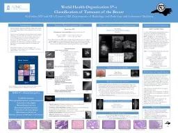
th ed Classification of Tumours of the Breast SG Jordan MD and SB OConnor MD Departments of Radiology and Pathology and Laboratory Medicine Introduction Invasive Breast Carcinoma IBC ID: 908210
Download Presentation The PPT/PDF document "World Health Organization 5" is the property of its rightful owner. Permission is granted to download and print the materials on this web site for personal, non-commercial use only, and to display it on your personal computer provided you do not modify the materials and that you retain all copyright notices contained in the materials. By downloading content from our website, you accept the terms of this agreement.


Slide1
World Health Organization 5thed Classification of Tumours of the BreastSG Jordan MD and SB O’Connor MD Departments of Radiology and Pathology and Laboratory Medicine
Introduction
Invasive Breast Carcinoma (IBC)
Genetic
tumour
syndromes
References
Acknowledgements
Fibroepithelial
tumours
and hamartomas
The World Health Organization (WHO) establishes the standard for histopathologic diagnoses, defining diagnoses on a per organ system basis.
The most recent classification of breast tumors is the 5
th
edition published in November 2019. The publication reflects the views of the WHO Classification of
Tumours
Editorial Board that convened at MD Anderson Cancer Center, Houston, USA December 9-11, 2018. 153 authors from 21 countries contributed. The end result is an authoritative reference book that serves as the international standard for oncologists and pathologists.
This exhibit is designed to increase radiologists’ and technologists’ understanding of breast pathology,
ie
to enhance CME and CEU at this conference.
Breast Cancer 2019
Estimated new cases and deaths
from breast cancer in the US
New cases: 268,600 15.2 % of all new cancer cases
Deaths: 41,760 6.9 % of all cancer deaths Invasive Breast Carcinoma (IBC) refers to a large and heterogeneous group of malignant epithelial neoplasms of breast glandular elements. IBCs are classified by morphology (below). All IBCs are grouped into biomarker-defined subtypes for treatment, based on estrogen receptor (ER) and human epidermal growth factor receptor 2 (HER2).
Epithelial TumoursInvasive breast carcinomaInvasive carcinoma of no special type Microinvasive carcinomaInvasive lobular carcinomaTubular carcinomaCribriform carcinomaMucinous carcinomaMucinous cystadenocarcinomaInvasive micropapillary carcinomaCarcinoma with apocrine differentiationMetaplastic carcinomasRare and salivary gland-type tumoursAcinic cell carcinomaAdenoid cystic carcinomaSecretory carcinomaMucoepidermoid carcinomaPolymorphous adenocarcinomaTall cell carcinoma with reversed polarityNeuroendocrine neoplasmsDuctal carcinoma in situNon-invasive lobular neoplasia Papillary neoplasmsEpithelial-myoepithelial tumoursAdenomasTubular adenomaLactating adenomaDuctal adenomaAdenosis and benign sclerosing lesionsBenign epithelial proliferations
Invasive NST
Invasive Lobular
Tubular
DCIS
Medullary
Metaplastic
Mucinous
Fibroadenoma
Phyllodes
tumour
: Benign, Borderline, Malignant
Hamartoma
WHO Classification of
Tumours
Editorial Board. Breast
tumours
. Lyon, (France): International Agency for Research on Cancer, 2019.
Mesenchymal
tumours
WHO 5
thed Broad CategoriesEpithelial tumoursFibroepithelial tumours and hamartomasTumours of the nippleMesenchymal tumoursHaematolymphoid tumoursTumours of the male breastMetastases to the breastGenetic tumour syndromes of the breast
Vascular tumours: haemangioma, angiomatosis, atypical vascular lesions, postradiation angiosarcoma, primary angiosarcomaFibroblastic and myofibroblastic tumours: nodular fasciitis, myofibroblastoma, desmoid fibromatosis, inflammatory myofibroblastic tumourPeripheral nerve sheath tumours: schwannoma, neurofibroma, granular cell tumourSmooth muscle tumours: leiomyoma, leimyosarcomaAdipocytic tumours: lipoma, angiolipoma, liposarcomaPseudoangiomatous stromal hyperplasia
PASH
Mucinous Carcinoma
Myofibroblastoma
Lipoma
Fibroadenoma
NEW! in this WHO edition
is a section delineating the familial predisposition to breast cancer, specifically the established and emergent genes that are a source of discussion. BRCA1 and BRCA2 are well-established, and increasingly PALB2, as important predisposition genes that merit testing in all patients with suspicion of familial predisposition. Many other genes (two examples are ATM, CHEK2) have been identified in familial syndromes though there is limited data on frequency and absent data on ethnic variations. Detailed familial syndromes with breast CA relevance are included:
BRCA 1/2-associated hereditary breast and ovarian cancer syndrome
Cowden syndrome
Ataxia-telangiectasiaLi-Fraumeni syndrome, TP53-associatedLi-Fraumeni syndrome, CHEK2-associatedCDH1-associated breast cancerPALB2-associated cancersPeutz-Jeghers syndromeNeurofibromatosis type 1The polygenic component of breast cancer susceptibility
Tumours
of the nipple
Paget Disease
Nipple adenomaSyringomatous tumor of the nipple (SyT)
Joanna Schneider MD UNC SOM Class of 2019Thomas J. Lawton MD UCLA Department of Pathology and Laboratory MedicineNorth Carolina Radiological Society
Paget
Nipple adenoma
Metastastes
to the breast
Tumours
of the male breast
Gynecomastia
Ductal carcinoma in situ
Invasive carcinoma
Defined: malignant
tumours
originating from an extramammary organ or siteMammography and ultrasound most commonly shows relatively small, irregular superficial masses, rarely spiculated. These require CNB for diagnosis, and comparison of mammary and extramammary malignant tissue is essentialMost common sources of breast metastases In adults: lymphoma/leukemia, melanoma, pulmonary, ovarian, gastric, prostatic, renal cell, colorectal, mesothelioma, neuroendocrine, cervical CA. In children: rhabdomyosarcoma
Gynecomastia
Male breast CA
Phyllodes
Invasive Lobular
DCIS
2020
https://seer.cancer.gov/statfacts/html/breast.html
WHO Classification of
Tumours
Editorial Board. Breast
tumours
. Lyon, (France): International Agency for Research on Cancer, 2019.
Calhoun BC. Core needle biopsy of the breast: an evaluation of contemporary data. Surg Pathol Clin 2018 Mar; 11(1):1-16. PMID 29413652.
Medullary Carcinoma
Invasive Micropapillary
Papilloma with DCIS
Invasive Carcinoma NST
GCT
Notes on core needle biopsy
5% of patients with screen-detected abnormalities typically undergo immediate excision, with 3:1 benign-to-malignant final diagnoses. Most patients with atypical breast lesions diagnosed on core biopsy are referred for surgical excision. However, recent studies indicate the rate of upgrade to carcinoma is lower than initially reported. Assure careful clinical-pathologic and
radpath
correlation in:
ADH: All patients should be referred for surgical consultation and excision
ALH: Surveillance may be appropriate if incidental
LCIS: Surveillance may be appropriate if incidental classic LCIS
FEA: Surveillance may be appropriate for FEA if all calcifications removed
Papilloma: Surveillance may be appropriate for benign solitary and incidental
papillomas
Radial scar: Surveillance may be appropriate for incidental radial scars without atypia