
By Dr Mohamed Abd Almoneim Attia Ischemic heart disease IHD includes Angina pectoris there is myocardial ischemia but the myocardium is still free and the condition is correctable ID: 908842
Download Presentation The PPT/PDF document "Ischaemic heart diseases" is the property of its rightful owner. Permission is granted to download and print the materials on this web site for personal, non-commercial use only, and to display it on your personal computer provided you do not modify the materials and that you retain all copyright notices contained in the materials. By downloading content from our website, you accept the terms of this agreement.


Slide1
Ischaemic heart diseases
By
Dr. Mohamed
Abd
Almoneim
Attia
Slide2Ischemic heart disease (IHD) includes:
Angina pectoris:
there is myocardial ischemia but the myocardium is still free and the condition is correctable.
Myocardial infarction (MI):
sudden persistent cessation of blood flow to certain area of the myocardium leading to irreversible
coagulative
necrosis.
Slide3Myocardial metabolism:
60%
of the myocardial energy comes from aerobic oxidation of
fatty acids
(gives more energy but utilizes more O
2
).
35%
of the myocardial energy comes from aerobic oxidation of
carbohydrates
(gives less energy but utilizes less O
2
).
5%
of energy comes from oxidation of
amino acids
,
ketone
bodies, etc.
Slide4Definition :
Ischemic heart disease is a condition in which there is imbalance between O
2
supply (
coronary blood flow
) and O
2
demand
(heart work)
.
ANGINA:
It is a clinical syndrome due to temporary myocardial ischemia due to imbalance between myocardial oxygen supply and oxygen needs, characterized by episodes of chest pain typically precipitated by exertion and relieved by rest
.
Slide5Pathophysiology
Angina occurs when the myocardial oxygen demands exceed the supply. The myocardial metabolism becomes anaerobic. This leads to shift in the metabolism of the myocardium from fat to carbohydrate with increase in the rate of
glycolysis
and increased formation of lactate and
pyruvate
. In the same time there is accumulation of substance P which is probably a
kinin
and which irritates the sympathetic nerve endings in the heart causing cardiac pain.
Slide6Etiology :
1-Atherosclerosis of coronary artery is the most common cause.
2-Thrombi, spasm, and coronary emboli cause limitation of coronary blood flow.
3-Hypertension where the myocardial O
2
demands is increased due to ventricular hypertrophy.
4-Reduction in the O2 carrying capacity of the blood as in severe anemia.
Slide7Types of Angina:
1-Stable angina: (Effort, typical angina, classic):
The pain is commonly induced by exercise, emotion or heavy meals. It is due to atherosclerosis.
Pain is induced by effort and disappears with rest.
2-Variant angina: (
Prinzmetal's
angina,
-receptor–mediated
vasoconst-riction
):
the coronary artery undergoes severe
spasm
due to
overactivity
of α
1
receptors.
May or may not be associated with atherosclerosis. Patient developed pain even at rest and exhibit elevation of ST segment.
The patient develops pain at rest.
3-Unstable angina: (accelerated angina =
preinfarction
syndrome ):
There is a change in pattern, increase in frequency, severity and/or duration of pain.
Any type of angina that developed recent changes in the character, duration, or frequency of pain. The patient must be hospitalized.
Slide8Clinical manifestations:
1) Chronic stable angina
Symptoms:
Pain
is the cardinal symptom it has a 4 important features:
Site and radiation:
retrosternal
may radiate up to neck or down to
epigastrium
commonly referred to both arms or left only (
ulnar
side of elbow and wrist down to the fingers)
Character:
Tight, band like, crushing, aching or bursting, but never pricking or stabbing (never sharp). Occasionally it is burning or likened to intense indigestion.
what increases it?:
Typically, the pain always follows a certain amount of effort and increase gradually in intensity as the effort continues, until the patient is forced to stop. The pain is also provoked by cold weather, heavy meals and emotions.
Duration:
commonly lasts 2-5min. never more than 5 min. If more than 5 min → myocardial infarction.
Slide9Angina
Slide10Signs:
In majority of cases there is no abnormality, some times there is tachycardia and hypotension. Occasionally there is a condition favoring angina such as D.M. and hypertension. In between the attack there is no signs.
2)
Prinzmetal's
angina (variant):
Symptoms
The same as in chronic stable angina but the chest discomfort occurs at rest or awakens the patient from sleep. It may also be brought by effort. It is caused by focal
spasm of proximal coronary artery.
Signs:
Deep Q wave usually present
3) Unstable angina:
The episodes of angina occurs at rest
Slide11Investigations:
1-ECG:
Inbetween
the attacks:
the ECG may be completely free
(so we can’t depend on ECG only for diagnosis).
During ischemia:
there is ST segment depression and T-wave inversion.
In myocardial infarction:
there is deep Q-wave.
2-Stress ECG testing:
recording ECG under controlled physical effort.
The normal response to exercise includes progressive, increase in heart rate and blood pressure, a positive test would indicated by development of ECG signs of ischemia, arrhythmia or abnormal heart rate or blood pressure.
3-Continuous 24-hour ECG recording (
Holter
monitoring).
4-Coronary angiography:
Catheterization of aorta and injection of contrast dye can show the site of coronary obstruction. In
prinzmetal
angina, spasm of the coronary may appear.
Slide12Drug therapy of angina pectoris:
Non-drug therapy = life style modification:
Dietary
s
odium
and
fat
restriction.
Stop
s
moking
and coffee.
Avoid
s
tress
.
Weight
reduction.
Encourage mild physical
exercise.
Control of
risk factors
: e.g. diabetes mellitus,
hyperlipidemia
, and obesity.
Avoid
vasoconstrictor drugs
e.g.
sympathomimetics
, nasal decongestants.
Slide13Drug therapy (
antianginal
drugs):
1-During the acute attack:
Short-acting nitrates and nitrites.
Sedatives and tranquilizers to calm the patient.
2-In-between the attacks (prophylaxis):
Long-acting nitrates.
Beta-blockers.
CCBs.
Cytoprotective
drugs:
trimetazidine
Antiplatelet
drugs:
aspirin,
dipyridamol
, etc.
3-Surgical treatment (myocardial revascularization).
Unstable angina:……………………………..
Must be hospitalized
Add anticoagulant(heparin) with aspirin.
Slide14Organic nitrates and nitrites:
Inorganic nitrates are ineffective
Classification:
1-Short-acting nitrates:
Amyl nitrite (inhalation)
Nitroglycerine (SL)
Isosorbide
dinitrate
(SL)
i.v
. nitroglycerin
2-Intermediate-acting nitrates:
Isosorbide
dinitrate
(Oral)
3-Long-acting nitrates:
Isosorbide
mononitrate
(Oral)
4-Transdermal patches
Slide15Pharmacokinetics:
Absorption:
nitrates are rapidly absorbed from
buccal
membrane, GIT, and bronchioles.
Metabolism:
in the liver:
If given oral
→ extensive first-pass hepatic metabolism (oral bioavailability <10%).
If given sublingual
→ no first-pass metabolism → high bioavailability.
Mononitrate
:
has no hepatic metabolism → high bioavailability.
Excretion:
via the kidney.
Slide16Dosing
Oral dosage is rapidly metabolized in the liver and only small doses reach the systemic circulatory system
For more effective absorption drug is given:
Sublingually
– under the tongue –
acts in 1 to 3 minutes
sustained-released tablet
Transdermal ointments – applied on hairless area on back, chest or upper arm
Patches – takes 40 minutes to an hour to start working
Slide17Sublingual Administration
Tablet should be held under tongue until dissolved. Avoid eating, drinking, or smoking until tablet is dissolved.
Acute
anginal
attacks:
Advise patient to sit down.
Relief should occur in
1 to 3 minutes
May be repeated every 5 minutes
for 3 doses.
If no relief ……………………..?...........................
Slide18Sustained Released Tablet
Administer dose 1 hour before or 2 hours after meal with a full glass of water for faster absorption
.
Slide19Nitroglycerine Patch
Slide20Nitroglycerin Patch
Place the patch on a hairless area of chest or upper arm each day.
Move patch to a different place on your body each day to prevent skin irritation.
Remove the patch for 8 to 12 hours each night
and put on a fresh patch each day.
Do not leave on all the time
.
Slide21Nitroglycerine Ointment
Slide22Nitroglycerine Ointment
Comes with paper with a ruled line for measuring the dose
Squeeze ointment onto the paper, carefully measuring the amount specified on the prescription label
Use the paper to spread ointment in a thin layer on a hair-free area of skin (2 by 3 inches)
Keep paper in place with bandage or tape
Ointment is applied three or four times a day
Slide23Mechanism of action:
Nitrates cause liberation of the free radical
nitric oxide (NO)
which is identical to the endothelial derived relaxing factor (EDRF) → ↑
cGMP
→ ↓ Ca
2+
influx into the vascular
sm
ms → VD (
more on the veins
).
They also ↑ formation of PGE
2
and PGI
2
→ VD.
Slide24Pharmacological effects:
CVS:
Blood vessels:
VD of the venous (and to lesser extent to the arterial) side leading to ↓ preload .
VD of coronary arteries leading to increased coronary blood flow.
VD of the arteries in the face and neck leading to
flushing.
VD of
meningeal
arteries leading to
throbbing headache
.
Heart:
Reflex tachycardia (in high dose) 2ry to ↓ BP.
BP:
High doses cause ↓ in both systolic and diastolic BP.
Slide25Smooth ms:
relaxation
of bronchial, GIT, uterine, and
biliary
sm
ms.
Respiration:
reflex
tachypnea
(↑
resp
rate) due to hypotension.
Blood:
Methemoglobinemia
in high doses due to oxidation of
Hb
into met-
Hb
.
Slide26Therapeutic uses:
1-Angina pectoris:
Nitrates are used for treatment of all types of angina both for relieving the
acute attack
and for
prophylaxis
.
These effects will lead to:
1-Reduction of myocardial O
2
demand through:
Venodilatation
→ ↓ venous return (preload) .
Arteriolodilatation
→ ↓ peripheral resistance (
afterload
).
2-Enhancement of myocardial perfusion through:
Coronary VD.
Redistribution of blood from large
epicordial
vessels to ischemic
subendo-cardial
vessels.
Slide272-Myocardial infarction:
to ↓ the area of myocardial damage.
3-Acute heart failure:
to ↓ preload and
afterload
.
4-Treatment of cyanide poisoning:
cyanide has high affinity for
metHb
more than normal
Hb
.
5-Biliary colic.
6-Management of constriction ring during labor.
Slide28Side effects:
1-Hypotension and reflex tachycardia:
may aggravate angina.
2-Throbbing headache:
due to VD of
meningeal
arteries.
3-Flushing
of the face.
4-Tolerance to its effect:
due to:
Depletion of
sulfhydryl
(-SH) containing enzymes necessary for liberation of NO from nitrates → ↓ formation of NO → ↓ VD effect.
Reflex sympathetic activation (due to hypotension), this will cause VC.
►
Prevention of tolerance:
make a daily
nitrate-free interval
(10–12 h) to give chance for the SH- containing enzymes to regenerate.
During this period, give another anti-
anginal
drug (e.g. beta-blocker or CCBs).
5-Methemoglobinemia
in high doses (rare).
Never to be combined with other drugs causing
vasodilation
(Viagra®) or hypotension
Slide29Precautions during nitrate therapy:
Use the
smallest effective dose
to avoid hypotension and reflex tachycardia.
The patient should
consult his doctor
if
anginal
pain does not improve after taking 3 SL tablets of NG during 15 min (may be MI).
Nitroglycerine tablets should not be put in
direct sunlight
or with
cotton
.
The
expiry date
(60 days) should be checked (active tablets have
burning
taste).
Sudden withdrawal
may cause acute MI.
Slide30Beta-blockers
Mechanism in angina:
1-Reduction of myocardial O
2
demand
through
↓
both heart work and systemic BP.
Bradycardia
leads to
↑ diastolic filling time
and improvement of myocardial perfusion
Redistribution of blood from normal to ischemic areas.
2-Cytoprotective
effect through
improving
glucose utilization by shifting tissue metabolism toward a greater use of carbohydrate and a lesser use of free fatty acids
3-Decreases platelets aggregation.
Slide31Combination of BBs and nitrates
↑ their efficiency and ↓ their side effects
:
Slide32N.B.
Beta-blockers
e.g
(propranolol) are contraindicated in
Prinzmetal’s
(variant) angina
because they block the β2-mediated coronary dilatation leaving the α1 receptors unopposed → ↑ coronary spasm.
Slide33Calcium channel blockers (CCBs):
1-They ↓ myocardial contractility and myocardial O
2
demand
.
2-They ↓ coronary vascular resistance and increase coronary blood flow.
3-They dilate
epicardial
coronary vessels.
4-↓ myocardial cell necrosis.
N.B.
Although
nifedipine
is coronary dilator, it has some disadvantages in angina:
It may cause hypotension and reflex tachycardia.
It may cause “
steal phenomenon
” .
Slide34Cytoprotective
drugs:
Trimetazidine
1-It
produces a metabolic switch via inhibition of fatty acid oxidation toward activation of glucose oxidation during
ischaemia
2-↓ intracellular lactic acid accumulation and intracellular ion disturbance.
3-↓ intracellular Ca
2+
and Na
+
accumulation and preserve contractile function.
4-↓ membrane damage through antioxidant effect.
Slide35Antiplatelet
drugs:
See blood.
Aspirin inhibits platelet aggregation by:
It causes irreversible inhibition of COX enzyme → decrease TXA
2
→ decrease platelet
aggregation.
Irreversible
acetylation
of platelet cell membranes → decrease platelet
adhesions.
Decrease platelet ADP synthesis → decrease platelet
accumulation.
The action of aspirin lasts for
7-9 days
, the lifetime of platelets.
A dose of
75-150 mg/day
is sufficient to reduce TXA
2
without significant effect on
prostacyclin
(PGI
2
) formation.
Slide36Management of angina with other concomitant diseases:
Hypertension and angina:
1-Control of blood pressure by using medication useful for angina e.g.
blockers or CCB e.g.
sustaimed
release
nifedipine
or
amlodepine
.
2-Low sodium and low fat diet.
3-Mild exercise
4-Avoid stress.
5-Control of
hyperlipidemia
.
Slide37Heart failure and angina:
1-Heart failure usually aggravates angina.
2-Treat H. F. with ACEIs + mild diuretics +
Digoxin
.
3-Use
antianginal
agents which don’t aggravate H.F e.g. nitrates.
4-Avoid
blockers and CCBs, having negative
inotropic
effect, except in cases of high output heart failure.
Slide38Angina and peripheral
vascular disease (PVD):
1-Use CCBs.
2-Avoid
blockers or use
cardoselective
blockers if PVD is mild.
Angina and bronchial asthma:
1-Give CCBs + nitrates.
2-Avoid
blockers
Angina and DM:
Use nitrates CCBs or
cardioselective
blockers
Slide39Slide40MYOCARDIAL INFARCTION (MI):
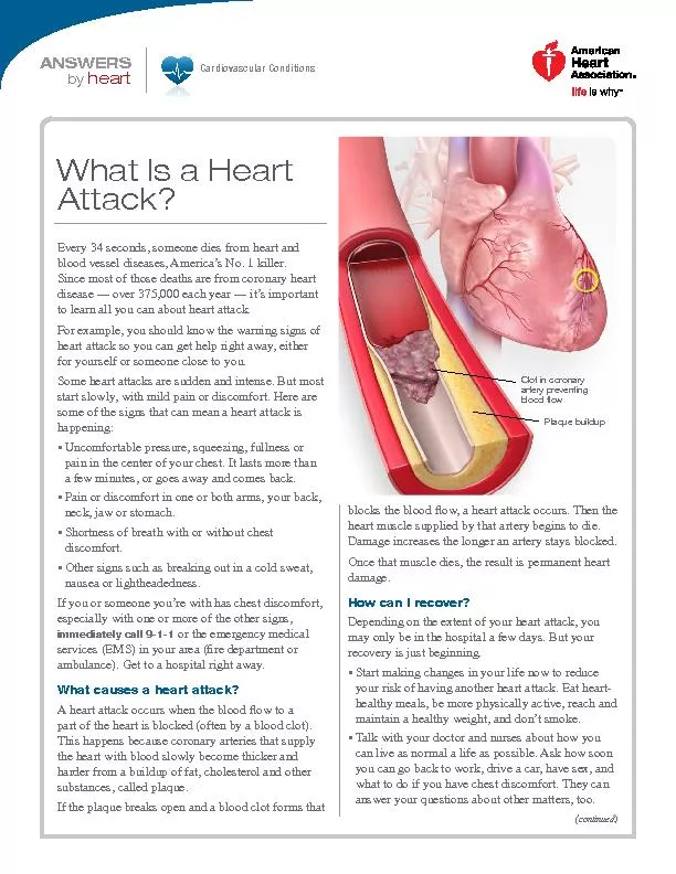
It is necrosis of myocardium due to acute obstruction of blood flow to the dependent tissue.
Pathophysiology
:
The majority of MI results from total occlusion of a coronary artery secondary to thrombus formation.
Slide41Clinical picture:
Symptoms:
Incidence of AMI is increased during morning hours between 6 am and 11 a.m. Patient complains of prolonged
substernal
chest pain or pressure in some patients the symptoms may be confused with indigestion. The pain may be stabbing or knife like and it may occur in arms, shoulder, neck, jaw or back. It has been estimated that 20% of AMIs are silent and tend to occur in elderly and diabetics.
Slide42So, Myocardial infarction clinical picture : same pain as angina but
:
1-more severe and persistent .
2-patient become breathless and vomiting is common.
3-fall in blood pressure with weak pulse.
Signs
:
signs of sever left ventricular or right ventricular dysfunction may be present. Patient may have severe hypertension or may have hypotension.
Slide43Diagnosis:
In addition to patient history and presentation, the diagnosis of AMI is based upon ECG and lab tests:
1-ECG:
the 12-lead ECG is helpful in determining the location of infarction.
The presence of a new Q wave is consistent with AMI. also show ST segment elevation.
2-Enzymes:
the measurement of cardiac enzymes is routine in making the diagnosis of AMI.
Creatine
kinase
(CK).
The most sensitive enzyme appear in serum within 3-4 hours after myocardial damage and peak 12-24 hours.
Lactate
dehydrogenase
(LDH):
Seen 24-48 hours after myocardial damage and peak 3-6 days.
3-Radionucltide technique:
e.g. technetium-99 for diagnosis of site of MI.
Slide44Complications of myocardial infarction:
1-Heart failure
2-Shock
3-Arrhythmias
4-Thromboembolism
Slide45Treatment of AMI:
1-Hospitalization (coronary care unit):
to control vital signs (respiration, HR, BP, pulse, etc.
)
and
continuous monitoring of ECG.
2-Oxygen:
To increase PO2 and hence increases the diffusion of oxygen in to ischemic
myocardia
.
Oxygen given
by nasal
cannula
or mask.
Slide463-Relief of pain: Morphine sulfate (5 mg
i.v
.): can
be repeated after 5 min
.
*To produce analgesia.
*To ↓ stress of the patient → ↓ sympathetic discharge and heart work.
*Morphine causes
venodilatation
→ ↓ venous return and ↓ cardiac work.
Morphine is usually given
s.c
.
except
in AMI (
5 mg
i.v
.).
Morphine is
contraindicated in cases of MI involving the
inferior wall
of the heart (inferior MI) because in this case, the patient has
bradycardia
and morphine causes
vagal
stimulation
→ aggravation of
bradycardia
.
So,What
another
opioid
analgesic could be used in inferior MI?
Meperidine
because it has
atropine-like action
→ counteract
bradycardia
.
What other
opioid
analgesics are contraindicated in AMI?
Pentazocin
and
butorphanol
because they ↑ pulmonary and systemic vascular resistance → more strain on the heart .
Slide474-Fibrinolytic therapy:
e.g. streptokinase,
urokinase
,
acylated
plasminogen
streptokinas
activator complex "APSAC", Pro-
urokinase
...etc.
Fibrinolytic
must be used within first 6 hours I.V (early
early
early
…..).
5-Measures to limit the size of infarction:
Beta-blockers, NG or CCBs.
Beta blockers decrease myocardial damage and decrease complication of myocardial infraction especially ventricular fibrillation. .
Slide486-Anticoagulants and
antiplatelet
: heparin
10,000 IU
i.v
. then 5000 IU/8h
s.c
. especially when the patient is obese or if there is history of previous MI.
7- Treatment of complications:
Cardiogenic
shock → give
dobutamine
i.v.i
Arrhythmia → give
lidocaine
i.v
.
8-Control of risk factors:
Stop smoking; physical activity, control hypertension, obesity, diabetes mellitus and correction of plasma lipids.
9-
Sedatives:
diazepam
5 mg
i.v
.
Slide49N.B.
Oral ACEI therapy beginning early in post MI period is beneficial.
Captopril could be given on post infarction day 2 or
3.
Used to:
1-
↓ mass and wall thickness of the left ventricle through:
2-↓
arterial BP.
3-↓
myocyte
cell proliferation and collagen synthesis.
4-↓
apoptosis.
Slide50Case 1:
A 62 years old male about 3 weeks before, he complained of
substernal
chest pain brought on by lifting heavy object on walking. He describes a crushing pain that never occurred at rest or particular time of day. When he stopped walking the pain subsides in 5 minutes. He is hospitalized for evaluation of chest pain, he suffers now from the same pain his resting vital signs include: B.P180/110 mm Hg, pulse 100/min, R.R22/min. ECG reveals ST segment depression without evidence of M.I.
Slide51What signs and symptoms would be significant for probable diagnosis?
Is there other diagnostic procedure, which would be helpful in confirming the diagnosis?
What will you give to the patient for the relieve of
anginal
pain? In what route and dose?
What instructions should the patient receive with regard to uses of the above drug? How rapid will it relieve chest pain?
What are the mechanisms by which the above treatment will relieve the pain?
Would you give him any drugs after relieve of pain to protect against attacks?
How can you evaluate the efficacy of the drug selected in question 6?
Slide52Case 2:
57 years old male is brought to the emergency room complaining of sever chest pain that occur 2 hours before and not relieved by sublingual NTG tablet. On Physical examination B.P was 90 / 60 mm, pulse 65/min RR.16 /; min ECG shows deep Q wave and ST segment elevation.
1-What is the possible diagnosis? What signs and symptoms are relevant to diagnosis?
2-What are the immediate lines of treatment?
3-What is the best analgesic drug, in what dose and why?
4-What is the other alternative analgesic? In which situation it is used?
5-What are the differences between the above alternative drugs? What are the possible side effects?
6-What are the
opioid
analgesics that are contraindicated and why?
7-What are other non-
opioid
analgesics that can be given?
8-How to give O2 and for how long?
Slide53