
Dr Kaushal Kumar Assistant Professor amp Head Department of Veterinary Pathology Bihar Veterinary College BASU Patna14 Subject Veterinary Pathology PaperI UnitI Introduction There are two ID: 915022
Download Presentation The PPT/PDF document "Necrosis & It`s Classification" is the property of its rightful owner. Permission is granted to download and print the materials on this web site for personal, non-commercial use only, and to display it on your personal computer provided you do not modify the materials and that you retain all copyright notices contained in the materials. By downloading content from our website, you accept the terms of this agreement.


Slide1
Necrosis & It`s Classification
Dr. Kaushal KumarAssistant Professor & HeadDepartment of Veterinary PathologyBihar Veterinary CollegeBASU Patna-14
(Subject: Veterinary Pathology, Paper-I ; Unit:-I)
Slide2Introduction
There are two different ways by which a cells can die: Apoptosis (Programmed cell death)Necrosis
(
U
nplanned cell death)
Slide3Necrosis
Necrosis is death of cells and tissues in the living animal. Focal/ Multifocal necrosis- terms used for one or more, small, clearly defined areas of necrosis. Diffuse necrosis- term used when necrosis affects a large area or the entire tissue or organ.
Slide4General causes of necrosis:
Poisons and toxins: Chemical: Strong acids, alkalies, insecticides, mercury etc. Infectious agents: Bacteria (Salmonella, Staphylococcus), viruses, fungi, protozoa etc.
Plant poisons
-
hepatotoxic alkaloids e.g. Senesce. •Circulatory disturbance: Anemia, congestion and ischemia.
Mechanical injuries:
Cutting, crushing and rubbing types.
Physical
: Extreme temperature, electricity, free radical.
Slide5Gross appearance:
Affected areas white, gray or yellow in colour. Have a cooked meat appearance. Sharply demarcated (by red zone) from healthy tissue. In case of gangrene the area is green, orange or black ( iron sulphide)
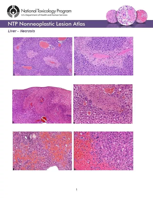
Slide6Microscopic appearance:
The microscopic changes of necrosis vary with the type of necrosis. Some general changes of necrosis in the cytoplasm are: Eosinophilia: The cytoplasm stains darker red in colour. Swelling and vacuolation: The cells are swollen and contain different types of vacuoles. Changes in the nucleus:
The nucleus may show condensation (
Pyknosis
), fragmentation (karyorrhexis) and may disappear (
karyolysis
)
Slide7Necrosis & Autolysis
Autolysis: Autolysis is death of cells and tissues after the death of the animal (somatic death) and it should be distinguished from necrosis.The enzymatic digestion of cells by enzymes present within them. The cells most susceptible to autolysis tend to be dying or dead cells. No sharp line of demarcation between affected and healthy tissue.Circulatory changes like congestion and haemorrhage
are not present.
Slide8NECROSIS
AUTOLYSISA line of demarcation is usually present Circulatory changes are presentInflammatory changes like infiltration of leukocytes are present.
Saprophytic growth not seen, but pathogenic bacteria maybe present
.
No sharp line of demarcation between affected and healthy tissue
Circulatory changes like congestion and
haemorrhage
are not present.
Inflammatory changes are not present.
Growth of saprophytic bacteria, bacillary rods in long chains are often
present.
Slide9Types of necrosis:
Different types of necrosis are recognized according to the causes, pathogenesis and the tissue involved. These include Coagulative, liquefactive, caseous and fat necrosis. •
Slide10Coagulative necrosis:
Most common type of necrosis. Architectural outlines persist but cellular details are lost. Type of tissue can be recognized. Denaturation (coagulation) of structural and enzymic proteins blocks proteolysis.
Kidney Infarct
(Source: Google)
Slide11Causes of Coagulative Necrosis
Ischemia due to thrombosis/ embolism as in infarcts.Bacterial toxins e.g. Fusobacterium necrophorum in livers in cattle. Muscular dystrophy due to deficiency of selenium and Vit.-E in cattle and sheep.
Necrosis of renal epithelium due to poisoning from mercuric salts.
Slide12Coagulative Necrosis Contd
…. Gross appearance: Necrotic area is firm, opaque with cooked meat appearance. It is sharply demarcated from the healthy areas. Microscopic appearance:
Architectural outlines are present; cellular details are lacking.
Result:
Dead tissues remain in the body for a long period, ultimately removed by macrophages.
Slide13Liquefactive necrosis:
There is digestion and liquefaction of necrotic tissue.Causes: Pyogenic bacterial infections attract neutrophils. Bacterial and leukocytic enzymes liquefy dead cells and tissues.Some chemicals like turpentine oil also attract
neutrophils
and cause pus formation and results into liquefaction of the tissue.
The necrosis in the nervous tissue is mostly
liquefactive
due to high content of lipids and water.
Abcess
in neck
Slide14Gross Appearance of Liquefactive
NecrosisThe necrotic tissue is liquefied and filled with semisolid creamy liquid called pus. Pus: It is a thick, white or yellow, creamy liquid consisting of exudate of leukocytes, tissue debris and microorganisms. Proteolytic enzymes released from neutrophils cause liquefaction of dead cells.
Abscess
: It is a localized collection of pus, surrounded by fibrous capsule.
Empyema
: It is accumulation of pus in a body cavity.
Slide15Microscopic appearance of Liquefactive
NecrosisNo architectural or cellular details are visible in the area of necrosis.The necrotic area usually appears as a cavity containing a mass of necrotic neutrophils, bacteria and tissue debris.The entire necrotic mass is surrounded by a fibrous connective tissue capsule..
Slide16Caseous necrosis
Dead tissue is converted into a homogenous, granular mass resembling cottage cheese. Cause: Associated with lesions of Mycobacterium tuberculosis and Arcanobacterium ovis, the cause of caseous lymphadinitis.
Bovine tuberculosis
( Source Google.com)
Slide17Caseous necrosis
Gross appearance: • The area of necrosis is amorphous, granular, friable, white-gray resembling cottage cheese. The caseous mass is enclosed within a connective tissue capsule. Microscopic appearance: No architectural or cellular details are seen. Calcification commonly occurs in the necrotic areas.The necrotic tissue is amorphous, granular mass enclosed inside a zone of granulomatous
inflammation, containing macrophages.
Slide18Thanks