/
PRODUCT DATA SHEET PRODUCT DATA SHEET

PRODUCT DATA SHEET - PDF document

jalin
jalin . @jalin
Follow
343 views
Uploaded On 2021-06-28

PRODUCT DATA SHEET - PPT Presentation

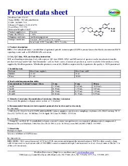
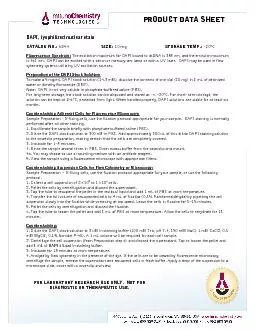
DAPI lyophilized nuclear stain CATALOG NO 6244 CATALOG NO 6244 SIZE 10 mg STORAGE TEMP 20 ID: 848227

dapi cells solution sample cells dapi sample solution pbs staining preparation stock suspension pellet buffer fluorescence add fixative cell

Share:

Link:

Embed:

Download Presentation from below link

Download Pdf The PPT/PDF document "PRODUCT DATA SHEET" is the property of its rightful owner. Permission is granted to download and print the materials on this web site for personal, non-commercial use only, and to display it on your personal computer provided you do not modify the materials and that you retain all copyright notices contained in the materials. By downloading content from our website, you accept the terms of this agreement.


Presentation Transcript

1 PRODUCT DATA SHEET DAPI, lyo
PRODUCT DATA SHEET DAPI, lyophilized nuclear stain CATALOG NO.: 6244 CATALOG NO.: 6244 SIZE: 10 mg STORAGE TEMP.: - 20 ° C Fluorescence Spectrum: The excitation maximum for DAPI bound to dsDNA is 358 nm, and the emission maximum is 461 nm. DAPI can be excited with a xenon or mercury - arc lamp or with a UV laser. DAPI may be used in flow cytometry systems utilizing UV excitation sources. Preparation of the DAPI Stock Solution: To make a 5 mg/mL DAPI stock solution (14.3 mM), dissolve the contents of one vial (10 mg) in 2 mL of deionized water or dimethylformamide (DMF). Note: DAPI is not very soluble in phosphate - buffered saline (PBS). For long - term storage, the stock solution can be aliquoted and stored at - – 20°C. For short - term storage, the solution can be kept at 2 – 6°C, protected from light. When handled properly, DAPI solutions are stable for at least six months. Counterstaining Adherent Cells for Fluorescence Microscopy: Sample Preparation - If fixing cells, use the fixation protocol appropriate for your sample. DAPI staining is normally performed after all other staining. 1. Equilibrate the sample briefly with phosphate - buffered saline (PBS). 2. Dilute the DAPI stock solution to 300 nM in PBS. Add approximately 300 uL of this dilute DAPI staining solution to the coverslip preparation, making certain that the cells are completely covered. 3. Incubate for 1 – 5 minu tes. 4. Rinse the sample several times in PBS. Drain excess buffer from the coverslip and mount. 4a. You may choose to use a mounting medium with an antifade reagent. 5. View the sample using a fluorescence microscope with appropriate filters. Counterstai ning Suspension Cells for Flow Cytometry or Microscopy: Sample Preparation – If fixing cells, use the fixation protocol appropriate for your sample, or use the following protocol. 1. Collect a cell suspension of 2×10 5 to 1×10 6 cells. 2. Pellet the cells by centrifugation and discard the supernatant. 3. Tap the tube to resuspend the pellet in the residual liquid and add 1 mL of PBS at room temperature. 4. Transfer the full volume of resuspended cells to 4 mL of fixative (0.5% Paraformaldehyde) by pipetting t he cell suspension slowly into the fixative while vortexing at top speed. Leave the cells in fixative for 5 – 15 minutes. 5. Pellet the cells by centrifugation and discard the fixative. 6. Tap the tube to loosen the pellet and add 5 mL of PBS at room tempera ture. Allow the cells to rehydrate for 15 minutes. Counterstaining: 1. Dilute the DAPI stock solution to 3 uM in staining buffer (100 mM Tris, pH 7.4, 150 mM NaCl, 1 mM CaCl2, 0.5 mM MgCl2, 0.1% Nonidet P - 40). A 1 mL volume will be required for each cell sample. 2. Centrifuge the cell suspension (from Preparation step 6) and discard the supernatant. Tap to loosen the pellet and add 1 mL of DAPI diluted in staining buffer . 3. Incubate for 15 minutes at room temperature. 4 . Analyze by flow cytometry in the p resence of the dye. If the cells are to be viewed by fluorescence microscopy, centrifuge the sample, remove the supernatant and resuspend cells in fresh buffer. Apply a drop of the suspension to a microscope slide, cover with a coverslip and view. FOR LABORATORY RESEARCH USE ONLY. NOT FOR DIAGNOSTIC OR THERAPEUTIC USE.