

Animal and Food Science Department Brigham Young University Idaho Scalpel types and sizes Scalpels come in various sizes with various types and sizes of blades Our program uses a 4 scalpel with a 20 blade ID: 1019047
Download Presentation The PPT/PDF document "Scalpel Training Anatomy and Physiology..." is the property of its rightful owner. Permission is granted to download and print the materials on this web site for personal, non-commercial use only, and to display it on your personal computer provided you do not modify the materials and that you retain all copyright notices contained in the materials. By downloading content from our website, you accept the terms of this agreement.


1. Scalpel TrainingAnatomy and Physiology LabAnimal and Food Science DepartmentBrigham Young University Idaho
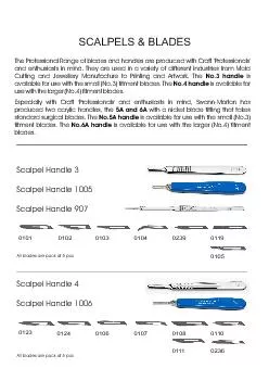
2. Scalpel types and sizesScalpels come in various sizes with various types and sizes of blades. Our program uses a #4 scalpel with a #20 blade.
3. Mounting a Scalpel BladeOpen the scalpel blade wrapper. Look for the end with a split in the wrapper to ensure you open the dull end. Once you have peeled back the foil, use your tweezers or your hemostats to remove the blade from the foil. You should never handle the scalpel blade with your fingers.
4. With your hemostats align the scalpel blade with to the edge of the scalpel handle. The blade should be held at a slight angle to allow it to slide into the groves on the handle. Once the blade is in the grooves flex the blade with your hemostats and slide it back towards the handle.
5. As you slide the blade down, it continue to fall in the small groves in the scalpel handle. Keep moving the blade down until it clicks into position. Notice where your hemostats are on the blade. Once locked into place your scalpel is then ready for use.
6. Removing a Scalpel BladeAt the sharps container, clamp your hemostats at the base of the blade. Lift the blade base slightly and begin to push the blade off the handle along the grooves. The blade should be pointed towards the sharps container. Notice where your hemostats are placed on the blade for removal.
7. Once the blade is removed from the handle, unclamp your hemostat and discard the blade in the sharps container. Blades should not be saved for reuse. Clean your scalpel handle and hemostats as directed.
8. Holding and using a scalpel bladePencil GripFinger-tip gripPalm grip
9. Pencil Gripthis grip limits amount of the blade’s cutting surface contacting the tissue. useful for short, precise incisionsjagged cut occurs when used to make longer incisionshandle is held in dominant hand like a pencilhandle is held between the thumb and index fingerhandle rests on remaining fingersfingers are moved to make the incision rather than armblade can be reversed for improved visualization and control as shownImage from https://wcvm.usask.ca/vsac205/Lab1/scalpels.php#Holds
10. Finger-tip gripmost common gripused to make a long, smooth incision provides the best accuracy and blade stability when making long incisionsmaximizes contact of blade's cutting surface with the tissuehandle held in the dominant handcutting pressure applied by fingers (primarily index finger)entire arm, not just the hand, moves to make the incisionImage from https://wcvm.usask.ca/vsac205/Lab1/scalpels.php#Holds
11. Palm Gripstrongest gripused when need strong pressure to incise the tissueuses entire armhold handle in palm of dominant handplace thumb on one side of handlerest index finger on top of handle or blade (optional)wrap remaining fingers around the handlecutting pressure applied by palm and fingersentire arm moves to make the incisionImage from https://wcvm.usask.ca/vsac205/Lab1/scalpels.php#Holds
12. Types of cuttingSlide CuttingPress CuttingScrape Cutting
13. Slide CutTo perform a slide cut, the scalpel is generally grasped in the fingertip grip. The cut is made by sliding the blade on its cutting edge, while exerting a sub-bursting pressure on the tissue. In the slide cut, the cutting motion is at a right angle to the direction of the scalpel pressure. The depth of the incision is determined by the amount of pressure exerted, the length of blade distributing that pressure, and the resistance to cutting of the tissue being incised. Light pressure, combined with an increased blade surface-area-to-skin ratio, results in a more superficial cut.
14. Slide cut advantages and disadvantagesIn slide cutting, depth control is precise since the bursting pressure is never exceeded. The slide cut is the most applicable cut for most veterinary scalpel applications. In particular, it is well suited to skin incisions because it allows accurate depth control and precise direction and length control. The main limitation of the slide cut is that it does not allow for short, deep incisions in tissue.
15. Press CuttingTo incise tissue with a press cut, begin by grasping the scalpel in a pencil grip. With the blade positioned over the tissue, slowly increase downward pressure on the blade tip. When the bursting strength threshold of the tissue is exceeded, the blade will suddenly “pop” through the targeted tissue. In press cutting, the direction of the pressure exerted is the same as the direction of the blade motion as it cuts through the tissue. A stab incision is a classic example of a press cut
16. Press Cutting advantages and disadvantagesWith press cutting, the wound is well controlled in both length and direction, as the length of the wound is exactly the width of the scalpel blade and the direction is in line with the plane of the blade. Press cutting is useful for making stab incisions in hollow structures. Once the wall is penetrated, the hollow space below the blade provides space for the blade to decelerate and stop without damaging deeper structures. The press cut is often used in veterinary surgery to drain abscesses and to open the bladder, stomach, and intestines. The usefulness of press cutting is limited by the all-or-none quality of tissue depth penetration. It is important to realize that depth control is not precise with press cutting; the pressure needed to overcome starting friction is greater than the pressure needed to continue the path of the scalpel once the tissue begins to part. Additionally, the outer layer of tissue generally has greater bursting threshold than deeper layers, which further accentuates this depth control problem. To mitigate this problem, position the sharp edge facing your palm, and use your index finger as a “bumper” to expose only a limited amount of the blade tip. Then rest your knife hand against the patient during the stab incision to limit the depth of the blade plunge.
17. Scrape CuttingTo perform a scrape cut, the scalpel can be grasped in either a pencil or fingertip grip. When scraping delicate tissue, such as an anal-sac wall, the pencil grip is generally preferred. The cutting motion in the scrape cut involves exerting sub-bursting pressure while moving the scalpel perpendicular to the edge of the blade and the direction of the pressure. Scrape cutting is exactly the same motion used when shaving hair with a razor blade.
18. Scrape Cutting advantages and disadvantagesScrape cutting is a precise way to separate layers of tissues without cutting the deeper layers beneath the blade, known as “buttonholing.” Button-holing can easily occur with push cutting, and sometimes with slide cutting. Scrape cutting is not an efficient method for incising tissue. This cutting technique is also more traumatic to tissues than sharp dissection with a slide cut.