
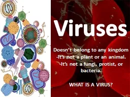
Assistant Professor College of Fisheries Kishjanganj BASU Patna Characteristics of viruses Viruses may be regarded as exceptionally complex aggregations of nonliving chemicals or exceptionally simple living microbes ID: 913464
Download Presentation The PPT/PDF document "Viruses Dr. Abhishek Thakur" is the property of its rightful owner. Permission is granted to download and print the materials on this web site for personal, non-commercial use only, and to display it on your personal computer provided you do not modify the materials and that you retain all copyright notices contained in the materials. By downloading content from our website, you accept the terms of this agreement.


Slide1
Viruses
Dr. Abhishek Thakur
(Assistant Professor)
College of Fisheries,
Kishjanganj
BASU, Patna
Slide2Characteristics of viruses
Viruses may be regarded as
exceptionally complex aggregations
of
nonliving chemicals
or
exceptionally simple living microbes
.
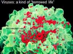
Because viruses are
inert outside living host cells
but
once viruses enter a host cell
, they become active and starts multiplication occurs.
Therefore viruses can be termed as
obligatory intracellular parasites.
Viruses contain a
single type of nucleic acid
(either DNA or RNA) &
a
protein coat
, sometimes enclosed by an envelope.
A complete, fully developed viral particle composed of nucleic acid surrounded by a coat is called a
Virion
.
Slide3Characteristics of viruses
Host range:
It refers to the
spectrum of host cells
in which a virus can multiply.
They are
host specific
,
that
infect
invertebrates, vertebrates, plants, fungi and bacteria.
Most viruses
infect specific types of cells
of only one host species.
Host range is determined by
the specific attachment site on the host cells’ surface.
Size:
Viral size is measured by
electron microscopy
.
Range from
20 nm to 300 nm
in length.
Slide4Viral Structure
Nucleic acid
Single kind of nucleic acid
(either DNA or RNA)
Can be single stranded
or double stranded
Can be
linear or circularCapsid and Envelope
Slide5Viral Structure
Capsid and Envelope
The nucleic acid of a virus is
surrounded by a protein coat
called the capsid.
Each capsid is
composed of protein subunits
called capsomeres, which can be a single type of protein or several types. The capsid of some viruses is enclosed by an envelope consisting of lipids, proteins and carbohydrates. Some envelopes are covered with
carbohydrate
–
protein complexes called spikes
.
Slide6Morphology
On the basis of their capsid architecture
Helical viruses
Polyhedral viruses
Enveloped viruses
Complex viruses
Slide7Morphology
Helical viruses
-
These resemble
long rods
, and their
capsids are hollow cylinders surrounding the nucleic acid
. Eg.Tobacco mosaic virus.
Slide8Morphology
2. Polyhedral viruses
–
These
have many sides
and usually the
capsid is an icosahedron
. Eg. Adenoviruses.
Slide9AdenovirusAdenovirus are medium sized (70-90nm) unenveloped, icosahedral, double stranded virus .
There are more than 50 serotype of human adenoviruses which are divided into six groups (A-F) on the basis of on the basis of genomic homology.
Slide10Morphology
3.Enveloped viruses
-
covered by an envelope
and are
roughly spherical but highly pleomorphic
. There are also enveloped helical viruses (influenza virus) and enveloped polyhedral viruses (herpes virus).
Slide11Morphology
4.Complex viruses
-
These have
complex structures
, eg. Many bacteriophages have a polyhedral capsid with a helical tail.
Slide12Slide13Virus Symmetry
The capsids of
virions
have one of two symmetries –
helical or cuboid
Helical Symmetry:
Nucleocapsids
form rigid, highly elongated rods or flexible filaments.In addition to classification as flexible or rigid and as naked or enveloped, helical nucleocapsids are characterized by length, width, pitch of the helix, and number of protomers per helical turn. The most extensively studied helical virus is tobacco mosaic.
Slide14Virus Symmetry
Icosahedral Symmetry:
An icosahedron is a polyhedron having 20 equilateral triangular faces, 30 edges and 12 vertices .
Slide15Slide16Taxonomy of Viruses
Viruses are classified on the basis of type of nucleic acid, morphological class and presence or, absence of an envelope
Virus family names end in --------- viridae and genus names end in ------- virus
Slide17Slide18Growth of viruses in the laboratory
Bacteriophages can be cultivated by plaque assay.
Plaque assay mixes bacteriophages with host bacteria and nutrient agar.
After several viral multiplication cycles, the bacteria in the area surrounding the original virus are destroyed. The
area of lysis
is called a
plaque
. Each plaque originates with a single viral particle. The concentration of viruses is expressed as plaque-forming units (pfu)
Slide19Growth of viruses in the laboratory
Cultivation of some animal viruses requires whole animals
Some of them cultivated in embryonated eggs
cell cultures
Slide20Viral identification
Serological or Immunological tests
are used to identify viruses.
Viruses may also be identified by techniques like
polymerase chain reaction (PCR) methods
,
restriction enzyme fragments
, appearance of host cells following infection and electron microscopy.
Slide21Multiplication of viruses
Viruses
invade
a host cell and
direct
the host’s metabolic machinery
to produce viral enzymes and components
.Multiplication cycle of viruses can be divided into five distinct stages, namely:AttachmentPenetrationBiosynthesis.Maturation and
Release.
Phages can multiply by two mechanisms
:
Lytic cycle - results with the lysis and death of the host cell
Lysogenic cycle - host cell remains alive in the lysogenic cycle.
Slide22Lytic cycle of T – even bacteriophage
Attachment
Penetration
Biosynthesis
Maturation
Release
The time from phage attachment to
release is known as
burst time
and is usually
from
20 to 40 min
.
Slide23Slide24Lysogenic cycle of Bacteriophage
phages begin a lysogenic cycle by incorporating their DNA into the host cell’s DNA
During this state, called
lysogeny,
the phage remains latent.
Upon penetration into a bacterial cell, the linear phage DNA becomes a circle. The circular DNA of the phage may recombine with and become part of the circular bacterial DNA. The inserted phage DNA is called a
prophage. Every time the host cell replicates the bacterial chromosome, the prophage DNA also gets replicated. The prophage remains latent within the progeny cells. Under some circumstances, or due to the action of UV light or certain chemicals excision of phage DNA occurs which initiates the lytic cycle.
Slide25Slide26Multiplication of Animal viruses
Animal viruses attach to the plasma membrane of the host cell and penetration occurs by endocytosis
uncoated by either viral or host cell enzymes
DNA is released into the copies of DNA are synthesised
Capsid protein is synthesised
After maturation, viruses are released
Budding, Naked viruses are released through ruptures in the host cell membrane
Slide27Slide28Thank You