
discolouration of the skin or mucous membranes due to the tissues near the skin surface having low oxygen saturation D efined historically as the presence of 5 gm dL of deoxygenated hemoglobin ID: 909243
Download Presentation The PPT/PDF document "Cyanosis is the bluish or purplish" is the property of its rightful owner. Permission is granted to download and print the materials on this web site for personal, non-commercial use only, and to display it on your personal computer provided you do not modify the materials and that you retain all copyright notices contained in the materials. By downloading content from our website, you accept the terms of this agreement.


Slide1
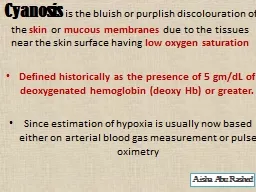
Cyanosis is the bluish or purplish discolouration of the skin or mucous membranes due to the tissues near the skin surface having low oxygen saturationDefined historically as the presence of 5 gm/dL of deoxygenated hemoglobin (deoxy Hb) or greater.Since estimation of hypoxia is usually now based either on arterial blood gas measurement or pulse oximetry
Aisha Abu
Rashed
Slide2the presence of cyanosis is dependent upon there being an absolute quantity of deoxyhemoglobin, the bluish color is more readily apparent in those with high hemoglobin counts than it is with those with anemia.When signs of cyanosis first appear, such as on the lips or fingers, intervention should be made within 3–5 minutes because a severe hypoxia or severe circulatory failure may have induced the cyanosis.The name cyanosis literally means the blue disease or the blue condition
Slide3An oxygen saturation of 66% corresponds to an arterial oxygen tension (paO2)= of approximately 35 mmHg ., the oxygen tension (PaO,) at which cyanosis is detected will be even lower. Assume for example that the patient's Hb is now 10 gm/dL. The saturation at which we will detect cyanosis (ie., when the DeoxyHb = 5 gm/dL) EX.: SaO, = OxyHb / (OxyHb + DeoxyHb) SaO, = 5 / (5 + 5) = 50% An oxygen saturation of 50% corre
-
sponds
to an oxygen tension (PaOJ of only 27 mmHg
Slide4under optimal lighting conditions with no excessive skin pigmentation and a normal hemo- globin level, the earliest that cyanosis can be appreciated is at an oxygen saturation of approximately 85%. This corresponds to a PaO, of 55 mmHg. At a SaO, of 70% most clinicians will be able to detect cyanosis (PaO, of apptoximately 40 mmHg). Cyanosis has been. When the hemoglobin level is 15 gm/dL and 5 gm/dL of this Hb releases oxygen to the tissues, the oxygenated hemo- globin portion (OxyHb) is 10 gm/dL. Hence the oxygen saturation is: SaO, = OxyHb/ (
OxyHb
+
DeoxyHb) SaO, = 10 / (10 + 5) = 66%
Slide5In severely anemic patients, oxygen saturation at which cyanosis is detectable will be lower than in normal patients. - Hb level = 10 gm/dL, 5 gm/dL release O2 -SaO2 = OxyHb / (oxyHb + DeoxyHb) = 5 / (5 + 5) = 50% -SAO2 of 50% corresponds to PaO2 of only 27 mmHg Under optimal conditions, the earliest that cyanosis can be appreciated is at an oxygen saturation of 85%(PaO2 of 55mmHg).
Slide6Slide72,3-diphosphoglyceric acid (conjugate base) is present in human red blood cells (RBC; erythrocyte) at approximately 5 mmol/L. It binds with greater affinity to deoxygenated hemoglobin When PaO2 is low ( PaO2<60%), the hemoglobin affinity to oxygen falls rapidly, explaining the sharp sloping. • Shifting in the curve is a normal process depending on the site of circulation.: • Right shift : Hb releases oxygen to tissues; muscles and placenta, more rapidly. • Left shift: Hb has a higher affinity for oxygen in the lungs • Causes of shifting Include changes in PaCO2, Ph, temperature and [2,3 DPG]. Key Values:
At PO
2
100 mmHg,
Hb
100% saturation.
At PO
2
40 mmHg,
Hb
75% saturation.
At PO
2
27 mmHg,
Hb
50% saturation
.
The lowest acceptable O
2
saturation level is 90%.
Slide8Remember that : left shift =less oxygen to tissue =tissue hypoxiaRight shift =more oxygen to tissue
Slide9Slide10Slide11BY: Aisha Abu Rashed Thank you