

Regions of the Face Forehead Extending from the eyebrows to the hairline Temples Anterior to the eyes Orbital Eye area that is covered by the eyelids External nose Regions of the Face cont ID: 917763
Download Presentation The PPT/PDF document "Chapter 10 Landmarks of the Face and Or..." is the property of its rightful owner. Permission is granted to download and print the materials on this web site for personal, non-commercial use only, and to display it on your personal computer provided you do not modify the materials and that you retain all copyright notices contained in the materials. By downloading content from our website, you accept the terms of this agreement.


Slide1
Chapter 10
Landmarks of the Face and Oral Cavity
Slide2Regions of the Face
Forehead: Extending from the eyebrows to the hairline
Temples: Anterior to the eyes
Orbital: Eye area that is covered by the eyelids
External nose
Slide3Regions of the Face cont.
Zygomatic (malar): Prominence of the cheek
Mouth and lips
Cheeks
Chin
External ear
Slide4Features of the Face
The dental assistant should be able to point out the following facial features:
Outer and inner canthus of the eye
Ala of the nose
Philtrum
Tragus of the ear
Nasion
Glabella
Root or “bridge” of the nose
Slide5Features of the Face cont.
The dental assistant should be able to point out the
following
facial features:
Septum of the nasal cavity
Anterior naris of the nostril
Mental protuberance of the mandible
Angle of the mandible
Zygomatic arch
Slide6Regions of the Face
Slide7Features of the Face
Slide8Skin
The skin of the face is thin to medium in relative thickness
It is soft and movable over a layer of loose connective tissue
The skin around the external ear and the ala of the nose is fixed to underlying cartilage
Facial skin contains many sweat and sebaceous glands
Slide9Lips
The lips are also known as
labia
The lips are outlined by the vermilion border
The labial commissure is the angle at the corner of the mouth where the upper and lower lips join
The nasolabial sulcus is the groove extending upward between each labial commissure and the ala of the nose
Slide10Frontal View of the Lips
Grasp your lip between your thumb and forefinger to feel the pulsations of the labial branches of the facial artery. The upper and lower lips are continuous at the angles of the mouth and blend with the cheeks.
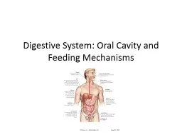
Slide11The Oral Cavity
Lined with mucous membrane tissue
Consists of two areas:
The vestibule is the space between the teeth and the inner mucosal lining of the lips and cheeks
The oral cavity proper is the space contained within the upper and lower dental arches
Slide12The Vestibule
The intraoral vestibule begins on the inside of the lips and then extends from the lips onto the alveolar process of both arches
The vestibular mucosa is thin, red, and loosely bound to underlying alveolar bone
The base of each vestibule, where the buccal mucosa meets the alveolar mucosa, is called the
mucobuccal fold
The mucogingival junction is a distinct line of color change where the alveolar membrane meets with attached gingiva
Slide13Vestibule and Vestibular Tissue of the Oral Cavity
Slide14Labial and Other Frenula
A
frenum
is a narrow band of tissue that connects two structures
The labial
frenum
passes from the midline of the maxillary or mandibular arch to the midline of the inner surface of the lip
The buccal
frenum
passes from the oral mucosa near the maxillary or mandibular first molars to the inner surface of the cheek
Slide15Gingiva
The gingivae, commonly referred to as the
gums
, are masticatory mucosae that cover the alveolar processes of the jaws and surround the necks of the teeth
Slide16View of Gingivae and Associated Anatomic Landmarks
Slide17Characteristics of Normal Gingiva
Normal gingivae surround the tooth like a collar and are self-cleansing
They are firm and resistant and tightly adapted to the tooth and bone
The surfaces of the attached gingivae and interdental papillae are stippled and similar in appearance to the rind of an orange
Surface color varies according to the individual's pigmentation
Slide18Color of the Gingivae Varies
Slide19Unattached Gingiva
Unattached gingiva, which is also known as
marginal gingiva
or
free gingiva
, is the border of the gingiva surrounding the teeth in collar-like fashion
It consists of the tissues from the top of the gingival margin to the base of the gingival sulcus
The unattached gingiva is usually about 1 mm wide and forms the soft wall of the gingival sulcus
Slide20Gingiva
Interdental gingiva (also called
gingival papilla
)
Extension of the free gingiva that fills the interproximal embrasure between two adjacent teeth
Gingival groove
The gingival groove is a shallow groove that runs parallel to the margin of the unattached gingiva and marks the beginning of the attached gingiva
Attached gingiva
The attached gingiva extends from the base of the sulcus to the mucogingival junction
Slide21The Oral Cavity Proper
The oral cavity proper is the area inside the dental arches
In back of the last molar on each side is a space that links the vestibule and the oral cavity proper
Slide22Hard Palate
The hard palate separates the nasal cavity above from the oral cavity below
The nasal surfaces are covered with respiratory mucosa, and the oral surfaces are covered with oral mucosa
The mucosa of the hard palate is tightly bound to the underlying bone, and therefore submucosal injections into the palatal area can be extremely painful
Slide23Landmarks on the Hard Palate
The incisive papilla is a pear-shaped pad of tissue that covers the incisive foramen
The palatal
rugae
are irregular ridges of masticatory mucosa extending laterally from the incisive papilla
The palatine raphe runs posteriorly from the incisive papilla at the midline
The palatal glands are numerous small glands that open onto the palatal mucosa as small pits
Slide24Soft Palate
The soft palate is the movable posterior third of the palate
It has no bony skeleton and hangs like a limp curtain into the pharynx behind it
The soft palate ends posteriorly as a free edge with a hanging projection called the
uvula
Slide25Soft Palate (Cont.)
The soft palate is supported posteriorly by two arches, the
fauces
The anterior arch runs from the soft palate down to the lateral aspects of the tongue as the palatoglossal arch
The posterior arch, the free posterior border of the soft palate, is called the
palatopharyngeal
arch
The opening between the two arches is called the
isthmus of
fauces
and contains the
palatine tonsil
Slide26Tongue
The tongue is an important organ, responsible for several functions:
Speech
Manipulation and positioning of food
Sense of taste
Swallowing
Cleansing of the oral cavity
Slide27Parts and Surfaces of the Tongue
Body: anterior two thirds of the tongue
Root: posterior portion that turns downward toward the pharynx
Dorsum: upper and posterior roughened surface
Sublingual surface: covered with smooth, transparent mucosa
Lingual frenulum: a thin fold of mucous membrane that extends from the floor of the mouth to the underside of the tongue
Slide28Taste Buds
Located on the fungiform papillae and in the trough of the large
vallate
papillae, which form a V on the posterior portion of the tongue
The sense of touch is provided by numerous filiform papillae that cover the entire surface of the tongue
Slide29Teeth
Teeth are either single-rooted or multirooted
The teeth sit in bony sockets, or alveoli, within the alveolar process of the maxilla and mandible
In the mouth, a cuff of gingival tissue surrounds each tooth
The portion of the tooth that is visible in the oral cavity is called
the crown