
14 ABSTRACT Patie nts with cleft lip and palate usually face a multitude of problems esthetic compromise being the most noticeable malocclusion missing teeth oronasal fistula speech and hea ID: 940516
Download Pdf The PPT/PDF document "Smile Dental Journal Volume 7 Issue 4 ..." is the property of its rightful owner. Permission is granted to download and print the materials on this web site for personal, non-commercial use only, and to display it on your personal computer provided you do not modify the materials and that you retain all copyright notices contained in the materials. By downloading content from our website, you accept the terms of this agreement.


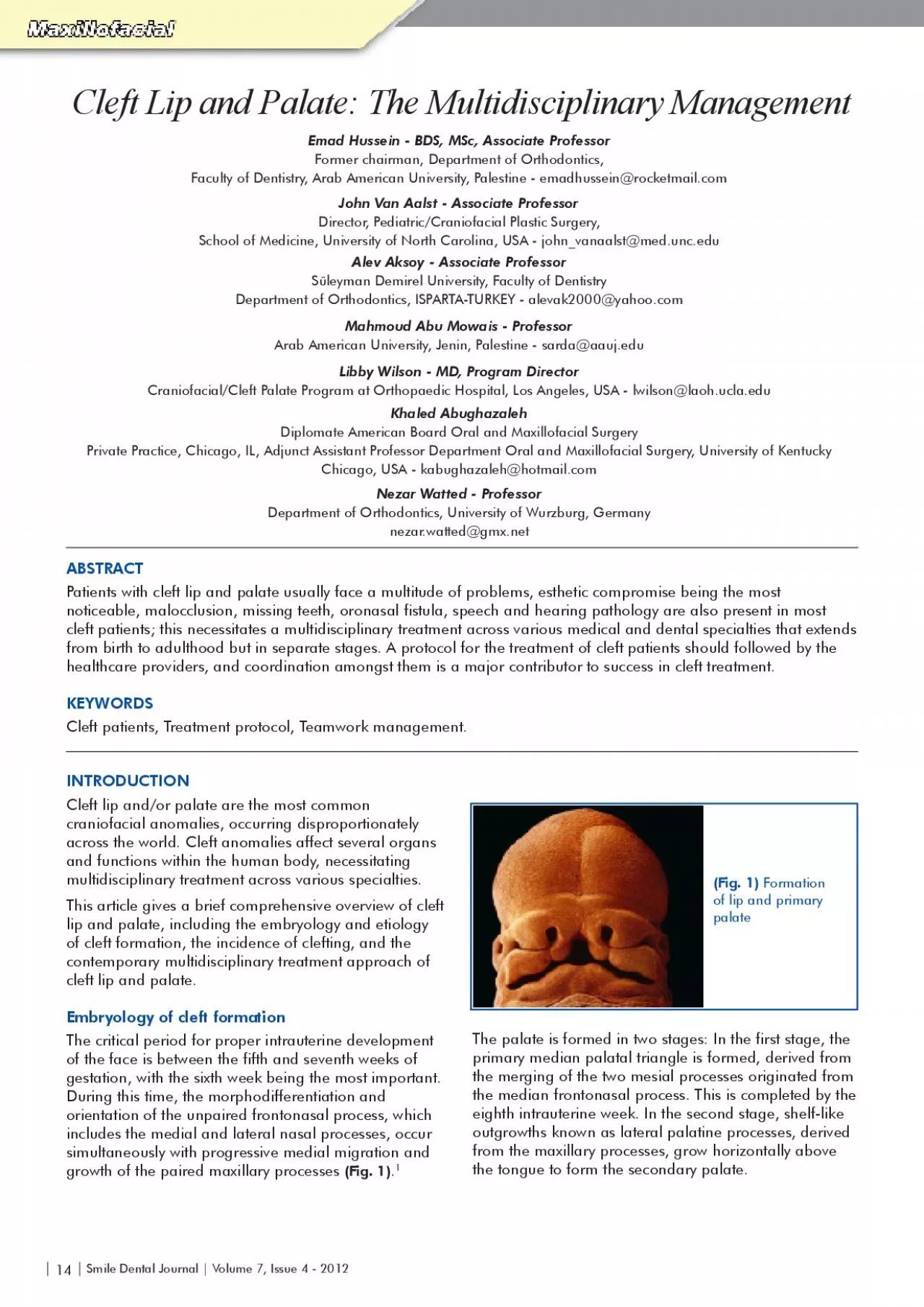
| 14 | Smile Dental Journal | Volume 7, Issue 4 - 2012 ABSTRACT Patie nts with cleft lip and palate usually face a multitude of problems, esthetic compromise being the most noticeable, malocclusion, missing teeth, oronasal fistula, speech and hearing pathology are also present in most cleft patients; this necessitates a multidisciplinary treatment across various medical and dental specialties that extends from birth to adulthood but in separate stages. A protocol for the treatment of cleft patients should followed by the healthcare providers, and coordination amongst them is a major contributor to success in cleft treatment. KEYWORDS Cleft patients, Treatment protocol, Teamwork management. Cleft Lip and Palate: The Multidisciplinary Management Emad Hussein - BDS, MSc, Associate Professor Former chairman, Department of Orthodontics, Faculty of Dentistry, Arab American University, Palestine - emadhussein@rocketmail.com John Van Aalst - Associate Professor Director, Pediatric/Craniofacial Plastic Surgery, School of Medicine, University of North Carolina, USA - john_vanaalst@med.unc.edu Alev Aksoy - Associate Professor Süleyman Demirel University, Faculty of Dentistry Department of Orthodontics, ISPARTA-TURKEY - alevak2000@yahoo.com Mahmoud Abu Mowais - Professor Khaled Abughazaleh Diplomate American Board Oral and Maxillofacial Surgery Private Practice, Chicago, IL, Adjunct Assistant Professor Department Oral and Maxillofacial Surgery, University of Kentucky Chicago, USA - kabughazaleh@hotmail.com Nezar Watted - Professor Department of Orthodontics, University of Wurzburg, Germany nezar.watted@gmx.net INTRODUCTION Cl craniofacial anomalies, occurring disproportionately across the world. Cleft anomalies affect several organs and functions within the human body, necessitating multidisciplinary treatment across various specialties. This article gives a brief comprehensive overview of cleft lip and palate, including the embryology and etiology of cleft formation, the incidence of clefting, and the contemporary multidisciplinary treatment approach of cleft lip and palate. Embryology of cleft formation Th e critical period for proper intrauterine development of the face is between the fifth and seventh weeks of gestation, with the sixth week being the most important. During this time, the morphodifferentiation and orientation of the unpaired frontonasal process, which includes the medial and lateral nasal processes, occur simultaneously with progressive medial migration and (Fig. 1) . 1 The palate is formed in two stages: In the first stage, the primary median palatal triangle is formed, derived from the merging of the two mesial processes originated from the median frontonasal process. This is completed by the eighth intrauterine week. In the second stage, shelf-like outgrowths known as lateral palatine processes, derived from the maxillary processes, grow horizontally above the tongue to form the secondary palate. (Fig. 1) Formation of lip and primary palate Libby Wilson -
MD, Program Director Craniofacial/Cleft Palate Program at Orthopaedic Hospital, Los Angeles, USA - lwilson@laoh.ucla.edu Smile Dental Journal | Volume 7, Issue 4 - 2012 | 15 | Additionally, adult patients with cleft anomalies may also suffer from marital problems. These and other scenarios all require psychosocial support for the cleft patient as well as his/her family. 5 By improving a child’s social skills, educational pursuit, both the child and his/her parents may be able to overcome the issues that stem from the child’s physical appearance. 5 Other associated abnormalities include oronasal fistula, speech and hearing pathology, malocclusion, which is always present in cleft lip and palate patients. Anterior and posterior crossbites due to anteroposterior and transverse deficiency of the maxilla, rotation of the incisors due to muscle pull, lateral incisors at the cleft site are frequently missing and supernumerary teeth may be present at the non-cleft side. Carious teeth and periodontal inflammation are often present due to dental neglect (Figs. 3-5). 6,7 The merging or fusion of these processes is completed by the twelfth intrauterine week (Fig. 2) . Any insult to the fusion process between the fifth and seventh intrauterine weeks leads to the formation of clefts. This may lead to irritation of the nasal septum, causing repeated respiratory tract and middle ear infections. Additionally, the tongue may be postured in the cleft space during swallowing, which may further widen the cleft space. Etiology of cleft Lip and Palate Th e etiology of cleft remains unclear. It is presumed to be multifactorial, with various contributing environmental and genetic factors. 2 Toxin exposure during the antenatal period is more likely to be present in developing countries due to poor sanitation, inadequate infrastructure, and political instability. For many of the same reasons, metabolic disorders and malnutrition are also more likely to be an issue for pregnant women in developing countries. Genetic factors and consanguinity are also major potential risk factors for birth defects. 3 Problems Associated With Clefts Pa tients with cleft lip and palate usually face a multitude of problems, esthetic compromise being the most noticeable. The esthetic obstacle in children with cleft lip and palate may often lead to various types of psychosocial distress. For example, patients may feel that there is a stigma attached to their appearance. They often perceive themselves as unattractive, which may influence their psychological behavior and lead to problems with communication. 4 Esthetic issues can also limit a child’s life prospects. Some affected children may not attend school regularly, due to teasing, bullying, speech difficulty, and hearing problems. 6 (Fig. 2) Formation of secondary palate (Fig. 3) Skeletal and Dentoalveolar irregularities associate with cleft lip and palate (Fig. 4) Oronasal fistula associated with cleft palate (Fig. 5) Repaired oronas
al fistula during alveolar bone graft | 16 | Smile Dental Journal | Volume 7, Issue 4 - 2012 TREATMENT TIMING & PROTOCOL The First Closure: Lip Th e first procedure that cleft patients receive is surgical closure of the lip. Following the “rule of 10s,” a patient admitted for lip closure should be 10 weeks of age, weigh at least 10 pounds, have a hemoglobin ≥10mg/100ml, and have aWBC count withstand surgery and anesthesia (Figs. 6,7). 8, 9 Neonatal orthopedic correction is still used in several cleft lip and palate centers around the world, despite several reports questioning its effectiveness. In patients with wide bilateral cleft lip and/or a severely protruding premaxilla, surgical taping as an alternative method for neonatal orthopedics has many benefits: it is easy to use, does not require several visits to pediatric dentists or orthodontists over a period of months, and the results are often successful. When used, this taping is worn for two weeks prior to the Von Langenbeck lip orMillard forked flap surgery (Fig. 8) . 9 In this approach, physiological forces will enhance palatal molding and will bring the premaxilla backwards. This in turn shortens the time for lip closure. When surgical taping is used rather than lengthy neonatal orthopedics, lip closure surgery can be performed 10 weeks later, thereby decreasing the psychological distress of the family as they witness the correction of their child’s cleft. Hard & Soft Palate Surgical Repair Sp eech is learned by hearing, which can be aided by surgical repair of the hard and soft palate. This surgery is performed around 12-18 months of age, when children first begin to talk. The surgery is also performed at this age with airway considerations in mind, as it benefits dentition eruption. 9 Palatal repair is done in a single stage using the Vomer flap for anterior palate closure, in addition to the Bardach two-flap palatoplasty with soft closure into both alveolar clefts (Fig. 9) . Treatment in the Primary Dentition Stage In the primary dentition stage, focus is on restoration of the primary dentition using mainly fillings, stainless steel crowns, and oral hygiene rather than orthodontics or surgery (Fig. 10). Sometimes, expansion by a removable appliance is used in cross bites with functional shifts of the mandible upon closure. At this young age, cooperation of the patient remains a factor. This further necessitates delay of treatment to the mixed dentition stage. 7 (Fig. 6) Bilateral Complete Cleft lip and palate before lip repair (Fig. 7) Bilateral Complete Cleft lip and palate after lip repair (Fig. 10) Dental management in the primary dentition stage (Fig. 9) Surgical repair of the palate (Fig. 8) Surgical lip repair Smile Dental Journal | Volume 7, Issue 4 - 2012 | 17 | The central incisor is usually distolabially rotated and inclined due to muscular pull (Fig.14) . Care should be taken while leveling this incisor and other teeth, so as not to push the roots of t
hese teeth into the cleft space. Following a successful graft placement, it is preferable to keep the incisor roots invested in bone at the pre- grafting stage while the correct tooth angulation is achieved at least 2 months after grafting (Figs.15,16) . 7 Treatment Principles in the Mixed Dentition Stage The main objective during the mixed dentition stage is to prepare the cleft patient for a bone graft. The collapsed (overlapped) alveolar segments may be impeded in their growth. It is important that these segments be unlocked by expansion during the early stages of development when growth is most rapid. Orthodontic expansion of collapsed buccal segments will also facilitate the push- back of the premaxilla to restore a favorable arch form, which was initially interrupted by the lack of alveolar bone continuity on the cleft side. Expansion is carried out successfully by a quadhelix expansion appliance (Fig. 11) , giving the surgeon a more favorable surgical field to perform the bone graft. Expansion devices should be used for at least four months after the bone graft, as freshly grafted bone is unable to maintain the expansion (Fig. 12) . 7 Crowding is usually present on the non-cleft side in unilateral cleft patients, and should also be relieved during the mixed dentition stage. At the crowding site, serial extractions can sometimes be carried out to provide space for eruption of the canine, while lateral incisors are usually missing at the cleft side. Supernumerary teeth should be extracted during the bone graft surgery and primary teeth adjacent to cleft should be extracted at least two months prior to surgery (Fig. 13) . 10 (Fig. 13) Post orthodontic expansion of collapsed buccal segments (Fig. 12) Collapsed buccal segments in unilateral cleft palate patient before expansion (Fig. 11) A quadhelix expansion appliance (Fig. 15) Corrected position of the central incisor (Fig. 16) A successful bone graft placement appearing by a periapical X-ray (Fig. 14) The central incisor is usually distolabially rotated and inclined in a cleft palate patient | 18 | Smile Dental Journal | Volume 7, Issue 4 - 2012 Bone Graft at the Cleft Side Bone grafting has several advantages. It is performed to support the long-term expansion of the dental arch, maintaining arch continuity and form while also providing bone for the passage of the erupting canine through the graft. 6 A bone graft will also support the teeth adjacent to the cleft site, ensuring orthodontic movement of these teeth without periodontally compromising them. The bone graft will also improve facial esthetics by providing support to the base of the nose, which is lacking due to the cleft space (Figs. 17-19) . The bone graft is usually taken from the cancellous bone of the iliac crest. Other donor sites that provide cancellous bone include the mandibular symphysis and the skull. Timing of alveolar bone graft relates to tooth development and is carried out between the ages of 6-9 years, using t
he stage of root formation of the maxillary canine as a guide for the proper timing for bone graft is better than just following chronological age, when one third of the canine is formed viewed by a periapical x-ray is the best time to consider bone graft. . This may lead to spontaneous canine eruption through the graft and healthy gingiva surrounding the graft, with normal bone height. 6, 9 Orthopedic Mid Face Growth Enhancement Maxill ary hypoplasia is a well-known feature of cleft lip and palate patients. It may be due to scarring from the surgical repair of the cleft lip and/or palate, which may cause lip tightness and palatal scarring that restricts the growth of the maxilla. Studies by Ross et al. showed that untreated cleft patients exhibited normal maxillary growth, further supporting this hypothesis. Orthopedics applies interrupted forces which can aid maxillary sutures. However, the success of maxillary orthopedic advancement remains limited due to the tightness of the lip and palatal scarring. The decision to use a face mask or to undergo orthognathic surgery should be made early during treatment planning for patients with mild to moderate skeletal discrepancies (Fig. 20) . 11,12 Orthognathic surgery Cl eft patients will usually have jaw disharmony at the end of their facial growth period, manifested by mid-face deficiency. Growth of the maxilla is impaired in sagittal, transverse and vertical dimensions, evident in the form of anterior and posterior cross bites. Mandibular over- closure is also apparent due to maxillary deficiency in the vertical dimension. Orthognathic surgery is required in about 25% of adult cleft patients according to the severity of skeletal features. The most commonly affected jaw in cleft patients is the upper jaw. Thus, in most cases, (Fig. 17) Alveolar bone exposed at cleft side (Fig. 18) Cancellous bone packed into cleft (Fig. 19) Incisor roots invested in bone at the pregrafting stage while the correct tooth angulation can be achieved after successful graft placement (Fig. 20) Orthopedic treatment using a facemask Smile Dental Journal | Volume 7, Issue 4 - 2012 | 19 | Orthodontic preparation for the orthognathic surgery aims to correct dental compensation due to the skeletal discrepancy. Other goals of pre-surgical orthodontics include arch coordination and removal of any occlusal prematurity. Relapse in the upper jaw position is possible due to the scar tissue resulting from the upper lip and palate repair. Distraction osteogenisis enables the gradual advancement of the hypoplastic maxilla with corticotomy cuts that will allow expansion of the scar tissue (Figs. 21,22) . This decreases the possibility of relapse and allows for a longer-term correction over orthognathic surgery. Distraction ostegenesis has recently benefited from major advances in instrumentation, especially regarding the materials used to create instruments and the creation of bidirectional instruments. This has improved results and further
decreased the need for more orthognathic surgery. 12 SUMMARY Pa tients with cleft lip and palate require a team approach for their treatment, comprised of several specialists. This multidisciplinary care starts from birth and continues into adulthood, and coordination amongst specialists is a major contributor to success in cleft treatment. ACKNOWLEDGMENTS The Au thors thank Sonya Patel and Hala Borno from the medical school at The University of North Carolina for their participation to the editing of this article REFERENCES 1. Mo ore KL. The Developing Human: Clinically Oriented Embryology. Philadelphia, PA: Saunders, 1977. 2. Malcolm C, Johnston P. Embryogenesis of cleft lip and palate, In: McCarty JG, editor. Plastic Surgery, vol 4. Cleft Lip and Palate and Craniofacial Anomalies. Saunders, Philadelphia, PA, 1990:2532. 3. Persaud TVN, Chudley AE, Skalko BG. Basic Concepts in Teratology. New York: Alan R. Liss, 1985. 4. Marcusson A. Adult patients with treated complete cleft lip and palate. Methodological and clinical studies. Swed Dent J Suppl 2001;145:1– 57. 5. Mars M, Sell D, Habel A (2008) Management of cleft lip and palate in the developing world. GBR, Chichester. 6. Peter D. Waite and Daniel E. Waite, Bone Grafting for the Alveolar Cleft Defect Seminars in Orthodontics, Vol 2, No 3 (September), 1996:192-6. 7. Christos C. Vlachos. Orthodontic Treatment for the Cleft Palate Patient Seminars in Orthodontics, Vol 2, No 3 (September), 1996:197-204. 8. Black PW, Scheflan M. Bilateral cleft lip repair: ‘‘putting it all together’’. Ann Plast Surg. 1984;12:118–127 23. 9. Samuel Berkowitz, A Comparison of Treatment Results in Complete Bilateral Cleft Lip and Palate Using a Conservative Approach Versus Millard-Latham PSOT Procedure Seminars in Orthodontics, Vol 2, No 3 (September), 1996:169-18. 10. Carla A. Evans, Orthodontic treatment for patients with clefts Clin Plastic Surg 31 (2004;271–90. 11. Rygh P, Tindlund R. Orthopedic expansion and protraction of the maxilla in cleft palate patients—a new treatment rationale. Cleft Palate J 1982;19:104-112. 12. Graber L , Vanarsdall R, Vig K. Orthodontics Current principles and Techniques, 5th edition, Elsevier. 2011;965-89. the orthognathic procedure performed involves the advancement of the upper jaw (Le Fort I) rather than setting the mandible back. When the reverse overjet is severe, a two-jaw surgery may be required, impacting breathing and esthetics (Figs. 21-24) . 7 The proper timing of orthognathic surgery is when facial growth has ended. A hand and wrist radiograph can help determine the end of facial bone growth. (Fig. 21) Cephalometric X-ray for adult cleft patient before surgery (Fig. 23) Lateral facial view of a cleft patient before orthognathic surgery (Fig. 22) Surgical advancement of the upper jaw (Le Fort I) for the same patient in Figure 21 (Fig. 24) Lateral facial view of the same cleft patient in Figure 23 after maxillary advancement and mandibular set