

CREATININE CLEARANCE URIC ACID Mohammed AlZubaidi PhD Laboratory Training Introduction The kidneys are two bean shaped organs lying retroperitoneally on each side of the vertebral column slightly above ID: 920906
Download Presentation The PPT/PDF document "BLOOD UREA BLOOD CREATININE" is the property of its rightful owner. Permission is granted to download and print the materials on this web site for personal, non-commercial use only, and to display it on your personal computer provided you do not modify the materials and that you retain all copyright notices contained in the materials. By downloading content from our website, you accept the terms of this agreement.


Slide1
BLOOD UREABLOOD CREATININECREATININE CLEARANCEURIC ACID
Mohammed Al-Zubaidi, PhD
Laboratory Training
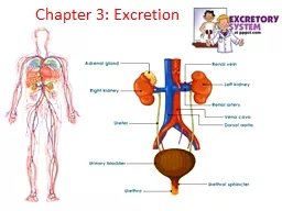
Slide2IntroductionThe kidneys are two bean shaped organs lying retroperitoneally on each side of the vertebral column slightly above the level of umbilicus.The range in length & weight, respectively, from approximately 6cm
& 24gms in a full term infant to more than/equal to 12cm & 150gms in an adult
Slide3IntroductionNephronEach kidney contains approx. 1 million nephrons.In humans, formation of nephron is complete at 36-40 weeks
of gestation, but functional maturation with tubular growth & elongation continues during the 1st decade of life
Slide4IntroductionNephronBecause new nephrons can’t be formed after birth, so any disease that results in progressive loss of nephrons can lead to renal insufficiency.A decreased number of nephrons secondary to low birth weight, prematurity and/or unknown genetic or environmental factor is hypothesised
to be a risk factor for the development of primary hypertension & Progressive Renal Dysfunction in adulthood.
Slide5IntroductionA Nephron consist :-OUTER LAYER(the cortex)-glomeruli-PCT & DCT-CD
INNER LAYER(the medulla)-Straight portion of tubules-LOH-vasa recta-terminal CD
Slide6Blood urea & serum creatinineEstimation of blood urea & serum creatinine are useful.These tests are less sensitive than the clearance tests.
Serum creatinine is a better indicator than urea.But a markedly increased blood urea is conclusive evidence of severe impaired glomerular function. In chronic renal disease, the blood urea level correlates better with symptoms of uremia than does the serum creatinine.As a kidney function test, serum urea is inferior to serum creatinine because:High protein diet increases urea formation.Any condition of proteins catabolism (Cushing syndrome, diabetes mellitus, starvation, thyrotoxicosis) urea formation. 50 % or more of urea filtered at the glomerulus is passively reabsorbed by the renal tubules. The amount reabsorbed depends on flow rate. Therefore urea is less accurate than creatinine as an estimate of GFR.
Slide7Blood ureaUrea forms in the liver and, along with CO2, constitutes the final product of protein metabolism. The amount of excreted urea varies directly with dietary protein intake, increased excretion in fever, diabetes, and increased adrenal gland activity
.In the US and a few other countries, plasma or serum urea concentration is expressed as the amount of urea nitrogen, blood urea nitrogen (BUN).The test for BUN, which measures the nitrogen portion of urea, is used as an index of glomerular function in the production and excretion of urea.Chemical formula of urea is ( NH2 )2 >C=O i.e. Molecular weight of urea is 60 while nitrogen in it is 28 . 60/28 = 2.14 , so the conversion is blood urea nitrogen × 2.14 = blood urea , a calculative value . Blood urea nitrogen normal value is between 6 and 20 mg per 100 ml while normal value of blood urea is between 15 and 40 mg per 100 ml.
Slide8Blood ureaA normal level of blood urea is often mistakenly regarded to indicate normal kidney function.In a steady state the blood urea may not rise beyond the upper range of normal(40mg/dl) even when 75% of the renal function is lost.On
the other hand, prerenal factors that decreases renal perfusion & GFR, such as dehydration, causes an increase in blood urea levels.There may be transient rise in blood urea level due to :- high protein intake & excessive protein catabolism ( e.g with severe infections, tissue breakdown, trauma, use of large doses of corticosteroids or tetracyclines)- gastrointestinal bleeding & inhibition of anabolism.
Slide9Blood ureaClinical implicationIncreased BUN levels (azotemia) occur in the following conditions:a. Impaired renal function caused by the following conditions:(1) Congestive heart failure(2) Salt and water depletion
(3) Shock(4) Stress(5) Acute MIb. Chronic renal disease such as glomerulonephritis and pyelonephritisc. Urinary tract obstructiond. Hemorrhage into GI tracte. Diabetes mellitus with ketoacidosisf. Excessive protein intake or protein catabolism as occurs in burns or cancerg. Anabolic steroid use
Slide10Blood ureaDecreased BUN levels are associated with the following conditions, although Clinically of little significance.a. Liver failure (severe liver disease), such as that resulting from hepatitis, drugs, or poisoningb. Acromegalyc. Malnutrition, low-protein dietsd. Impaired absorption (celiac disease)
e. Nephrotic syndrome (occasional)f. Syndrome of inappropriate antidiuretic hormone (SIADH)
Slide11Blood ureaInterfering Factors1. A combination of a low-protein and high-carbohydrate diet can cause a decreased BUN level.2. The BUN is normally lower in children and women because they have less muscle mass than adult men.3. Decreased BUN values normally occur in late pregnancy because of increased plasma volume (physiologic hydremia
).4. Older persons may have an increased BUN when their kidneys are not able to concentrate urine adequately.5. IV feedings only may result in overhydration and decreased BUN levels.6. Many drugs may cause increased or decreased BUN levels.Azotemia:Increase in the blood levels of NPN (creatinine, urea, uric acid) is referred to as azotemia & is the hallmark of kidney failure.
Slide12Blood ureaInterventionsPretest Patient Care1. Explain test purpose and blood-drawing procedure. Assess dietary history.Posttest Patient Care1. Interpret test outcome and monitor as appropriate for impaired kidney function.
2. In patients with an elevated BUN level, fluid and electrolyte regulation may be impaired.
Slide13Blood ureaCONTROL OF BLOOD UREACorrect intake of protein: If kidneys can’t do their work properly, extra protein will increase the workload on
kidneys. On the contrary, lack of protein may lead to malnutrition. Under this circumstance, it is necessary to restrict the amount of protein intake to 0.6-0.8g/kg every day.Supplement enough calories: This can reduce the consumption of protein in the body. Generally, it is suitable to take in 30Kca/Kg of calories every day.
Slide14Serum Creatinine & Creatinine ClearanceCreatinine is derived from the metabolism of creatine and phosphocreatine, the bulk of which is in muscle.
Creatine and creatine phosphate exist in a reversible equilibrium in skeletal muscle.In skeletal muscle, approximately one-fourth of creatine exists as free creatine and three-fourth exists as creatine phosphate.Since
creatinine
is chiefly excreted by
glomerular
filteration
,
S.creatinine
levels reflects changes in GFR.
S.creatinine
values are low when the muscle mass
is decreased
, as in malnutrition.
Bilirubin
interferes with
creatinine
m
easurements
.
Slide15Serum CreatinineSerum creatinine affected partly by:The
amount of muscle tissue you have. Men tend to have higher levels of blood creatinine because they have more skeletal muscle tissues than women.Protein in diet
.
Vegetarians
have
been
shown to
have
lower
creatinine
levels
in blood.
This test diagnoses impaired renal function. It is a more specific and sensitive indicator of
kidney disease
than BUN, although in chronic renal disease, both BUN and
creatinine
are ordered to
evaluate renal
problems because the BUN-to-
creatinine
ratio provides more information.
Slide16Serum CreatinineReference ValuesNormalAdult men: 0.9–1.3 mg/dL Adult women: 0.6–1.1 mg/dL
BUN-to-creatinine ratio: 10:1 to 20:1Clinical alert: Critical value is 10 mg/dL in nondialysis patients.Procedure1. Obtain a 5-mL venous blood sample. Serum is preferred, but heparinized blood can be used. Place specimen in a biohazard bag.2. Observe standard precautions.
Slide17Serum CreatinineClinical Implications1. Increased blood creatinine levels occur in the following conditions: a. Impaired renal function
b. Chronic nephritis c. Obstruction of urinary tract d. Muscle disease (1) Gigantism (2) Acromegaly (3) Myasthenia gravis (4) Muscular dystrophy (5) Poliomyelitis e. Congestive heart failure f. Shock g. Dehydration h. Rhabdomyolysis (skeletal muscle tissue breakdown) i. Hyperthyroidism
Slide18Serum Creatinine2. Decreased creatinine levels occur in the following conditions: a. Small stature b. Decreased muscle mass
c. Advanced and severe liver disease d. Inadequate dietary protein e. Pregnancy (0.4–0.6 mg/dL is normal; 0.8 mg/dL is abnormal and should be noted)3. Increased ratio (>20:1) with normal creatinine occurs in the following conditions: a. Increased BUN (prerenal azotemia), heart failure, salt depletion, dehydration b. Catabolic states with tissue breakdown c. GI hemorrhage d. Impaired renal function plus excess protein intake, production, or tissue breakdown
Slide19Serum Creatinine4. Increased ratio (>20:1) with elevated creatinine occurs in the following conditions: a. Obstruction of urinary tract
b. Prerenal azotemia with renal disease5. Decreased ratio (<10:1) with decreased BUN occurs in the following conditions: a. Acute tubular necrosis b. Decreased urea synthesis as in severe liver disease or starvation c. Repeated dialysis d. SIADH e. Pregnancy6. Decreased ratio (<10:1) with increased creatinine occurs in the following conditions: a. Phenacemide therapy (accelerates conversion of creatine to creatinine) b. Rhabdomyolysis (releases muscle creatinine) c. Muscular patients who develop renal failure
Slide20Serum CreatinineInterfering Factors1. High levels of ascorbic acid and cephalosporin antibiotics can cause a falsely increased creatinine level; these agents also interfere with the BUN-to-creatinine ratio.2. Drugs that influence kidney function plus other medications can cause a change in the
blood creatinine level.3. A diet high in meat can cause increased creatinine levels.4. Creatinine is falsely decreased by bilirubin, glucose, histidine, and quinidine compounds.5. Ketoacidosis may increase serum creatinine substantially.
Slide21Serum CreatinineInterventionsPretest Patient Care1. Explain test purpose and procedure.2. Assess diet for meat and protein intake.Posttest Patient Care
1. Interpret test results and monitor as appropriate for impaired renal function.2. Possible treatment includes hemodialysis and renal replacement therapy, including kidney transplant.
Slide22Clearance testTest for glomerular filtration rate (GFR)Useful index for the assessment of severity of kidney damageThe concept of clearance is based upon the fact that the rate of removal of a substance from the plasma must equal its simultaneous rate of excretion in urine.Thus, ‘
Clearance is defined as the quantity of blood or plasma that is completely cleared of a substance per unit time’Alternatively >>>> ml of plasma which contains the amount of that substance excreted by kidney within a minuteUnits: ml/min
Slide23Clearance testCalculation: (U X V)/PWhere:U = concentration of substance in urine (mg/dl)
V = volume of urine excreted per minute (ml/min)P = concentration of substance in plasma/serum (mg/dl)Types of Clearance testsEndogenousExogenousCreatinineInulinCystatin CPara-amino hippuric acid (PAHA)Diodrast (di-iodo pyridone acetic acid)
Slide24Clearance testThe best substance to use for glomerular clearance:It is filtered completely through the glomerulus and not reabsorbed through the nephron tubuleIt is not affected by endogenous and exogenous factors
Renal Clearance of INULIN is the Gold Standard for determination of GFR. However, inulin does not occur naturally in the body and requires several hours of infusion to reach steady state concentration.51Cr-EDTA clearance closely resembles Inulin clearance & it is the radionuclide of choice for GFR estimation in Europe.However, 99m Tc-DTPA is often the preferred agent, because 99m Tc-DTPA is inexpensive, easily available & renal imaging can be simultaneously performed.GFR can be estimated based upon either plasma clearance or upon the tracer uptake by the kidneys.
Slide25Clearance testCYSTATIN C : It is a LMW nonglycosylated protein produced at a constant rate by all nucleated cells in the body, freely filtered by the glomeruli, not secreted, but totally reabsorbed by the renal tubules.
Little or no cystatin is excreted in urine.Normal adults have circulating level of approx. 1mg/l.This is better indicator of renal function as compared to creatinine in early stages of GFR impairment as it is independent of age, gender, body composition & muscle mass.Cystatin C can be estimated in blood by enzyme immunoassays or immunoturbidometry. Both techniques are currently kit based & expensive.
Slide26Creatinine clearance testCreatinine clearance test measures the rate at which the kidneys are able to remove (to clear) a filterable substance from the blood
.creatinine clearance is a rough measure of the glomerular filtration ratea) Serum creatinine conc (Crs)b) Urine creatinine conc ( Cru)c) Urine VolumeCreatinine clearance=(Cru x V/t) / CrsNormal range male = 85 – 125 ml/min female = 75 – 115 ml/min
Slide27Creatinine clearance testIn clinical practice creatinine clearance may be used to estimate GFR for the following reasons:
Inulin is not produced endogenously.Therefore, it must be infused intravenously if it is to be used in renal function tests. So, it is much more convenient to use a substance that is normally present in plasma that is freely filtered, but neither secreted, nor reabsorbed.2. Creatinine, a normal breakdown product of creatine, is an endogenous compound that fulfills these criteria.Endogenous creatinine production is constant as long as the muscle mass remains constant. Because all creatinine filtered by the kidneys in a given time interval is excreted into the urine, creatinine levels are equivalent to the glomerular filtration rate (GFR). Disorders of kidney function prevent maximum excretion of creatinine. The creatinine clearance test is a specific measurement of kidney function, primarily glomerular filtration. It measures the rate at which the kidneys clear creatinine from the blood.However, in humans, a small amount of creatinine is secreted into the urine in the proximal tubules.Consequently, the rate of excretion of creatinine exceeds its rate of filtration by 5 to 10%. The clearance of creatinine thus exceeds the true GFR by 5 to 10%.
Slide28Creatinine clearance testIn renal failure the kidney will not be able
to excrete creatinine in urine leading to an elevation in serum creatinine level.Both serum Cr. and creatinine clearance are used as kidney function tests to : Confirm the diagnosis of renal disease.Give an idea about the severity of the disease. Follow up the treatment.
A raised serum
creatinine
is
a good indicator of impaired renal function
But normal serum
creatinine
does not necessarily indicate normal renal function as serum
creatinine
may not be elevated until GFR has fallen by as much as 50%
Slide29Creatinine clearance testIn the traditional method, creatinine content of a 24 hr urine collection & the plasma concentration in this period are estimated.
Try this: calculate the creatinine clearanceUsing urine creatinine of 120mg/dl, serum creatinine of 1.0 mg/dl and urine volume of 1440 ml obtained from 24 –hour specimen. Calculate the GFRAnswer ?Creatinine clearance = (Cru x V/t) / Crs = [120mg/dl x 1440ml / (24h x 60min)]/1.0mg/dl =[120 x 1440ml / 1440min]/1.0 =120ml/min
Slide30Creatinine clearance testClinical Implications1. Decreased creatinine clearance is found in any condition that decreases renal blood flow: a. Impaired kidney function, intrinsic renal disease, glomerulonephritis
, pyelonephritis, nephrotic syndrome, acute tubular dysfunction, amyloidosis, interstitial nephritis b. Shock, dehydration c. Hemorrhage d. Chronic obstructive lung disease e. Congestive heart failure2. Increased creatinine clearance is found in: a. State of high cardiac output b. Pregnancy c. Burns d. Carbon monoxide poisoning
Slide31Creatinine clearance test3. Increased urine creatinine is found in: a. Acromegaly b. Gigantism
c. Diabetes mellitus d. Hypothyroidism4. Decreased urine creatinine is found in: a. Hyperthyroidism b. Anemia c. Muscular dystrophy d. Polymyositis, neurogenic atrophy e. Inflammatory muscle disease f. Advanced renal disease, renal stenosis g. Leukemia
Slide32Creatinine clearance testInterfering Factors1. Exercise may increase creatinine clearance and urine creatinine.2. Pregnancy substantially increases creatinine clearance.
3. Many drugs decrease creatinine clearance.4. The creatinine clearance overestimates the GFR when there is severe renal impairment. The serum creatinine is more indicative of the GFR in this situation.5. A diet high in meat may elevate the urine creatinine concentration.6. Proteinuria and advanced renal failure make creatinine clearance an unreliable method for determining GFR.
Slide33Creatinine clearance testInterventionsPretest Patient Care1. Instruct the patient about the purpose and procedure of the test and urine specimen collection.A written reminder may be helpful.2. Allow food and encourage fluids for good hydration. Large urine volumes ensure optimal
test results. Avoid tea and coffee (diuretics).3. Avoid vigorous exercise during the test.4. Drugs affecting the results should be stopped beforehand (especially adrenocorticotropic hormone [ACTH], cortisone, or thyroxine). 5. Avoid eating large amounts of meat.Posttest Patient Care1. The patient may resume normal food, fluids, and activity.2. Interpret test outcomes and monitor appropriately.
Slide34Creatinine clearance testLimitations of creatinine clearanceIncludes both GFR and secretionMay overestimate GFR when GFR is low (due to secretion)Excellent for estimation of GFR
in healthy individuals
Slide35Uric acidUric acid is formed from the breakdown of nucleic acids and is an end product of purine (adenosine and guanine) metabolism in the liver. Uric acid is transported by the plasma from the liver to the kidney, where it is filtered and where about 70% is excreted in urine.The remainder of uric acid is excreted into the GI tract.
Slide36Uric acidRenal handling of uric acid is complex and involves four sequential steps1.Glomerular filtration of virtually all the uric acid in capillary plasma entering the glomerulus.2.Reabsorption in the proximal convoluted tubule of about 98% to
100% of filtered uric acid.3.Subsequent secretion of uric acid into the lumen in the distal portion of the proximal tubule.4.Further reabsorption in the distal tubule. The net urinary excretion of uric acid is 6% to 12% of the amount filtered.Uric acid in urine exists as mono- and di-sodium, potassium, ammonium and calcium urates.
Slide37Uric acidClinical applicationsUric acid is measured to :Diagnosis and monitor treatment of gout.Diagnosis of renal calculiDetect kidney function.Assess inherited
disorders of purine metabolismMonitor if uric acid levels are too high after chemotherapy or radiation.
Slide38Uric acidReference ValuesNormalMen: 3.4–7.0 mg/dLWomen: 2.4–6.0 mg/
dLClinical Significance1. Disease states with increased plasma uric acid (Hyperuricemia)GoutIncreased catabolism of nucleic acidsrenal diseaseMetabolic acidosis, diabetic ketoacidosisLeukemia, multiple myeloma, lymphoma
Slide39Uric acid2. Decreased levels of uric acid (Hypouricemia) occur in the following conditions:Fanconi’s syndrome (disease of the proximal renal
tubules)Wilson’s disease (autosomal recessive disorder resulting in the accumulation of copper in tissues)SIADHSome malignancies (e.g., Hodgkin’s disease, multiple myeloma)Xanthinuria (deficiency of xanthine oxidase)Interfering Factors1. Stress and strenuous exercise will falsely elevate uric acid.2. Many drugs cause increase or decrease of uric acid.3. Purine-rich diet (e.g., liver, kidney, sweetbreads) increases uric acid levels.4. High levels of aspirin decrease uric acid levels.5. Low purine intake, coffee, and tea decrease uric acid levels.
Slide40Uric acidInterventionsPretest Patient Care1. Advise patient of test purpose and blood-drawing procedure; fasting is preferred.2. Promote relaxation; avoid strenuous
exercise.Posttest Patient Care1. Have patient resume normal activities.2. Interpret test results and monitor appropriately for renal failure, gout, or leukemia. Uric acid level should fall in patients who are treated with uricosuric drugs such as allopurinol, probenecid, and sulfinpyrazone.
Slide41Uric acidGoutGout is due to elevated levels of uric acid in the blood. This occurs due to a combination of diet and genetic factors.In Gout increased serum levels of uric acid lead to formation of
monosodium urate crystals around the joints.Uric acid test is useful to assess for gout and to monitor patients with renal failurePrimary gout: is a condition in which uric acid is synthesized in excess and decreased ability of plasma to retain uric acid in solution. The cause for primary gout is unknown , but there is a metabolic disorder.Secondary gout: is accumulation of uric acid in plasma, than other tissues, due to increased purines catabolism, it is not due to excessive synthesis of uric acid
Slide42Uric acidUric acid stone formationSaturation levels of uric acid in blood may result in one form of kidney stones when the urate crystallizes in the kidney. These uric acid stones are radiolucent and so do not appear on an abdominal plain X-ray, and thus their presence must be diagnosed by
ultrasound for this reason or stone protocol CT. Very large stones may be detected on X-ray by their displacement of the surrounding kidney tissues
Slide43Uric acidCONTROL OF URIC ACIDAdjust Diet: To gain control of uric acid levels, avoid eating foods high in purine.Limit Alcohol: Because alcohol dehydrates the body, it is advisable to limit consumption, particularly when
consumed with foods high in purine.Water: Keep your body hydrated.
Slide44Thank you