![Case W arr [GRCh37] 5q23.2 (125989631_126295396) x 3](https://www.docslides.com/dplay.png)
![Case W arr [GRCh37] 5q23.2 (125989631_126295396) x 3](https://thumbs.docslides.com/920871/case-w-arr-grch37-5q23-2-125989631-126295396-x-3-l.jpg)
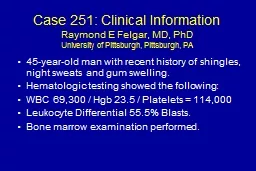
45yearold male with gait abnormalities Clinical Information arr GRCh37 5q232 125989631126295396 x 3 45yearold male referred for gait abnormalities Inheritance is unknown parents are deceased Patient reports father with history of ataxia and tremor ID: 920871
Download Presentation The PPT/PDF document "Case W arr [GRCh37] 5q23.2 (125989631_12..." is the property of its rightful owner. Permission is granted to download and print the materials on this web site for personal, non-commercial use only, and to display it on your personal computer provided you do not modify the materials and that you retain all copyright notices contained in the materials. By downloading content from our website, you accept the terms of this agreement.


Slide1
Case Warr[GRCh37] 5q23.2 (125989631_126295396) x 3
45-year-old male with gait abnormalities
Slide2Clinical Information
arr
[GRCh37] 5q23.2 (125989631_126295396) x 3
45-year-old male referred for gait abnormalities
Inheritance is unknown, parents are deceased. Patient reports father with history of ataxia and tremor.
Use the GAIN scoring metric
Slide3Section 1: Initial Assessment of Genomic Content
Case W
Genes contained
Would apply category 1A (contains protein-coding or other known functionally important elements), as this duplication includes protein-coding genes.
0 points; continue evaluation
Total: 0 points
Slide4This duplication does include an established TS gene. In a typical evaluation, this could lead you to a classification of Pathogenic.
However, for the sake of this example, we will be ignoring this in order to focus on how to evaluate the case-level data in the event this gene had not already been curated by ClinGen Dosage Sensitivity.
Section 2: Overlap with Established TS, HI, or Benign Genes/Genomic Regions
Established TS Gene
Total: 0 points
Slide5Section 3: Evaluation of Gene Number
c
There are only 2 protein-coding genes in the interval (category 3A, 0 points).
Total: 0 points
Slide6Where to start?LMNB1 is an OMIM Morbid gene associated with autosomal dominant leukodystrophy (ADLD), so this is a logical first place to start.
Section 4: Detailed Evaluation of Genomic Content
Slide7Autosomal Dominant Leukodystrophy
From
GeneReviews
–
Nahas
et al.
“Autosomal Dominant Leukodystrophy with Autonomic Disease”
https://www.ncbi.nlm.nih.gov/books/NBK338165/
Slide8Slide9Slide10Giorgio et al. 2013 (PMID:23649844)
Describes detailed molecular analysis of the largest collection of ADLD families studied to date (31 individuals from 20 independent families)
Families from countries around the world: USA, Italy, Sweden, Germany, France, India, Canada, Israel, Brazil
Reassuring to see individuals coming from various ethnic groups, unlikely to be related
9 had been described previously
Pay attention to this type of information to make sure you are not counting cases twice
Though duplications involving
LMNB1 had all been identified previously in these individuals through various methods, samples were reanalyzed by the authors for the purposes of defining the boundaries of the duplicationsNo comment was made on previous testing on any of the individuals to rule out other genetic causes of leukodystrophy16 unique duplications were identified
Slide11Our case is very similar in genomic content to several of those reported in Giorgio.Any of these (with the possible, conservative exception of BR1) would be appropriate to use as case evidence
This paper does not provide detailed information on family structure; the 6 previously unpublished cases here could be used in Category 4E (inheritance unknown).
Giorgio
et al
. 2013, Figure 1A
Case W
*
*
*
*
*
*
*
*
*
*
Previously published
Slide12Schuster et al. 2011 (A2, A3, A10, A11 in Giorgio et al.)
4 non-related families with ADLD with autonomic symptoms
2 Swedish, 1 German, 1 Israeli (of Arab descent)
Samples from one affected patient from each family were analyzed by genome-wide SNP array
Western blot analyses of
lamin
B1 were done on 5 individuals from the 2 Swedish families (including both probands)
Showed significantly increased (~twofold) lamin B1 protein levelsLevels of MARCH3 mRNA were similar between patients and controls
Case W
Slide13= genotype +
= sig. increased
lamin
B1 protein levels
Slide14Dos Santos et al. 2011 (G1 in Giorgio et al.)
47-year-old male with 2-year history of:
Gait disturbance
Micturition problems
Personality changes
Brain MRI: bilateral T2-hyperintense lesions in the subcortical and deep cerebral white matter
Normal: lumbar puncture, nerve conduction studies, electromyography, AAs, LCFAs, and lysosomal enzymes
5q32.2 duplication involving LMNB1 gene identified on array in proband and 44-year-old sisterSister asymptomatic, but was also found to have hyperintense lesions of the subcortical and deep cerebral white matter on brain MRI
PMID:21909802
= genotype +
Our Case (W)
Case G1
Slide15Other cases: Padiath et al
. 2006
Describe 3 unique duplications from 4 families involving
LMNB1
with variable involvement of the nearby
MARCH3
gene
2 Irish-American families with identical duplications – thought to arise by a common founderCandidate gene evaluation prompted by linkage analysisFunctional studies show:Lamin B1 is overexpressed in brain tissue of affected individualsIncreased expression of lamin B1 in flies resulted in a degenerative phenotype
No difference in MARCH3 expression on Northern blot in brain tissue of affected individual vs. controlPaper does not specify who was tested in each family/which individuals confirmed to have variant
PMID:16951681
Slide16Other cases: Potic et al.
2013
PMID:23681646
Serbian family presenting with progressive pyramidal and cerebellar signs, slow cognitive decline, and late-stage autonomic dysfunction
MRIs show bilateral T2-hyperintense lesions in the subcortical and deep cerebral white matter
LMNB1 copy number assessed by quantitative RT-PCR
_
_
_
_
Genotype -
Genotype +
…and many other cases available in the literature
Slide17Putting it all together…
We have a wealth of information indicating that duplications of
LMNB1
cause autosomal dominant leukodystrophy (ADLD)
Genetic evidence from at least 15 probands (with more in the literature)
Functional evidence showing increased
lamin B1 protein levels in probands (leukocytes and brain tissue); overexpression in flies causes a degenerative phenotype; normal mRNA levels of frequently involved neighbor gene
MARCH3 (likely more in literature) However, this scenario doesn’t neatly fit our “rules”Testing not consistently done on affected family members – can’t demonstrate genotype+ status for segregationNo known de novo cases
Hitting category maximums
Slide18A note about category maximums
Evidence Type
Evidence
Suggested Points/Case
Max Score
Category maximums were put in place to prevent certain evidence types from taking a case all the way to Pathogenic on their own without other, supportive information
Goal is to encourage the collection of diverse pieces of information if available/appropriate
In some circumstances, however, this is not possible
Consider carefully if your situation warrants override of category maximum
Slide19Trying to avoid scenarios like this:
If all of these people were genotype+, this family would have 11 segregations
If we allowed increasing segregation data to score up to 1.0 points, a single family could drive the classification of a variant.
This would be inappropriate, as segregation implicates a locus, not a variant.
Slide20In our case, we have:
15 different probands, all with positive family history, some with documentable segregation
Supportive functional data
Because the phenotype is adult-onset and does not appear to impact reproductive fitness, we have families with numerous affected individuals and no (documented)
de novo
cases
Achieving a variety of genetic evidence types is not possible in this case. This is a well-studied gene-disease relationship with extensive evidence, and an example of when it would be appropriate to override category maximums.
Slide21Segregations (being conservative)
Dos Santos
et al.
2011 - 1
Potic
et al.
2013 (counting intervening obligate carriers)
–
4
Total: 5 segregations (0.30 points)
Slide22What about the rest of the cases?
Could count the other 13 cases at the most conservative level (0.10 points each): > 1.0 points in addition to the segregation data
This is the same amount of points assigned for assumed
de novo
, non-specific phenotype (4C) and specific phenotype, unknown inheritance (4E)
The number of observed cases, not the number of segregations, is driving this classification
The specificity of the phenotype, the large amount of data, and the supportive functional data can all serve as rationale for this scoring change
Slide23Category 5: Incorporating our patient’s data
Our patient is a 45-year-old man with “gait abnormalities.” His parents are deceased, but he reports a history of a father with ataxia and tremor.
This is consistent with ADLD, but could also be indicative of a number of other things.
Quick search for “gait abnormalities” among Clinical Synopses in OMIM returns over 1000 matches
With this information alone, consider scoring with Category 5G (nonspecific but consistent phenotype)
When you call the ordering clinician to discuss the case, they reveal that the patient has had a brain MRI, which demonstrated findings consistent with leukodystrophy.
OMIM Clinical Synopses search for “leukodystrophy” = 47 results
With this more specific information, consider awarding full points within Category 5H (up to 0.30).
Slide24Summary
PATHOGENIC
Evidence supporting pathogenicity includes:
numerous (13+) probands with similar duplications reported in the literature with diagnoses of ADLD (scoring at 0.10 points each = >1.0 points)
At least two families with documented segregation of the duplication among affected family members (0.30 points)
Patient under evaluation has a phenotype consistent with what has been previously reported (0.30 points with MRI evidence of leukodystrophy)
Supportive functional data