
sharda Assistant professor General surgery Email prateeksharda2006gmailcom Clinical vignette 72 years old man presented with jaundice for 7 days with dull abdominal discomfort for 2 months He gives HO loss appetite and loss of weight ID: 913377
Download Presentation The PPT/PDF document "Pancreatic carcinoma Dr. Prateek" is the property of its rightful owner. Permission is granted to download and print the materials on this web site for personal, non-commercial use only, and to display it on your personal computer provided you do not modify the materials and that you retain all copyright notices contained in the materials. By downloading content from our website, you accept the terms of this agreement.


Slide1
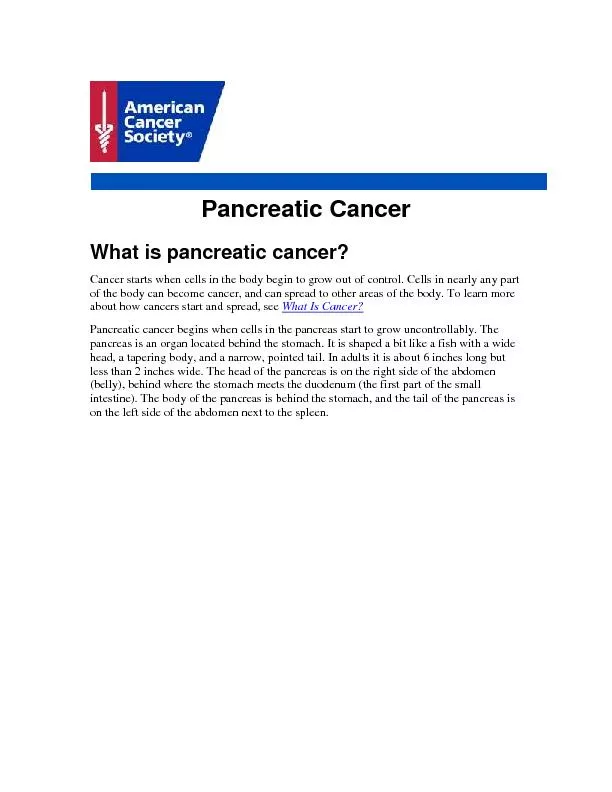
Pancreatic carcinoma
Dr. Prateek
sharda
Assistant professor
General surgery
Email:-
prateeksharda2006@gmail.com
Slide2Clinical vignette
72 years old man presented with jaundice for 7 days with dull abdominal discomfort for 2 months. He gives H/O loss appetite and loss of weight.
He is passing clay color stools.
He has a 50+ pack year smoking history before quitting last year.
He was recently diagnosed with type 2 diabetes, but has no other medical problem
Slide3O/E
: He has a yellow hue to his eye and tongue, along with scratch marks on his skin
A non-tender globular mass is palpable in right upper outer quadrant of the abdomen
Ix : Laboratory testing reveals total and direct bilirubin of 18 mg/dl(normal 0.2-1.3 mg/dL) and 17.2 mg/dL (<0.3 mg/dL), respectively.
Alkaline Phosphatase (ALP) elevated at 215 µ/L (33-131 µ/L). AST & ALT mildly elevated.
Slide4Anatomy of pancreas
Slide5Blood supply of pancreas
Slide6INTRODUCTION
3
rd
most common GIT cancer.
4
th
most common cause of cancer death
Male to female ratio 2:1
Peak age 65 to 75 years
More common in African-American males
Slide7Risk Factors
Cigarette smoking
Diabetes mellitus
Chronic pancreatitis
Family H/o Pancreatic cancer in more than 2 first degree relatives
Slide8Contd.
Increased fat intake
Chronic familial relapsing pancreatitis.
Familial breast cancer (BRCA-2)
Peutz
Jegher
syndrome
Slide9Contd.
HNPCC (Hereditary non polyposis colorectal cancer)
Gardener syndrome
Slide10Pathology
Site:-
55% head of pancreas; 25 % body; 15% tail; 5 % periampullary
Macroscopic :
Growth is hard & infiltrating
Histology:
90% ductal adeno ca
9% cystic neoplasms
1% endocrine neoplasms
Slide11Spread
:
Local Spread
To adjacent structure like duodenum, portal vein , superior mesenteric vein, retroperitoneum.
Spread is more likely in carcinoma head of pancreas than in periampullary carcinoma
Perineural spread is common
Slide12Nodal Spread:
Usually to
perihepatic nodes
around the duodenum and CBD,
subpyloric
, celiac nodes.
Hard dark greenish nodes are typical. Often nodal enlargement
Distant Spread:
To Liver as multiple secondaries
Occasionally to lungs, adrenals, brain and bone etc.
Slide13Slide14Clinical Features
Head & Periampullary : Painless progressive jaundice with palpable GB – “
Courvoisier’s Law
”;
Vomiting due to duodenal obstruction
Ampullary tumors
mainly present with jaundice and weight loss
CA head of pancreas
and neck present with weight loss and jaundice
Cystadenoadenoma
present with pain and weight loss and mass.
Slide15Jaundice
obstructive
progressive
A/w
pruritis ( due to deposition of bile salts in the skin which releases histamine).
Waxing and Waning (due to necrosis of tumor jaundice is relieved thus being intermittent).
Slide16Contd.
Pain
in the right hypochondrium, epigastrium
Back pain d/t involvement
reteropancreatic
nerves , pancreatic duct obstruction or stasis, disruption of nerve sheath
Diarrhoea
, steatorrhea, alcoholic stools, tea colored stools
Loss of appetite and weight
Scratch marks on back
Slide17Silvery stools
Loss of appetite and weight
Scratch marks on back
Left supraclavicular lymph node.
Migratory Superficial thrombophlebitis- Trousseau’s sign is due to release of platelet aggregating factors from tumor or its necrotic material.
Contd.
Slide18Ascitis
Secondaries in
reterovesical
pouch (
blummer
shelf)
Hydrohepatosis
Splenic vein thrombosis with splenomegaly
Contd.
Slide19INVESTIGATIONS
Liver function tests: Serum bilirubin, direct component (conjugated) is increased. Serum albumin is decreased
Prothrombin time is increased
Ultrasound Abdomen– findings
Slide20Contd.
Barium meal shows widened duodenal “C” loop – pad sign
reverse 3 sign is seen in carcinoma – periampullary region
Spiral CT Scan – shows portal vein
infilteration
,
reteroperitoneal
L.N and tumor size
Slide21ERCP
Slide22Slide23Slide24Slide25Endoscopic ultrasound technique
Slide26Slide27Slide28MRCP
CA19-9 : - more than 37 units/ml
Endosonography
Gastroduodenosocopy
Urine test
Contd.
Slide29Contd.
Trucut
biopsy is not advised
Diagnostic laparoscopy
CT angiogram
PTC – if ERCP fails if lesion is proximal
Slide30Staging
Slide31T – Tumor
N – Nodal status
M - Metastasis
Tx- Primary cannot be
assesed
Nx
- - Regional node cannot be
assesed
Mx- Cannot be
assesed
T0- No evidence of tumor
N1- No nodal spread
M0- No distant spread
Tis-carcinoma in situ
N2- Nodal spread present
M1- Distant metastasis present
T1- limited to pancreas <2
cms
T2-limited to pancreas >2
cms
T3- extension to duodenum or bile duct
T4- Extension to portal
vein,SMV,Stomach,spleen,colon
, celiac plexus
R0- No residual tumor found after resection
R1- Microscopic residual after resection
R2- Macroscopic residual after resection
Slide32Slide33Slide34Slide35Slide36Slide37Slide38S. no.
Differences between features of carcinoma head of pancreas & periampullary carcinoma of pancreas
Carcinoma of head of pancreas
Periampullary carcinoma
1
Pain and weight loss
Early features
late features
2
Jaundice
Persistent and progressive
Waxing and waning
3
Occult blood in stool
Absent
Present
stools are silvery
4
Endoscopic examination
Growth not visible
Growth visible
5
Prognosis
Not good
Good
Slide39Pre- operative preparation
Adequate hydration
Glycogen reserve in liver will be inadequate so preop glucose in given orally or intravenously
Pts are prone to hepatorenal syndrome so. Mannitol needs to be started before surgery
Inj. Vit. K to given to optimize PT-INR.
ERCP stenting- maybe done in severe obstructive jaundice
Slide40Contd.
Antibiotics
TPN can be given pre and post operatively
Improve pulmonary function
Respiratory physiotherapy
Slide41Treatment
Only 10 – 15
pancreatic carcinoma are operable.
40 -50% are locally advanced
40-50% will have distant spread
Slide42Criteria for resection
Tumour
size less than 3 cm
Periampullary tumors
Growth not adherent to portal system
Slide43In operable cases
Whipple operation
Areas removed :-
Head and neck of pancreas
C loop of duodenum
40% of distal stomach
Slide44Contd.
10 cm proximal jejunum
Lower end of bile duct
Gall bladder
Peripancreatic,
pericholedochal
,
paraduodenal
, perihepatic nodes
Slide45Anastomoses done :-
Choledochojejunostomy
Pancreaticojejunostomy
Gastrojejunostomy
jejunostomy
Slide46Normal Anatomy
Slide47Resected specimen
Slide48After
whipple
procedure
Slide49Other procedures
Transverso-longermire
pylorus preserving pancreaticoduodenectomy
Duodenum is cut 2
cms
distal to pylorus and then anastomoses with jejunum
Fortner’s regional pancreatectomy ( extended Whipple )
Whipple procedure + removal of segment of superior mesenteric vein and clearance of all regional lymph nodes and portal vein . Vascularity is maintained by vascular graft.
Slide50Contd.
Total pancreatectomy
Distal pancreatectomy or central pancreatectomy or total pancreatectomy for cystadenocarcinoma depending upon extent and size of tumor
Slide51Inoperable cases
For palliative obstructive jaundice
, duodenal obstruction and pain
Roux-en-Y
Choldechodchojejunostomy
along with gastrojejunostomy after doing cholecystectomy
ERCP and stenting is done to drain bile
Chemotherapy
Steatorrhea is treated with enzymes
Slide52Adjuvant therapy
Adjuvant chemotherapy :- using gemcitabine, 5 fluorouracil, mitomycin, vincristine, cisplatin, docetaxel oxaliplatin
Radioactive iodine seeds I
125
External Radiotherapy
Immunotherapy
Slide53Other endocrine tumors
Insulinoma
Commonest endocrine tumor arising from - cells of pancreas.
c/f:- Abdominal discomfort, discomfort, trembling, sweating, hunger, diplopia, hallucinations, weight gain, neurological deficit
Whipple triad
:-
Attack of hypoglycemia
Blood sugar 45 mg% during attack
Symptoms relived by glucose
Slide54Gastrinoma
Arising from non beta cells (G – cells) of pancreas
Associated with MEN syndrome
C/f
:- Multiple ulcer, resistant ulcer, jejunal ulcer, recurrent ulcer
Investigation
:- Gastrin assay , gastroscopy, Ultrasound MRI, Angiogram, Increased gastrin level
Treatment
:- Enucleation of tumor, distal
pancreatetctomy
,
Pancreaticoduodenectomy, subtotal pancreatectomy, often total gastrectomy
Slide55glucaginomas
Arising from alpha cells of pancreas
Commonly in body and tail
common in females
C/f:- necrolytic migratory erythema, Diabetes,
diarrehea
, stomatitis,
anaemia
Slide56Contd.
Investigations:- MRI, CT scan, Angiogram, Increased serum glucagon levels
Treatment:- distal pancreatectomy
Occasionally
whipple
procedure
Slide57