
Pelvis Models Rudiger Anatomia and 3B Scientific https3d4medical8K6xxapi Figure 12 Figure 11 Rudiger Anatomie Model Rectal Hiatus Anal Aperture Urogenital Hiatus Urogenital Lab 1 Station 2 ID: 1039585
Download Presentation The PPT/PDF document "Urogenital Lab 1, Station 2" is the property of its rightful owner. Permission is granted to download and print the materials on this web site for personal, non-commercial use only, and to display it on your personal computer provided you do not modify the materials and that you retain all copyright notices contained in the materials. By downloading content from our website, you accept the terms of this agreement.


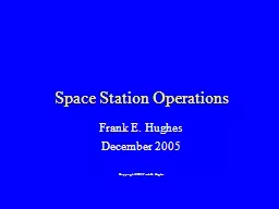
1. Urogenital Lab 1, Station 2Pelvis Models: Rudiger Anatomia and 3B Scientific
2. https://3d4medic.al/8K6xxapiFigure 1.2Figure 1.1: Rudiger Anatomie ModelRectal Hiatus (Anal Aperture)Urogenital HiatusUrogenital, Lab 1: Station 2Levator AniObturatorInternus m.Iliococcygeus m.Pubococcygeus m.Puborectalis m.CoccygeusPiriformisBladderUterusRectumVaginaTendinous archGreater Sciatic Foramen
3. Obturator internus m.Piriformis m.Coccygeus m.Levator aniTendinous archFigure 1.3:Figure 2.1Levator aniObturator internus m.Figure 1.43B Scientific ModelFigure 1.33B Scientific ModelUrogenital, Lab 1: Station 2
4. rectal hiatus(anal aperture)Figure 2.1Figure 2.1Urogenital triangleAnal triangleFigure 2.2Figure 2.3Superficial transverse perineal m.Urogenital, Lab 1: Station 2Sacrotuberous ligamentCoccyxIschiopubic ramus
5. Figure 2.4Figure 2.5Figure 2.1Ischioanal fossaAnal canal / AnusExternal anal sphincterInferior rectal n.Pudendal n.Urogenital, Lab 1: Station 2
6. Perineal MembraneFigure 3.2Figure 3.1Bulbospongiosus m.Perineal bodySuperficial transverse perineal m.Greater vestibular (Bartholin) glandBulb of VestibuleUrogenital, Lab 1: Station 2Figure 3.3Glans clitorisPerineal Membrane(green tape)Ischiocavernosus m.
7. Pelvic DiaphragmSuperficial PouchDeep Pouch (Fibromuscular Region)“Urogenital Diaphragm”Deep Pouch (A Recess Ischioanal Fossa)Perineal MembraneColles FasciaFigure 3.4Figure 3.6Figure 3.5Perineal Membrane(green string)Muscle in deep pouch(“fibromuscular” region or formerly named urogenital diaphragm)Urogenital, Lab 1: Station 2UrethraDeep perineal space (pouch)Anterior recess of ischioanal fossaGlans clitorisBody clitorisPerineal body
8. Sacral ventral rami (S1-S4)Lumbosacral trunkLumbosacral plexusFigure 4.1Pudendal n.Muscle in deep pouch(deep to removed perineal membrane)Anterior recess of ischioanal fossaPerineal n.Dorsal n. clitorisPudendal n.Figure 4.2Urogenital, Lab 1: Station 2