
Dr Sushma Tomar Associate Professor Department of Anatomy Lesson Plan Introduction Classification Relationship to the orbit Frontal Air Sinuses Introduction Drainage amp Nerve Supply ID: 912977
Download Presentation The PPT/PDF document "PARANASAL AIR SINUSES Presented by:-" is the property of its rightful owner. Permission is granted to download and print the materials on this web site for personal, non-commercial use only, and to display it on your personal computer provided you do not modify the materials and that you retain all copyright notices contained in the materials. By downloading content from our website, you accept the terms of this agreement.


Slide1
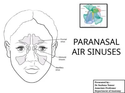
PARANASAL AIR SINUSES
Presented by:-Dr. Sushma TomarAssociate ProfessorDepartment of Anatomy
Slide2Lesson Plan
Introduction.Classification.Relationship to the orbit.
Frontal Air Sinuses:Introduction.Drainage & Nerve Supply.
Measurements.
Relations.
Applied Aspects.
Maxillary Air Sinuses:
Introduction.
Presenting Parts.
Drainage.
Arterial Supply.
Lymphatic drainage.
Nerve Supply.
Applied Aspects.
Ethmoidal Air Sinuses:
Introduction.
Drainage.
Applied Aspects.
Sphenoidal Air Sinuses:
Introduction.
Drainage.
Relations.
Applied Aspects.
Slide3Introduction
Air-containing cavities in the bones around nasal cavity.Paranasal air sinuses develop as mucosal diverticulae of nasal cavity, invading the adjacent bones.Paranasal air sinuses perform the following
functions:
Make the skull lighter.
Add resonance to the voice.
Act as air conditioning chambers by making the inspired air moist and warm.
Aid in growth of facial skeleton.
Paranasal air sinuses are lined by respiratory epithelium and respiratory mucosa is
highly vascular
and contains a large number of
cavernous spaces
and
sinusoids
to warm the air.
Respiratory mucosa also contains a large number of serous glands and secretion of these glands makes the air moist.
Slide4Classification
Paranasal air sinuses are named after the bones containing them, viz,Frontal air sinuses.
Ethmoidal air sinuses.
Maxillary
air sinuses.
Sphenoidal
air sinuses.
All paranasal air sinuses are present in rudimentary form at birth except frontal air sinuses
.
Slide5Relationship to the Orbit
Frontal air sinus- above.Ethmoidal air sinuses- medial.
Maxillary air sinus- below.
Sphenoidal air sinus-
behind.
Slide6Frontal Air Sinuses
Slide7Introduction
Frontal air sinuses are not present at birth.Start developing 2 or 3 years after birth.Number- 2.
Shape- Triangular.
Lie between inner and outer tables of frontal bone.
Right frontal air sinus is separated from the left by a septum.
Slide8Drainage & Nerve Supply
Drainage- Drains into anterior part of hiatus semilunaris of middle meatus through frontonasal duct.Nerve Supply-
Supraorbital nerve.
Hiatus
Semilunaris
Slide9Measurements
Height- ~ 3 cmWidth- 2.5 cmAnteroposterior- 1.8 cm
Slide10Relations of Frontal Air Sinus
Anterior-Superciliary arch of forehead.Posterior-Meninges and frontal lobe of brain.
Inferior-Roof of nose.
Roof of orbit (medial part).
Ethmoidal air cells.
Frontal air sinus
Frontal lobe
Slide11Applied Aspects
Frontal Headache (Office Headache)Headache from frontal sinusitis shows characteristic periodicity.It starts on waking, gradually increases and reaches its peak by about midday and then starts subsiding.
Slide12Applied Aspects
contd…Frontal Lobe Abscess-Infection of frontal air sinus may spread posteriorly into frontal lobe of brain causing
frontal lobe abscess.
Frontal air sinus
Slide13Applied Aspects
contd…Orbital Cellulitis-Infection of frontal air sinus may spread inferiorly into orbit causing orbital cellulitis.
Slide14Maxillary Sinus
(Antrum of Highmore)
Slide15Introduction
Largest paranasal air sinus.Present in body of maxilla.First to develop.Appears around 4th
month of intrauterine life.
Slide16Maxillary Sinus
contd…SHAPE- Pyramidal.Base-Directed medially.Formed by a part of lateral wall of nose.
Opening or
ostium of the sinus
is present in the upper part of base, close to the roof.
Apex-
Directed laterally.
Extends into zygomatic process of maxilla.Roof-Formed by the floor of orbital cavity.
Infraorbital nerve and artery traverse the roof
in a bony canal.
Floor-
Formed by the alveolar process of maxilla.
Lies
~1.25 cm
below the floor of nasal cavity.
Zygomatic bone
Ostium of Maxillary air sinus
Slide17Slide18Floor of Maxillary Sinus
contd…The level of floor corresponds to the ala of nose.Normally the roots of first and second molar teeth project into the floor.Sometimes roots of third molar, first and second premolars may project into the floor.
Rarely, root of canine may project into the floor.
Sometimes roots of teeth are separated from the sinus only by a thin layer of mucosa.
Slide19Base of Maxillary Sinus
It is formed by medial surface of body of maxilla and some other bones.In maxilla, medial surface of its body presents a large maxillary hiatus.In the skull, base of maxillary sinus presents a small opening ( ostium).
Maxillary Hiatus
Slide20Reduction of large maxillary hiatus to small ostium
It occurs by the following bones:Uncinate process of ethmoid.Descending process of
lacrimal.Ethmoidal process of
inferior nasal concha.
Perpendicular plate of
palatine.
Slide21Maxillary Sinus
contd…Anterior wall-Has a curved bony canal for anterior superior alveolar nerve – Canalis
Sinuosus
.
Posterior wall-
Separates the sinus from infratemporal and pterygopalatine fossae.
It is pierced by the
posterior superior alveolar nerves and vessels.
Slide22Drainage
In posterior part of hiatus semilunaris of middle meatus.
Hiatus Semilunaris
Opening of
Maxillary Sinus
Slide23Arterial Supply
Anterior superior alveolar artery.Middle superior alveolar artery.Posterior superior alveolar artery.
Slide24Lymphatic Drainage
Submandibular lymph nodes.
Slide25Nerve Supply
Anterior superior alveolar nerve.Middle superior alveolar nerve.Posterior superior alveolar nerve.
Slide26Applied Aspects
Maxillary Sinusitis-Maxillary sinus is the most commonly infected paranasal air sinus.The opening of maxillary sinus is in a disadvantageous position for natural drainage.Sources of infection:Infected nose.
Carious upper premolar and molar teeth.Infected frontal and anterior ethmoidal air sinuses.
Slide27Surgical Drainage of Maxillary Sinus
Antral puncture (Antrostomy)-Trocar and canula are passed below the inferior nasal concha in an outward and backward direction.Caldwell-Luc operation-Maxillary sinus is opened through gingiva-labial sulcus.
Slide28Applied Aspects
contd…Carcinoma of Maxillary Sinus-Arises from mucosa of the sinus.Clinical Features-
Due to upward invasion:Proptosis (protrusion of eyeball).Diplopia (double vision).
Pain and
anaesthesia
over the face below the orbit.
Due to downward invasion:
Swelling or even ulceration of palatal roof of oral cavity.Due to medial invasion:Nasal obstruction.Epistaxis.
Epiphora (overflow of tears).Due to lateral invasion:Swelling on the face and palpable mass in gingiva-labial sulcus.
Due to posterior invasion:
Referred pain to upper teeth.
Slide29Ethmoidal Sinuses
Slide30Introduction
Present within labyrinth of ethmoid bone.Between upper part of lateral nasal wall and orbit.3
groups:Anterior
(up to 11 air cells).
Middle
(1-3 air cells).
Posterior
(1-7 air cells).
Slide31Drainage
Anterior group drains into middle part of hiatus seminularis of middle meatus.Middle group drains on the surface of
bulla ethmoidalis
of middle meatus.
Posterior group
drains into posterior part of
superior meatus.
Slide32Applied Aspects
Ethmoidal Sinusitis-Often asoociated with infection of other sinuses.Clinical Features-
Localized pain over bridge of nose.
Due to invasion into the orbit-
Orbital cellulitis.
Slide33Sphenoidal Sinuses
Slide34Introduction
Number- 2 (right and left)Lie within the body of sphenoid bone.Separated from each other by a bony septum.
Bony Septum
Slide35Drainage
Into sphenoethmoidal recess.
Slide36Relations
Slide37Applied Aspects
Sphenoidal Sinusitis-One of The Most Dangerous Sinus Infection.It is rare in isolation.
It is usually a part of pansinusitis.
It may be associated with infection of posterior ethmoidal sinuses.
Slide38