PDF-RETINA TODAY
Author : liane-varnes | Published Date : 2017-01-25
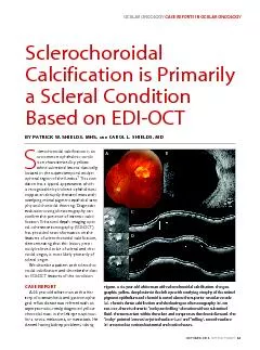
OCTOBER 2014 61 S clerochoroidal calcification is an uncommon ophthalmic condi tion characterized by yellow white subretinal lesions classically located in the
Presentation Embed Code
Download Presentation
Download Presentation The PPT/PDF document "RETINA TODAY" is the property of its rightful owner. Permission is granted to download and print the materials on this website for personal, non-commercial use only, and to display it on your personal computer provided you do not modify the materials and that you retain all copyright notices contained in the materials. By downloading content from our website, you accept the terms of this agreement.
RETINA TODAY: Transcript
Download Rules Of Document
"RETINA TODAY"The content belongs to its owner. You may download and print it for personal use, without modification, and keep all copyright notices. By downloading, you agree to these terms.
Related Documents