
285 286 288 289 291 292 294 295 296 297 298 299 302 303 Another design for 4MDCT detector arrays is illustrated in Fig 12 When small slices are desired only the central portion of thallow simultaneo ID: 891398
Download Pdf The PPT/PDF document "detector dimensions are normalized relat..." is the property of its rightful owner. Permission is granted to download and print the materials on this web site for personal, non-commercial use only, and to display it on your personal computer provided you do not modify the materials and that you retain all copyright notices contained in the materials. By downloading content from our website, you accept the terms of this agreement.


1 285 286 288 289 291 292 294 295 296 297
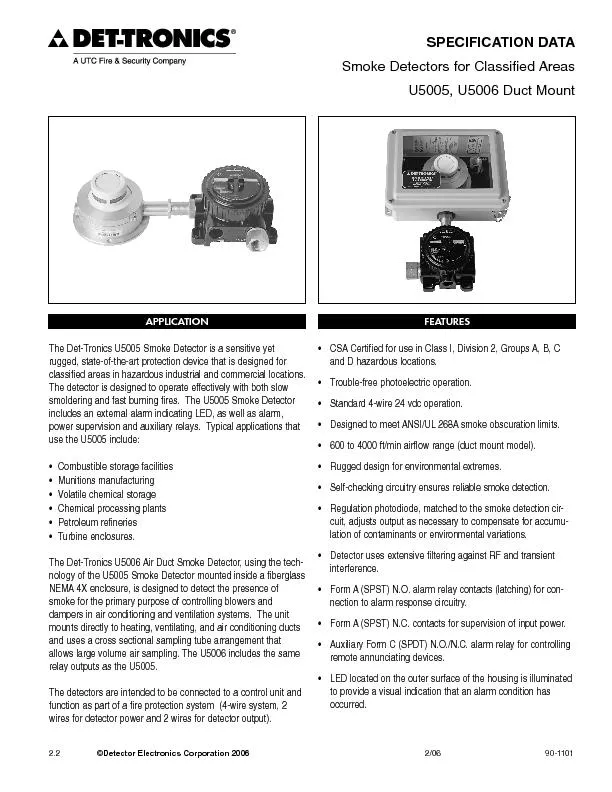
285 286 288 289 291 292 294 295 296 297 298 299 302 303 (detector dimensions are normalized relative togantry). The detector arrays are made from multiple rows, each approximately 1-mm wide (e.g. sixteen 1.25-mm wide (..) Another design for 4-MDCT detector arrays is illustrated in Fig. 1.2. When small slices are desired, only the central portion of thallow simultaneous acquisition of four slices each of 5 mm thickness. This design is somewhat less expensive and more geometrically efficient. Fig. 1.2 Diagram of the detector geometry used in a 4-MDCT from two major manufacturers. The detector aitudinal axis and uses eight rows of varyi
2 ng widths to allow simulta (..) Curren Curren")
ng widths to allow simulta (..) Currently, MDCT systems are capable of acquiring up to 64 slices simultaneously ree of the four manufacturers use 64 rows of either 0.625 mm or 0.5 mm detectors. The fourth manufacturreduction of spiral artifacts and improved spat 10 308 309 310 311 314 Fig. 1.3 Diagram of the detector geometries used in 64-MDCT from four major manufacturers. The Siemens 64-MDCT uses 32 sub-mm detectoverlapping slice measurements. (..) For sequential data acquisitions (e.g. the table is stationary during the rotation of the x - sufficient data to create one “slice” or image, so as many as 64 independent images a
3 long the z axis could theoretically be r
long the z axis could theoretically be reconstructed. For narrow slice widths, geometrical “cone-beam” considerations may limit the number of allowed images per rotation to less than 64. For example, one manufacturer’s 16-detector scanner allows scanning because of cone beam considerations (..) The primary attribute of MDCT systems is not the number of physical detectors rows, but the number of slices that are acquired simultaneously. The speed needed to cover a given volume is improved by a factor equivalent to number of slices included in the scan 11 1069 1070 1075 1076 1084 1085 1086 examination and the default settings of
4 image Automatic exposure control (AEC
image Automatic exposure control (AEC) systems do not protocols to be prescribed using measures related to image quality. If the image quality is the clinical task, then there is reduction in patients, the dose is increased to improve the image quality. AEC does not imply total freedom from operator selection of scan parameters. While CT systems , AEC systems require understanding of newer concepts such as noise index, reference mAs and reference images in order for AEC to be ing of some parameters e.g. the standard deviation of image 3.3.3. Image quality selection paradigms (..) The use of an image quality selection paradigm allows the
5 CT system to calculate the location, in
CT system to calculate the location, in order to deliver the desired image quality at the lower dose. This broad concept, implemented practically with some variation between manufacturers, is known as Automatic Exposure Cis relatively straightforward for the system to deliver the desired image quality (once defined). However, it can be quite difficult to determine the image quality requirement for various CT applications and patient sizes. (..) In defining the required image quality, the user needs to remember that “pretty” pictures er a level of quality will need to be chosen – diagnostic task. The CT system will then adjust the m
6 A either during the rotation (x,y), alon, alon")
A either during the rotation (x,y), along the z-direction, or in all three dimensions (x, y and z) according to the patient’s body habitus and the user’s image quality requirements. Thus one musttask of modulating the 41 1100 1101 1106 1107 mA to achieve a defined image quality (tube current modulation) and ththe user of the desired image quality. The selection of image quality parameters in AEC systems is not a straightforward process. There is a lack of consensus on how image quality is to be specified; with the result that there are significant differences in the way different companies achieve exposure control. It is important
7 that users are aware of the behaviour o
that users are aware of the behaviour of their system. (..) Each manufacturer of CT systems uses a different method of defining the image quality in the user interface. GE uses a concept known as the specific size water phantom. A “look-up-table” maps the patient attenuation measured from the CT localizer radiograph (Scout) image into mA values for each tube rotation according to a proprietary algorithm. This algorithm is designed to maintain the same image noise as the patient’s attenuation changes from one rotation to the next. A different noise index may be required for differe (..) Philips uses a concept to help users sel
8 ect the desired image quality that shoul
ect the desired image quality that should be matched. They refer to this as Automaacceptable patient exam, including the CT localizer radiograph (SurView), and the system saves the raw data. This information is saved as the Reference Case, on a protocol by protocol basis, to be matched on later exams using their proprietary algorithm. (..) Siemens uses a Quality Reference mAs for users to establish the desired image quality level. The user selects, on a prffective mAs (mAs/pitch) typically used for an approximately 80 kg patiepirical algorithm; hence image noise is not kept constant for all patient sizes but is adjusted according to the radiolog
9 ist’s impression of image quality.
ist’s impression of image quality. The CT localizer radiograph (Topogram) for individual patientsired image quality for the specific 42 1131 1132 1137 1138 1140 1141 1142 1158 1159 1160 patient size and anatomy. An on-line feedback system adjusts the actual mA value during the scan acquisition to match the precise patient attenuation at all angles, as opposed to the attenuation estimated by the one angle. (..) Toshiba offers the user two methods of selecting the desired image quality in their AEC product: Standard Deviation or Image Quality Level. Both are referenced to the xel values in an attenuation-equivalent water phantom, which i
10 s created from the CT localizer radiogra
s created from the CT localizer radiograph (Scanogram) data. The reference value, index, or image can be ll manufacturers’ implementations. Assumptions regarding optimal noise levels (..) Image quality is a non-specific measure of the subjective sense of “quality” of an image, which must be assessed by a trained observer. Objective measures such as image noise or contrast-to-noise ratio can be made relatively easily, but may not completely capture all of the features relevant to making a gnosis. Thus, determining “optimal” image quality can be a complex task, as both quantitative metrics (e.g., noise) and observer percepti
11 ons require a specific noise level for
ons require a specific noise level for a specific (..) Table 3.3 provides measured noise for a constant mAs (chosen to be 130) as the diameter of a water phantom was varied. Table 3.4 demonsimage noise (chosen to be 13.0 HU) as the diameter of a water phantom was varied. Tables 3.3 and 3.4 together demonstrate that it is not technically feasible to maintain a constant image noise over all patient sizes, even if this was clinically desired, because CT systems cannot reach these extremely low and high mAs values. The large range of mAs values required to maintain constant image noise as object size is varied is a co e diameter of a water phanto
12 m is varied (adapted from McCollugh 2002
m is varied (adapted from McCollugh 2002 and Boone 2003) Diameter (cm) 10 14 20 25 30 40 Noise (HU) 1.9 3.5 5.1 8.2 13.0 33.6 43 1161 1162 1163 1164 Table 3.4. Tube current time product (mAs) required for a constant image noise (13.0 HU) as the diameter of a water phantom is varied (from McCollugh 2006) Diameter (cm) 10 14 20 25 30 40 Tube current-time product (mAs) 2.9 6.2 19 50 130 869 1165 1166 1167 1168 1183 1184 1185 (..) With empirically determined technique charts (i.e., appropriate mA values are determined for each patient size by a trained observer), both the extreme low and high mAs requirements a
13 re noticeably absent (see Tables 3.1 onl
re noticeably absent (see Tables 3.1 only pragmatic with both a patient dose and image the criterion of having a fixed More aggressive dose reduction is not acceptable in children, and more aggressive dose increase is unnecessary. (Wilting et al., 2001). When Wilting et al. presented images with constant noise tient sizes, ranging from pediatric to obese patients, the pediatric images were found to be unacceptable, even though they contained the same level of image noise as normal and obese patients (Wilting et al., 2001). Kalra et al. observed a similar situation using the General Electric noise index paradigm, which for a given noise index attem
14 pts to deliver a constant noise across a
pts to deliver a constant noise across anatomic regions and patient sizes (Kalra et al., 2003). They found that readers required a lower noise index (less image noise) for smaller patients and a higher noise index (more image noise) for larger patients. Although lower image noise was found to be required for small patients, a dramatic level of mAs reduction is still appropriate to compensate 3.3.4. Temporal mA modulation (..) Temporal mA modulation alters the tube current according to a time-based is most-commonly used in CT examinations of the heart, reducing the dose for projections of limited interest, such as in early systole where the rap
15 id cardiac motion compromises image base
id cardiac motion compromises image based mA modulation scheme can reduce dose by up to 50% for a cardiac CT study for systems with more for dual-source systems (Flohr 44 1192 1193 1198 1199 1204 1205 1206 (..) Usually, the tube current required for acceptable image quality is used for a time window that is somewhat wider than the desired temporal resolution (e.g., 330 to 350 ms time window for a 250 ms temporal resolution) in order to allow for some flexibility in the case of irregular heart desired for image reconstruction. Outside this time window, the tube current is not completely switched off, but is reduced to a much lower that data is
16 available to perform dynamic studies ov
available to perform dynamic studies over the entire heart cycle, al outside of the time window selected for primary image reconstruction. However, in patients with higher heart rates (more than 60-65 beats per minute and irregular heart rates (premature ventricular contractions), where systolic or multiple reconstructions may be needed for primary interpretation, ECG based mA modulation will yield much noisier images indata window is fixed, the dose reduction achieved by this feature depends on the heart rate. 3.4. Tube potential (kVp) (..) Tube potential (kVp) determines the energybeam. Variation in the tube potential causes a substantial chang
17 e in CT dose as well as image noise and
e in CT dose as well as image noise and contrast. In children and small adults, reducing the kVp leadp values (Funama et al., 2005;2005; Nakayama et al., 2005; Siegel et al., 2004). Most MDCT examinations are performed at wer values. Recent reports suggest substantial kVp) for CT angiography. In the abdomen, compared to 120 kVp, use of 100 kVp resulteangiography of the abdominal aorta and iliac arteries (Winterspergerabdominal MDCT in newborn and infants (Siegel in a substantial increase in the image noise, it can impair image quality if the patient is too large or if the tube current is not appropriately increased to compensate for the lower tube
18 voltage. Thus, when implementing reduced
voltage. Thus, when implementing reduced kVp protocols, itare determined as a function of patient size. For very large patients, relais almost always needed to obtai 45 3.5. Pitch, beam collimation and slice width 1221 1222 1223 1236 1237 1238 1243 1244 1245 (..) These three factors are related to the deresults in more dose efficient examinations, as overbeaming constitutes a smaller proportion of the detected x-ray beam. However, a wider beam width can limit the thinnest reconstructed sections for MDCT systems with systems, narrow beam widths decrease dose efficiency due to overbeaming, but are needed to allow reconstruction of thinner slice w
19 idths. Hence, beam width must be careful
idths. Hence, beam width must be carefully selected to address the specific clinical requirements. (..) In single-detector CT, increasing pitch nd image width increase at higheincrease in pitch is associated with an increase in image noise. Hence, tube current must be adjusted upward to maintain adequate image noise. Thus there is no fundamental dose saving achieved in MDCT at increased pitch values unless lower tube currents are simultaneous employed. Most scanners allow the users to override the automatic adjustment of mA or mAs. 3.6. Scan mode (..) Overranging of the x-ray beam with spiral MDCT leads to some amount of unused of interest.
20 Due to this phenomenon the use of a sin
Due to this phenomenon the use of a single spiral acquisition (as opposed to multiple contiguous spiral clinical considerations. However, this may be unavoidable in multi-region studies such as simultaneous neck and chest CT (position of arm) or simultaneous chest and abdomen CT (different delay times for optimal contrast enhancement). 3.7. Scan coverage and indication (..) With the short scan acquisition times of MDCT, there is a tendency to increase the scan length to include multiple body regions either in part or completely (Kalra et al., 2004; Campbell ssential to inform the patient’s exams of inappropriate anatomy or for non-medically
21 -necessar 46 3.8. System Software: Im
-necessar 46 3.8. System Software: Image reconstruction 1249 1250 1251 1252 1267 1268 (..) Image-space (i.e., the reconstructed image) and sinogram-space (i.e., the raw projection data) smoothing filters can be used to reduce image noise and consequently allow the user to ned noise level. Such methods, however, reduce spatial resolution. Special “adaptive” noise reduction filters allow for reduced settings while preserving spatial resolution (Raupach et al., 2005). Such filters analyze the image or projection , and smooth regions where there is little edge information, while leaving intact the regions wsavings of 30% have been demonstr
22 ated with thal., 2005; Raupach et al., 2
ated with thal., 2005; Raupach et al., 2005). Similarly, ongoing work in the area of image reconstruction algorithms, presents substantiaalgorithms with noise properties superior to those in images reconstructed by the conventional fan-beam filtered back-projection algorithm have been reported, and 3D cone beam algorithms, ithms, and time-averaged Fourier methods for CT perfusion are all (..) A substantial decrease in detected mmon in high attenuation regions, such as shoulders, due to beam attenuaincreased image noise with impaired image quality. Projection space filters, available on most scanners, increase the filtration e reconstruction data
23 and thus minimize the loss of resolution
and thus minimize the loss of resolution. Although there is some loss of image resolution (less than 5%) with the use of these filters, these reconstruction filters avoid an otherwise diagnostically compromised image. These filters can allow a 30-60% reduction in image noise without an (..) Image post-processing filters have been designed to decrease image noise in scans acquired with reduced radiation dose. Unlike image reconstruction algorithms, these techniques challer et al., 2000). Different approaches have 47 1279 1280 1285 1286 1303 1304 1305 1308 been adopted for noise reduction in scan volume datasets, which include linear low pass
24 filter, three-dimensional filters. (
filter, three-dimensional filters. (..) Image post-processing filter the principle that in any image, a group of structural pixels representatiof non-structural regions in the image are both present. The filter technique involves isotropic filtering of non-structured regions with a low pass filter and directional filtering of the structured regions with a smoothing filter, operating parallel to the edges and with an enhancing filtthe edges. Two dimensional, non-linear filters decrease image noise in low-dose CT images but adversely affect the image contrast, sharpness and lesion conspicuity (Kalra et al., 2004). In addition, a three-dimensional
25 filtration method, which generalizes the
filtration method, which generalizes the two-dimensional non-linear smoothing tloss of contrast and sharpness of small structures, e filters may improve image noise without affecting image contrast and lesion conspicuity in low-dose CT (Rizzo et al., 2006). (..) Streak artifacts from high-attenuation metallic implants are a common problem in CT scanning and can occur from metallic implants such as joint replacement prosthesis, dental implants, or surgical clips. To reduce loss of information from streak artifacts caused by dental implants, particularly in facial CT, a second series of images may be acquired with gantry nal radiation exposure to the
26 patients. In order to reduce streak ect
patients. In order to reduce streak ects, linear interpolation of reprojected metal traces and multi-dimensional adaptive filtering of the raw dataWatzke and Kalender, 2004). These algorithms reduce streak artifacts form metallic implants and may help in reducing radiati 3.9. Modification of scan acquisition and reconstruction parameters (..) Where possible, CT should be obtained with the lowest achievable radiation dose to the patient. Multiphase examinations should be limited to the fewest phases necessary to make the of anatomy imaged. The image wicompensate for the increased noise levels. For children and small patients, the kVp should be as
27 48 1309 1310 1315 1316 tomated expos
48 1309 1310 1315 1316 tomated exposure control should be used almost universally. In the case where a CT system is not equipped with automated exposure control, ledgeable medical physicist, and consistently used for all patients. This is absolutely essential for pediatric CT, in particular. Finally, providers of CT imaging services should be required to compare their dose levels and image quality measures, by patient size and exam typestandards, in order to ensure that they are offering high quality examinati 49 4. DOSE MANAGEMENT IN CLINICAL SITUATIONS 1339 1340 1341 1342 1345 1346 1347 1348 1350
28 1351 1352 1353 1354 1355 1356 1357 1361
1351 1352 1353 1354 1355 1356 1357 1361 1362 (..) “One-size-fits-all” type protocols Justification is a shared responsibility between requesting clinicians and radiologists. It includes justification of CT study for a given indication, and classification of clinical indications into those requiring standard or high dose CT and those for which information can be obtained with low dose CT examination. There are indications that awareness on adapting exposure factors to manage patient study indication, patient age and body parameters. n be avoided and triaged to an and CT staff can help in optimization of scan 4.1. Justification
29 of examination (..) Justification is a Justification is a")
of examination (..) Justification is a shared responsibility (ICRP 2000b). With the continuous increasing data on suitability and efficacy of MDCT, it is important to ensure that only a qualified medical practitioner generates requests for CT examinations. The radiologist should be appropriately trained and skilled in optimization in CT to alternative imaging or laboratory techniques. Thus, each CT exam must be performed when the radiation dose is deemed to be justified by the potthe availability of resources alevel, to advise requesting clinicians and radiologists about appropriateness and acceptability of CT examinations. In the absence of natio
30 nal level agreement on these issues, loc
nal level agreement on these issues, local institutional e guidelines must help radiologists and clinicians to triage patients to ultrasound or magnetic resonance imaging (MRI) and management. Such guidelines can also help in eliminating unnecessary CT examinations and must include a list of clinical indications for CT pertaining to diagnosis, treatment (surgical guidance and biopsy, drainage or other interventional radiology 50 1371 1372 1377 1378 American College of Radiology Appropriateness Criteria provide evidence based medicine based ng an appropriate imaging test (ACR 2000).The European Commission and United Kingdom’s Royal College
31 of Radiologists (RCR) document titled & document titled &")
of Radiologists (RCR) document titled “Referral guidelines for imagoverview of clinical indications for imaging examinations including CT and other radiation and non-radiation based imaging (..) Justification of a CT examination may intion of clinical indications into thosand those for which information can be obtained with low dose CT examination. In this respect, the introduction of informed consent for patientradiation risks may help in creating greater awareness amongst pati do not take informed consent for radiation risks from the patients undergoing CT scanning. Introduction of informed ng, may help to increase awareness about CT rad
32 iation dose and perhaps decrease some &#
iation dose and perhaps decrease some “unnecessary” CT from being performed. Such informed consent may include discussion of potenful effects such as cancer. (..) According to the charter on Advisory Commission on Consumer Protection appointed by the former United States President emust “discuss all risks, benefits, and consequences to treatment or non-treatment” with the consumer or patient. In this coradiation based exam as well as the risk of radiation induced carcinogenesis from associated ) felt that information a(defined as critical organ damage, which are permanent, life threatening, require surgery or negatively affec
33 t quality of life) must be provided to t must be provided to t")
t quality of life) must be provided to them even if the risk is 0.1%. Interestingly, 51 1402 1403 1408 1409 1410 1424 1425 1426 so estimated a 0.18% risk of lifetime cancer mortality tion from CT scanning of abdomen. 15% (14/91) of the radiology departments currently inform patients about possible radiation risks and only 9% (8/88) of radiology departments inform 4.2. Training issues (..) Recent surveys suggest that there is a substantial lack of comprehension of CT radiation dose amongst requesting physicians (Lee et al., 2004; Thomas et al., 2006). Furthermore, there are considerable variations in McLean, 2006). Requesting physicians must
34 be informed about appropriate indicaimag
be informed about appropriate indicaimaging techniques for triage ng, so that they can justify benefits of CT examinations over potential harmful effects. The radiologists and CT technologists must be indications such as CT for liver metastases or low dose CT indications for screening CT studies, parameters. With the constant upgrade of MDCT technology it is important to become acquainted rameters from one scanner to another system. Interestingly, a Japanese survey recently reported that more CT centres are adapting parameters according to patient age and are more frequently using automatic exposure control techniques in order to manage radiation do
35 se (Miyazaki et al., 2005). 4.3. CT dos.
4.3. CT dos")
se (Miyazaki et al., 2005). 4.3. CT dose and risk for individual situations (..) Most studies on low radiation dose CT hacurrent, either with fixed tube current or with automatic exposure control techniques (Kalra et al., ation profile with automatic exposure control techniques), or study indications (lower tube current for screening CT studies, kidney stone CT, and chest CT). 52 1430 1431 1436 1437 1443 1444 1445 been assessed with use of hiuse of special techniques such as two- and three-dimensional non-linear noise reduction filters. Although this section provides some tabulated protocols for dose reduction with examples mostly from st
36 udies assessing 4 to 16 slice MDCT scann
udies assessing 4 to 16 slice MDCT scanners, the same principles of dose reductions apply to other MDCT scanners including 32, 40 and 64-slice MDCT scanners. The purpose of these which are likely to be variable for different for their scanners. At the time of writing of this document, there was less data on similar dose scanners. Further, the amination in demonstrating dose management does not imply that these are common clinical applicamanagement studies in these applications. 4.3.1. Chest CT (..) As described in preceding sections, image noise, a principle component of image quality, beam attenuation results in lower image noise for chest w
37 hen compared to abdomen or pelvis, compa
hen compared to abdomen or pelvis, compared to abdomen or pelvis CT, a lower obtain a similar image quality foemployed low tube current to reduce radiation dose with chest CT (Wormanns et al., 2005) n acceptable image quality for evaluating normal anatomic structures with 50% 140 mAs compared to 220-280 mAs tive of patient size. Studies have also employed different ation dose for chest CT based on patie2000; Picozzi et al., 2005), pulmonary nodules (Diederich et al., 1999; Leader et al., 2005), al., 2004), emphysema (Zaporozhan et al., 2006), high resolution chest CT (Ikura et al., 2004), patients with neutropenia 53 1460 1461 1466 1467 1468 14
38 69 1470 1471 1474 (Wendel et al., 2005)")
69 1470 1471 1474 (Wendel et al., 2005) and cystic fibrosis (Jimenezal., 2001). Recently, lower kVp (at 80 kVp compared to commonly used kVp of 120) has been described for CT angiography for pulmonary embolism to reduce radiaimage contrast (Sigal-Cinqualbre et al., 2004) (Table 4.1B). Use of automatic exposure control techniques for chest CT, combined modulation and angular modulation, has been reported to reduce radiation dose by 20 and 14% compared to Table 4.1A. Tube current adjustment is the most frequently documented method to optimize radiation dose. Low dose chest CT with reduced tube current is generally sufficient for evaluation of pulmo
39 nary abnormalities. This table summarize
nary abnormalities. This table summarizes the use of low tube current CT (20 mAs versus 100 mAs for 80% dose reduction) for evaluation of pulmonary nodules (Wormanns et al., 2005). Due to high air-soft tissue radiation dose. The data in all columns in the Table is from the CT units from a particular manufacturer. Scanning parameters Low tube current chest CT Standard dose chest CT Scanner 4-detector row MDCT 4-detector row MDCT mAs 20 mAs (effective) 100 mAs (effective) kVp 120 120 Rotation time 0.5 second 0.5 second Pitch 1.75 1.75 Detector configuration 4 x 1 mm 4 x 1 mm Scan coverage/area scanned Chest Chest Slice thick
40 ness 5 mm 5 mm CTDI vol 2.0 mGy 1
ness 5 mm 5 mm CTDI vol 2.0 mGy 10.1 mGy Effective dose 1.4 mSv 6.8 mSv 1475 1476 54 1477 1478 1479 1480 1481 1482 Table 4.1B. Compared to the use of low tube current to reduce radiation dose, kilovoltage adjustment has mmarizes the use of 80 kVp in patients undergoing contrast enhanced CT of the chest (Sigal-Cinqualbre et al., 2004). Compared to conventionally used kVp of 120-140, use of 80 kVp can allow 2- 4 folds dose reThe data in all columns in the Table is from the CT units from a particular manufacturer. Scanning parameters Low kVp chest CT Low kVp chest CT Standard dose chest CT Scanner 4-detector row MDCT 4-dete
41 ctor row MDCT 4-detector row MDCT mAs
ctor row MDCT 4-detector row MDCT mAs 135 effective mAs 180 effective mAs 90 effective mAs kVp 80 80 120 Rotation time 0.5 second 0.5 second 0.5 second Table speed 10 mm/rotation 10 mm/rotation 10 mm/rotation Pitch 1:1 1:1 1:1 Detector configuration 4 x2.5 mm 4 x 2.5 mm 4 x 2.5 mm Scan coverage/area scanned Chest Chest Chest Slice thickness - - - Effective dose (mSv) 1.54 (males), 1.88 (females) 2.05 (males), 2.51 (females) 2.05 (males), 2.51 (females) 1483 1484 1485 1486 1490 1491 4.3.2. CT for coronary calcium quantification (..) For coronary CT examinations, it is important to reconstruct images during
42 the phase of the cardiac cycle that will
the phase of the cardiac cycle that will be associated with least motion of the coronary arteries. Current multidetector CT technology allows ECG gating of scan acquisition and reconstruction of images at any desired phase of the cardiac cycle. This needs scan acquisition at small, overlapping pitch, which leads to a higher radiation dose despite smaller scan for some low dose coronary CT angiography and calcium scoring protocols are summarized in Table 4.2A, 4.2B & 4.2C. (..) CT for coronary calcium quantification can be performed with low-dose CT due to high inherent contrast between coronary calcium and adjoining soft tissue, which allow inte
43 rpretation even with high image noise. S
rpretation even with high image noise. Several strategies can be adopted for reducing dose with coronary cium scoring CT, which include 55 1497 1498 1503 1504 1506 1507 (Shemesh et al., 2005) (Table 4.2A) and tube potenbased adjustment of tube current for reducing CT angiography images of 30 patients. They noted that acceptable image quality and 17.9 (males)-26.3% (females) dose based adjustment of tube current. Table 4.2A. Radiation dose reduction for coronary calcium quantification can be accomplished with use of low fixed tube current or with ECG pulsing. In this study, there was excellent correlation between coronary calcium scores at 165
44 mAs and 55 mAs (r= 0.9, p.01) (Shemesh (Shemesh")
mAs and 55 mAs (r= 0.9, p.01) (Shemesh et al., 2005). The data in both columns in the Table is from the CT units from a particular manufacturer. Scanning parameters Coronary calcium quantification Low dose CT for coronary calcium Scanner 4-detector row MDCT 4-detector row MDCT mAs 165 mAs 55 effective mAs kVp 120 120 Rotation time 0.5 second 0.5 second Detector configuration 4 x 2.5 mm 4 x 2.5 mm Scan coverage/area scanned Heart (120 mm) Heart (120 mm) Slice thickness 2.5 mm 2.5 mm CTDI vol 12 mGy 4 mGy 1511 1512 56 1513 1514 1515 1516 1517 1518 1519 1520 1521 Table 4.2B. Radiation dose reduction for c
45 oronary CT angiography with use of lower
oronary CT angiography with use of lower kVp (80 kVp versus 120 kVp used in most centers) as well as ECG modulated tube current (ECG pulsing) in slim patients. Use of lower kVp may result in inadequate signal and disproportionate image noise if used in patients with greater size (Abada et al., 2006). Scanning parameters Low dose coronary CT angiography Scanner 64-detector row MDCT mAs 520 effective mAs (with ECG pulsing) kVp 80 Rotation time 0.33 second Detector configuration 64 x 0.6 mm Scan coverage/area scanned Heart Slice thickness 0.75 mm Effective dose (mSv) ~ 2 mSv 1522 1523 1524 1525 1526 1527 1528 1529 1530 1531 153
46 2 1533 1534 1535
2 1533 1534 1535 57 1536 1537 1538 1539 1540 1541 Table 4.2C. ECG modulated tube current helps to reduce radiation dose. ECG modulated tube current is more efficient at lower heart rates therefore, administration of beta blockers helps to reduce dose. This table summarizes dose savings (45% for females, 48% for males) with ECG modulated tube current compared to CT performed without modulation in size-matched patients (Jakobs et al., 2002). The data in both columns in the Table is from the CT units from a particular manufacturer. Scanning parameters Coronary calcium quantification Low dose CT for coronary calcium
47 Scanner 4-detector row MDCT 4-detector
Scanner 4-detector row MDCT 4-detector row MDCT Mean body mass index (kg/m 25.59 25.65 ECG modulated mA No Yes mAs 100 effective mAs 55 effective mAs kVp 120 120 Helical pitch 1.5:1 1.5:1 Table speed 7.5 mm/second 7.5 mm/second Rotation time 0.5 second 0.5 second Detector configuration 4 x 2.5 mm 4 x 2.5 mm Scan coverage/area Heart (120 mm) Heart (120 mm) Slice thickness 1.5 mm 1.5 mm CTDI vol 12 mGy 4 mGy Effective dose (mSv) 1.95 (male), 2.48 (female) 1.03 (male), 1.37 (female) 4.3.3. CT colonography 1542 1543 1544 (..) CT colonography is being cancer. In order to reduce the number of false positive lesions
48 and differentiate between a lesion fecal
and differentiate between a lesion fecal matter (st 58 1548 1549 1554 1555 1559 1560 offer a unique opportunity to reduce radiation dose for CT colonography. Effective doses for some low dos (..) Compared to routine abdominal CT st be performed at a much lower dose. In fact, several strategies have been adopted for reducing dose associated with able 4.3B) and kilovoltage (Capunay et al., 2005) (Table 4.3C). Recently, automatic exposure control technique has been reported to reduce Table 4.3A. High inherent contrast between air or contrast filled colon and colonic lesions or mucosa allow use of lower tube current as well as higher beam pitc
49 h values (compared to beam pitch of less
h values (compared to beam pitch of less than 1 Scanning parameters Low dose CT colonography Scanner 4-detector row MDCT mAs 10 effective mAs kVp 120 Rotation time 0.5 second Pitch 2:1 Detector configuration 4 x1 mm Scan coverage/area scanned Abdomen and pelvis Slice thickness 1.25 mm Number of acquisitions 2 (prone and supine) Total effective dose (mSv) 0.7 (males), 1 (females) 1563 1564 59 1565 1566 1567 1568 1569 1570 1571 ubstantial dose reduction for CT colonography despite two CT passes. This table illustrates use of a very low tube current (10 effective mAs) to reduce radiation dose for CT colonography (
50 Iannaccone et al., 2003). Thtotal combin. Thtotal combin")
Iannaccone et al., 2003). Thtotal combined dose for localizer radiographs and CT acquisition in both supine and prone positions. Scanning parameters Low dose CT colonography Scanner 4-detector row MDCT mAs 10 effective mAs kVp 140 Rotation time 0.5 second Pitch 0.875:1 Table speed 17.5 mm/second Detector configuration 4 x 2.5 mm Scan coverage/area scanned Abdomen and pelvis Slice thickness 3 mm Reconstruction kernel B 20 (smooth) Number of acquisitions 2 (prone and supine) Total effective dose (mSv) 2.15 (males), 2.75 (females) 1572 1573 1579 60 1580 1581 1582 Table 4.3C. For pediatric applications
51 of CT colonography, dose can be reduced
of CT colonography, dose can be reduced further with use of lower kVp as well as lower mAs (Capunay et al., 2005). Scanning parameters Low dose CT colonography Scanner 4-detector row MDCT mAs mAs kVp 90 Rotation time 0.5 second Pitch 1.5:1 Table speed 25 mm/second Scan coverage/area scanned Abdomen and pelvis Slice thickness 3.2 mm Number of acquisitions 2 (prone and supine) CTDI vol 0.3-0.7 mGy Total effective dose (mSv) 0.3- 0.6 mSv 4.3.4. CT for trauma 1583 1584 1585 (..) Trauma is a major cause of morbidity and mortality in young people throughout the world. It is also a major indication for CT smillion CT or MRI
52 examinations each year in the United St
examinations each year in the United States alone (Kalra et al., 2005; McCaig et al., 2004). Indeed, CT has become the imaging technique of choice for patients with trauma to head, neck, chest, abdomen and pelvis. However,reported protocols for trauma CT and raised concerns about overuse of CT in emergency settings (Hadley et al., 2006, Kortesniemi et al., 2006). Hadley et al have reported that use of American College of Radiology (ACR) appropriateness criteria on CT for trauma can help in reducing radiation dose by about 44% and imaging costs by 39%. The study also reported an estimated effective dose of 16 mSv to a typical trauma patient 61
53 1595 1596 1601 1602 1614 1615 1616 (..)")
1595 1596 1601 1602 1614 1615 1616 (..) The most important approach for reducing radiation dose associated with the use of CT in trauma is appropriate selection of patients for imnon-radiation based or low-radiation dose imagidose increases with number of acquisitions over the same area of interest. Therefore, efforts must be directed towards limiting the number of acquisitions and reducing radiation dose for the “less (..) Often, patients with trauma undergo scanning of contiguous chest, abdomen and pelvis or chest and abdomen, in the same imaging session. It is important to remember that due to cone beam shaped x-ray beam, there is a smal
54 l portion of the x-ray beam at the start
l portion of the x-ray beam at the start and end of each helical run which is not incident on the detectors. These unused x-rays result in radiation exposure to the patients and increase with increasing number of helices acquired during a CT examination. Furthermore, raoverlapping of helices between two anatomic areas of interest (at the level of diaphragm for chest and abdomen CT). Therefore unless there are oved during CT examinations must be limited. Indeed, Ptak et al , whole-body CT examination resulted in 17% dose reduction compared to the multi-helical, conventional segmented CT protocol for head, neck, chest, abdomen and pe 4.3.5. CT of the
55 urinary tract (..) CT has replaced conv CT has replaced conv")
urinary tract (..) CT has replaced conventional radiography and intravenous urography for evaluation of urinary tract calculi and urinary tract in many centres in the world, particularly in the United information pertaining to the urinary tract, it comes at the price of higher radiation dose to patients with benign disease, who (..) Several studies in patients and phantoms have documented that urinary tract calculi can be imaged with low dose CT, as “radio-opaque” or dense calculi offer high contrast against soft Kalra et al., 2005). Since all attempts must be made 62 1625 1626 1631 1632 and limit the number of CT examination pe
56 rformed for its evaluation. Radiation do
rformed for its evaluation. Radiation dose for stone wer tube current time product (Kluner et al., 2006) and automatic exposure cont Table 4.4A. Radiation dose can be reduced for CT for evaluation of suspected urinary tract calculi with low tube current. High contrast between most urinary calculi and soft tissues allow evaluation in relatively noisy images at low doses (Kluner et al., 2006). Scanning parameters Low dose CT for urinary calculi Scanner 16-row CT scanner mA 20 kVp 120 Rotation time 0.5 second Pitch 1.43:1 Detector configuration 16 x 1 mm Scan coverage/area scanned Abdomen and pelvis Slice thickness 5 mm Reco
57 nstruction kernel soft tissue kernel T
nstruction kernel soft tissue kernel Total effective dose (mSv) 0.5 (males), 0.7 (females) 1635 1636 63 1643 1644 1645 1646 1647 1648 Table 4.4B. This table summarizes use of z-axis (longitudinal) automatic tube current modulation (Auto mA) for dose reduction in patients with suspected urinary calculi (Kalra et al., 2005). Noise index is the noise in the center of an image of a water phantom. Higher noise index requires less tube current and therefore less radiation dose. The data in all columns in the Table is from the CT units from a particular Scanning parameters Regular dose CT Low dose CT Low dose CT Scanner 16-
58 row MDCT, 16-row MDCT 16-row MDCT N
row MDCT, 16-row MDCT 16-row MDCT Noise index - 14 20 Average mAs 182.4 Range, 160–240 104 range, 50–160 62.6 range, 37.5–186.9 kVp 140 140 140 Rotation time 0.5 second 0.5 second 0.5 second Pitch 0.938:1 0.938:1 0.938:1 Detector configuration 16 x 1.25 mm 16 x 1.25 mm 16 x 1.25 mm Scan coverage/area scanned Abdomen and pelvis Abdomen and pelvis Abdomen and pelvis Slice thickness 2.5 mm 2.5 mm 2.5 mm Reconstruction kernel Standard Standard Standard Effective dose (mSv) 25 15 8.8 64 4.3.6. CT guided interventions 1649 1650 1651 1659 1660 1661 1662 (..) CT guided interventions pose s
59 pecial issues to the radiology staff per
pecial issues to the radiology staff performing the procedure. Generally, two or more “passes” or scan acquisitions are obtained in the area of interest. With CT fluoroscopy, radiation exposure to the patient as well as the radiologist concern (Table 4.5A,B). There is by limiting the scan length, reducing mAs and fluoroscopic time, and use of alternative non-radiation based imaging guidance (such as ultr Table 4.5A. Efforts must be directed towards reducing radiation exposure from CT fluoroscopy to both patients and physicians. This table summarizes patient and physician doses from CT fluoroscopy (Buls et al., 2003). Physician doses are
60 average doses from CT fluoroscopy guide
average doses from CT fluoroscopy guided biopsy, aspiration and drainage, and radiofrequency. Scanning parameters CT fluoroscopy Scanner 4-detector row MDCT mA 90 kVp 120 Rotation time 0.75 second Scan coverage/area scanned Area of interest at the level of needle/catheter Slice thickness 8 mm CTDI vol 12 mGy Average effective dose (50% range) Patients: Biopsy Aspiration and drainage Radiofrequency ablation Overall Physicians: Overall median doses Eyes Thyroid Left hand Right hand 18.3 mSv (9.8-23.0) 15.8 mSv (12.6-26.9) 36.3 mSv (26.3-51.5) 19.7
61 mSv (10.8-27.1) 0.210 mSv (0.143-0.31
0.210 mSv (0.143-0.31")
mSv (10.8-27.1) 0.210 mSv (0.143-0.313) 0.240 mSv (0.155-0.406) 0.176 mSv (0.118-0.260) 0.759 mSv (0.445-1.41) 65 1663 1664 1665 1666 Table 4.5B. Radiation dose from CT guided biopsy can be reduced by reducing tube current or limiting the scan volume. This table summarizes application of low tube current for dose reduction in children undergoing CT guided biopsy (Heyer et al., 2005). Scanning parameters Low dose CT guided biopsy Clinical indications Chronic infectious interstitial pulmonary disease in Scanner 4-detector row MDCT mAs 20 effective mAs kVp 120 Detector configuration 5 x 2 mm Scan coverage/area scanned Regi
62 on of interest (10 mm) Maximum number o
Maximum number o")
on of interest (10 mm) Maximum number of images 4 Slice thickness 10 mm Effective dose 0.83 mSv (range, 0.38- 1.40 mSv) 4.3.7. CT in children 1668 1669 1670 1671 (..) Children are more susceptibced carcinogenesis compared to medical physicists, and technologists, must pay special attention ation dose when imaging children. small adults can be reduced wImage noise is proportional to the x-ray beam attenuation, which in turn is affected by the scanner. Scanning parameters Alternatively, automatic exposure co (..) In a recent review on radiation dose reduction in children, Vock recommends several strategies to accomplish this objective in
63 cluding rigorous justification of CT exa
cluding rigorous justification of CT examinations, 66 1681 1682 1687 1688 1689 1690 1691 1709 acceptance of images with greater noise if diagnostic information can be obtained, optimization minimum length as needed, and reduction of repeated scanning of evaluation of pediatric trauma suggests that more than one-half of the examinations were normal (Fenton et al., 2004). For follow-up CT studies, the scan volume can also be restricted deported substantial dose reduction (55%) by limiting the scan coverage to just 6 images per examination for follow-up CT of patients with cystic fibrosis (Jimmenz et al., 2006). 4.3.8. CT of the pregnant patients
64 (..) Common indications for CT scannin Common indications for CT scannin")
(..) Common indications for CT scanning inappendicitis, pulmonary embolism, and urinary tract calculi. To minimithe fetus, it is important to triage the patient appropriately if diagnostic information can be obtained from an alternative non-radiation based imaging. Radiologists and physicians must also decide if immediate scanning is reStrict x-ray beam collimation in modern CT scanners allows very little scattered radiation dose. For scanning body regions outside abdomen and pelvis, such as chest CT for suspected pulmonary embolism, shielding is not indicated as most scattered radiation comes from internal attering is minimal due to tight beam co
65 llimation. For abdominal-pelvic CT, scan
llimation. For abdominal-pelvic CT, scanning parameters must be selected tocollimation and pitch, and lower mA 4.6). For CT in a pregnant patient with suspected appendicitis, the scan volume must be restricted to the necessary anatomy, voided (Wagner and Huda, 2004; Ames Castro, 2001). A “step-and-scan protocol” may help in terminating the study when the (Wagner and Huda, 2004). renal calculi in a pregnant patient, fetal dose must be reduced with use of low mAs, high pitch and a limited scan volume, without substantially compromising th 67 1712 1713 1714 1715 1716 Table 4.6. Summary of typical scanning protocols and radiation d
66 oses for scanning used in a CT center fo
oses for scanning used in a CT center for imaging pregnant patients with suspected pulmonary embolism, appendicitis, and renal stones, which represent the commonest indications for CT in pregnancy (Hurwitz et al. 2006). The radiation dose values were estimated using anthropomorphic phantoms simulating a pregnant woman. Scanning parameters Pulmonary emobolism Appendicitis Renal stone Scanner 16-MDCT 16-MDCT 16-MDCT mA 380 340 160 Gantry rotation time 0.8 second 0.5 second 0.5 second kVp 140 140 140 Pitch 1.375:1 1.75:1 1.75:1 Detector configuration 16 x 1.25 mm 16 x 0.625 mm 16 x 0.625 mm Scan coverage/area scanned Ch
67 est Abdomen- pelvis Abdomen- pelvis S
est Abdomen- pelvis Abdomen- pelvis Slice thickness 2.5 mm 2.5 mm Fetal dose at 3 months 0.07 cGy 1.5 to 1.7 cGy 0.4 to 0.72 cGy Maternal effective dose (mean ± SD) 14.4 ± 2.1 mSv 13.3 ± 1.0 mSv 4.51 ± 0.45 mSv 4.4. Future directions 1717 1718 1719 1726 1727 (..) CT vendors have invested efforts towards the development of dose efficient technologies efforts at dose management should include the development of guiof patients, further optimization of automatic exposure control techniques and other dose management strategies, continued efforts of the international, national or regionalians and medical physicists to realms of rad
68 iation dose associated with MDCTradiatio
iation dose associated with MDCTradiation based imaging techniques which will be able to replace CT by providing equal information in a timely and appropriate manner. 68 APPENDIX A 1728 1729 1730 1731 1732 1733 HOW TO DESCRIBE DOSE IN CT A1. CT Dose Index (CTDI) (..) The CT Dose Index (CTDI) is the primary dose measurement concept in CT. It represents tiguous exposures. It is measured from one axial integrated absorbed dose by the total beam width. CTDI theoretically estimates the average dose within the central region of a scan volume, which is referred to as the Multiple Scan Average Dose (MSAD) (Shope et al., 1981), the direct measur
69 ement of which requires multiple exposur
ement of which requires multiple exposures. The CTDI offered a more convenient, yet nominally equivalent method of estimating this value, the early days of CT, saved a considerable amount of time. 1735 1746 1747 1751 1752 1753 (..) The equivalence of the MSAD and the CTDI requires that all contributions from the tails included in the CTDI dose measurement. The exact integration limits required to meet this the total beam width and the length of the scattering medium. To standardize CTDI measurements, the FDA introduced the integration limits of ± 7T, where T represented the nominal slice wiInterestingly, the original CT scanner, the EMI Mark
70 I, was a dual-detector-row system. Henc
I, was a dual-detector-row system. Hence, the nominal radiation beam width was equal to twice the nominal slice width (i.e., N x T mm). To account for this, the CTDI value, while integrated over the limits ± 7T, was normalized to 1/NT: CTDI = 1/NT • -7T D(z) dz (Eqn. 1) where D(z) represents the radiation dose profile along the z axis. However, the FDA definition integrate over a longer limit (±7NT). (..) The scattering media for CTDI measurements were also standardized by the FDA (United States FDA Code of Federal Regulations, 1984). These consist of two polymethylmethacrylate linders of 14-cm length. To estimate dose values
71 for head examinations, a diameter of 16
for head examinations, a diameter of 16 cm is used, and to estimate dose values for body examination, a 69 1759 1760 1762 1763 1764 1765 1766 1767 1768 1769 1779 1780 1786 1787 diameter of 32 cm is used. These are typically CTDI phantoms. (..) The CTDIdiation dose profile from a single axial scan over specific integration limits. In the case of CTDI, the integration limits are of the commercially available “pencil” ionization chamber (European Commission, 2000; Jucius and Kambic, 1977; Pavlicek et al., CTDI = 1/NT • -50mm +50mm D(z) dz (Eqn. 2) is acquired using a 100-mm long, 3 cm active volume CT “pencil” io
72 nization chamber and the two standard CT
nization chamber and the two standard CTDI acrylic phantoms. The measurement must be performed with a patient table. (..) The CTDI can vary across the field-of-view. For body imaging, the ace than at the centre of rotation. The average CTDI across the ) (European Commission, 2000; CTDI = 1/3 CTDI100,center100,edge. (Eqn. 3) (..) The values of 1/3 and 2/3 approximate the relative areas represented by the centre and specific kVp and mAs. One must use the f-factor (fkerma (mGy) or exposure (R) to absorbed dos must use CTDImGy/mGy) (European Commission, 2000; Interncal Commission, 2002). Volume CTDI (CTDIvol) (..) To represent dose for
73 a specific scan protocol, which almostsc
a specific scan protocol, which almostscans, it is essential to take into account any gaps or overlaps between the radiation dose profiles from consecutive rotations of the x-ray source. This is accomplished with use of a dose descriptor 70 1789 1790 1795 1796 1810 1811 1815 1816 1817 1818 known as the Volume CTDIechnical Commission, 2002), CTDI = (N•T/I) • CTDI (Eqn. 4) (..) In helical CT, the ratio of the table travel per rotation (I) to the total beam width (N•T) is CTDI/ pitch. (Eqn. 5) (..) So, whereas CTDIrepresents the average absorbed radiation dose over the x and y represents the average absorbed
74 radiatand z directions. It is conceptua
radiatand z directions. It is conceptually similar to the MSAD, but is standardized with respect to the integration limits (±50 mm) and the f-factor used to convert the exposure or air kerma measurement into dose to is the parameter that best represents the average dose at a point with the scan volume for a standardized phantom (International Electrotechnical Commission, 2002). The SI units are milli-Gray (mGy). It is a useful indicator of the dose for a specific exam protocol, because it takes into account protocol-specific information such as pitch. problem when measuring CTDI in MDCT is that occasionallybeyond the 100mm that the pencil chamb
75 er is designed. There are new chambers t
er is designed. There are new chambers that are designed to overcome this problem. (..) While CTDIestimates the average radiation dose within the irradiated volume of a CT acquisition for an object of similar attenuation to the CTDI phantom, it does not well represent the average dose for objects of substantially different size, shape, or attenuation. Additionally, it does not indicate the total energy deposited into the scan volume because is independent of the A2. Dose Length Product (DLP) (..) To better represent the overall energy DLP (mGy-cm) = CTDI(mGy) • scan length(cm) (European Commission, 2000) (Eqn. 6) 71 1819 1820 1821 18
76 24 1825 1826 1836 1837 1838 1839 1840 18
24 1825 1826 1836 1837 1838 1839 1840 1841 1842 1843 1844 1845 1846 1847 1848 1849 (..) The DLP reflects the total energy absorbed ological effect) from abdomenal CT might have the same CTDIabdominal and pelvic CT, the latter exam would have a greater anatomic coverage of the scan. A3. Organ dose and effective dose (..) The effective dose is a “dose” parameter thin terms of a whole body exposure.ed to normalize partia to enable comparisons of ly obtained from Monte Carlo modeling of absorbed organ doses within mathematical anthropomorphic phantoms, and recently also voxel phantoms based on real humans. EffmilliSieverts (mSv), and can
77 be compared to the effective dose from
be compared to the effective dose from other sources of ionizing radiation, such as that from background radiation level (e.g., radon, cosmic radiation, etc.) which 1 to 3 mSv depending upon the location. Typical values for common CT and non-CT exams are given in Table A.1 72 1850 1851 1852 Table A.1 Typical effective dose values in diagnostic radiological and nuclear medicine examinations (adapted from McCollough and Schueler 2000). Head CT 1 - 2 mSv Chest CT 5 - 7 mSv Abdomen CT 5 - 7 mSv Pelvis CT 3 - 4 mSv Abdomen and pelvis CT 8 - 11 mSv Coronary artery calcium CT 1 - 3 mSv Coronary CT angiogr
78 aphy 5 - 12 mSv Hand radiograph Sv D
aphy 5 - 12 mSv Hand radiograph Sv Dental bitewing Sv Chest radiograph 0.1 - 0.2 mSv Mammogram 0.3 - 0.6 mSv Lumbar spine radiograph 0.5 - 1.5 mSv Barium enema exam 3 - 6 mSv DiagnosticCoronary angiogram 5 - 10 mSv Sestamibi myocardial 13 - 16 mSv Thallium myocardial 35 - 40 mSv 1853 1854 1855 1859 1860 (..) Although effective dose calcucharacteristics, a reasonable estimate of effective dose, independent of scanner type, can be Effective Dose = k • DLP (Eqn 7) where k is a weighting factor (mSv mGy 73 1861 1862 1863 Table A.2: Head, neck, thorax, abdomen, or pelvis values of k (European Commission, 2000; G
79 eleijns et al., 1994) Region of body
Region of body")
eleijns et al., 1994) Region of body k (mSv Head 0.0023 Neck 0.0054 Chest 0.017 Abdomen 0.015 Pelvis 0.019 1864 1865 1866 1881 1882 1883 The Commission wishes to emphasize that effectivquantity on the basis of reference values and therefore should not be used for epidemiological ons of human exposure. Rather, absorbed dose should be used with the most alimitations. Effective dose can be of some value for comparing doses from different diagnostic and therapeutic procedures and for comparing the use of similar technologi as well as from use of different technologies for the same medical examinations. For planning the exposure of p
80 atients and risk-benefit assessments, ho
atients and risk-benefit assessments, however, the equivalent dose or the absorbed dose to irradiated tissues is the more relevant (..) Effective dose, however, does not tell the complete story with regard to the potential others. While this is reflected in effective dose, thalso an important consideration. A4. Dose estimation tools (..) Modern CT systems display the CTDI and DLP information for every scan acquisition. From these values, an estimate of effective dose may be obtained, as discussed above. For more complete calculations of organs dose, data from Monte Carlo dose calculations must be used. Kingdom (Hart et al., 1994; Shrimpton et
81 al., 1991)); the GSF of Germa 74 2126); the GSF of Germa
74
2126")
al., 1991)); the GSF of Germa 74 2126 2127 2129 2130 2131 2132 2133 2134 2135 2136 2137 2139 2140 2141 2144 2145 2146 2147 Measurements National conference on dose reduction in CT, with an emphasis on pediatric tch relationship for a particular multislice CT scanner. Mahnken, A.H., Raupach, R., Wildberger, J.E., et al. (2003) A new algorithm for metal McCaig, L.F., Burt, C.W. (2004) National Hospital ambulatory meEmergency department summary. Adv. Data. 340 1-34, 29. McCollough, C.H. (2002) Optimization of multidetector array CT acquisition parameters for CT colonography. Abdom. Imaging. 27(3), 253-259. iac computed tomography. Herz. 28(1),
82 1-6. McCollough, C.H., 2005. Automatic
1-6. McCollough, C.H., 2005. Automatic exposure cont -Gray, M.F., et al. (2004) The phantom portion of the American College of Radiology (ACR) computed tomography (CT) accreditation program: Practical tips, artifact examples, and pitfalls to avoid. Med. Phys. McCollough, C.H., Bruesewitz, M.R., Kofler management tools: Overview of avai McCollough, C.H., Primak, A., Saba, O., et al. (2005) Dose performance of a new 64-), Radiological Society of North America scientific assembly and annual meeting program [book online], onference/event_display.cfm?em_id=4425806 h America, Oak Brook, IL. 2149 2151 2152 2153 2154 2156 2157 2158 2159 2161 2162 2164 2
83 165 McCollough, C.H., Zink, F.E. (1999)")
165 McCollough, C.H., Zink, F.E. (1999) Performance evaluation of a multi-slice CT system. fler, J., et al. (2002) Dose optimization in CT: Creation, implementation and clinical ac Mettler Jr., F.A., Wiest, P.W., Locken, J.A., et al. (2000) CT scanning: Patterns of use and ., et al. (2001) An assessment CT in the detection of benign asbestos-related pleural abnormalities. J. Radiol. 82, 922- Miyazaki, O., Kitamura, M., Masaki, H., et al. (2005) Current practice of pediatric MDCT in Japan: Survey results of demographi Mori, S., Endo, M., Tsunoo, T., et al. (2004) Physical performance evaluation of a 256-slice CT-scanner for four-dimensional imagin
84 g. Med. Phys. 31(6), 1348-1356. 82 2, 1348-1356.
82
2")
g. Med. Phys. 31(6), 1348-1356. 82 2166 2167 2168 2169 2174 2175 2176 2179 2180 2182 2183 2185 2186 2189 2190 2192 2193 2194 2195 2196 2197 2199 2200 2202 2204 Moss, M., McLean, D. (2006) Paediatric and adult computed tomography practice and Mulkens TH, Bellinck P, Baeyaert M, Ghysen D, Van Dijck X, Mussen E, Venstermans C, Termote JL.(2005) Use of an automatic exposure control mechanism for dose optimization in multi-detector row CT examinations: clinical evaluation. Radiology 237(1):213-23 in Computed Tomography. Fundamentals, Influencing Parameters, Dose Assessment, Optimisation, Scanner Data Terminilogy. 4B Publications, Hamburg. K, Stam
85 m G, Stender HS, Suss C, Turkay S,has be
m G, Stender HS, Suss C, Turkay S,has been achieved and what remains to be done? Rofo. 176:1683-94. German Nagel, H.D. (2005) Significance of overbeaming and overranging effects of single- and multi-slice CT scanners, In: Nuremburg. Nakayama, Y., Awai, K., Funama, Y., et al. (2005) Abdominal CT with low tube voltage: Preliminary observations about radiation dose, contrast enhancement, image quality, and Origgi, D., Vigorito, S., Villa, G., et al. (2006)mparison between two methods to estimate the fulfilment of the diagnostic reference levels. Eur. Radiol. 16(1), 227-237. Papadimitriou, D., Perris, A., Manetou, A., tomography scanners in Greece
86 and 32 scanners in Italy. Examination f
and 32 scanners in Italy. Examination frequencies, dose ot. Dosim. 104(1), 47-53. adjusted for pediatric patients? Am. J. Roentgenol. 176, 297-301 chamber for direct beam CT measurements. Health Physics. 37, 773-774. udy and design of the randomised controlled trial ''Italung-CT''. Radiol. Med. Prasad, S.R., Wittram, C., Shepard, J.A., et al. (2002) Standard-dose chest CT: Comparing the effect on image quality. Am. J. Roentgenol. 179(2), 461- Prokop, M. (2003) General principles of MDCT. Eur. J. Radiol. 45 Suppl 1, S4-S10. whole-body multi-detector row CT trauma protocol compared with a conventional segmented method: Initial experience. Radiol