
American Board of Audiology Objectives By the end of this presentation students will be able to Recognize type degree and possible causes of hearing loss Interpret basic audiology testing results including pure tone ID: 1038577
Download Presentation The PPT/PDF document "Hearing Disorders Murad Al-momani, Ph.D..." is the property of its rightful owner. Permission is granted to download and print the materials on this web site for personal, non-commercial use only, and to display it on your personal computer provided you do not modify the materials and that you retain all copyright notices contained in the materials. By downloading content from our website, you accept the terms of this agreement.


1. Hearing Disorders Murad Al-momani, Ph.D., CCC-A, FAAAAmerican Board of Audiology
2. Objectives By the end of this presentation, students will be able toRecognize type, degree and possible causes of hearing loss.Interpret basic audiology testing results including pure tone audiometry, Tympanometry, acoustic reflexes, speech audiometry, otoacoustic emissions and auditory brainstem response.Read and interpret basic vestibular and balance testing results.
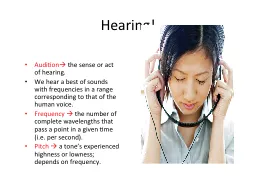
3. Hearing lossHearing loss is defined as having one or more frequencies out of the normal hearing range and it has degrees.The more sever the hearing loss is, the more effect will be on the overall functioning of the individual with hearing loss.But, even slight hearing loss can impede the development and acquisition of the normal language.
4. Types of Hearing lossConductive hearing loss: a failure in the efficient conduction of sound waves through the outer ear, tympanic membrane (eardrum) or middle ears (ossicles).
5. Causes of conductive hearing lossEarwax, also known by the medical term cerumen, is a yellowish, waxy substance secreted in the ear canal of humans and many other mammals. It plays an important role in the human ear canal, assisting in cleaning and lubrication, and also provides some protection from bacteria, fungi, and insects. Excess or impacted cerumen can press against the eardrum and/or occlude the external auditory canal and impair hearing
6. Otitis media is an inflammation of the middle ear: the space behind the ear drum. Otitis media is very common in childhood, and includes acute and chronic conditions; all of which involve inflammation of the ear drum (tympanic membrane), and are usually associated with a buildup of fluid in the space behind the ear drum (middle ear space).
7. Rupture or perforation (hole) of the eardrum can occur in infection, trauma (e.g. by trying to clean the ear with sharp instruments), explosion or loud noise.
8. Cholesteatoma is a destructive and expanding sac in the middle ear and/or mastoid process. There are two types: congenital and acquired.
9. Otosclerosis is a progressive degenerative condition of the temporal bone which can result in hearing.
10. Sensorineural Hearing lossSensorineural hearing loss is a type of hearing loss in which the root cause lies in the vestibulocochlear nerve (Cranial nerve VIII), the inner ear, or central processing centers of the brain.
11. Causes of Sensorineural Hearing LossCongenital.Acquired:1- Inflammatory Suppurative labyrinthitis Meningitis Mumps Measles Viral Syphilis
12. Causes of Sensorineural Hearing loss2- Ototoxic drugs.3- Physical trauma - either due to a fracture of the temporal bone affecting the cochlea and middle ear.4- Noise-induced - prolonged exposure to loud noises (>90 dB) causes hearing loss which begins at 4000Hz (high frequency). The normal hearing range is from 125 Hz to 20,000 Hz.5- Presbyacusis - age-related hearing loss that occurs in the high frequency range (4000Hz to 8000Hz). 6- Meniere's disease - causes sensorineural hearing loss in the low frequency range (125 Hz to 1000 Hz). Meniere's disesase is characterized by sudden attacks of vertigo lasting minutes to hours preceded by tinnitus, aural fullness, and fluctuating hearing loss.
13. Assessment ProtocolsOuter to inner.Objective and subjective.Determines hearing thresholds (air/bone).Determines site of lesion.Leads to diagnosis and intervention plan.
14. Otoscopic examination
15. Important LandmarksAn annulus fibrosus Lpi long process of incus - sometimes visible through a healthy translucent drumUm umbo - the end of the malleus handle and the centre of the drumLr light reflex - antero-inferioirlyLp Lateral process of the malleusAt Attic also known as pars flaccidaHm handle of the malleus
16. Abnormal TMMild retraction may be difficult to identify. The margin of the drum (annulus may become more pronounced as in this image
17. Abnormal TMAs the drum retracts so does the handle of the malleus and it may appear to be shortened on otoscopy. The lateral process will also become much more prominent than normal
18. ABNORMAL TMAs the drum becomes increasingly retracted, it drapes over the ossicular chain, and the incus and stapes head may be outlined
19. ABNORMAL TMEventually, nearly all the middle ear space may be lost and the drum comes into contact with the medial wall of the middle ear (this is known as atelectasis)
20. COLOR OF TMCompare this drum with the normal one. It is opaque and pale. There is slight injection of blood vessels. This is one appearance of glue ear
21. GLUE EARGlue ear may make the drum yellow or even darker than normal. Note also how retracted this drum is and how prominent the lateral process of the malleus appears. Blood in the middle ear will give the ear a blue or brown colour, this is called a haemotympanum
22. ABNORMAL TMwhite patches actually on the drum or within the drum itself are usually tympanosclerosis, which is deposition of calcium into the drum itself in response to trauma or infection. This is not normally of any consequence unless it is severe, which can lead to a mild conductive hearing loss.
23. ABNORMAL TMIf the white patch is behind the drum, this may represent cholesteatoma (keratin in the middle ear). This normally needs to be treated surgically.
24. ABNORMAL TMThis small red lesion at the tip of the malleus handle is a vascular lesion called a glomus tumour. This might cause pulsatile tinnitus, but is rare. This needs surgical treatment
25. ABNORMAL TMThis red bulge in the canal is another glomus tumour (glomus jugulare). this is the tip of a much larger lesion involving the temporal bone
26. PERFORATIONSafe perforation of the anterior part of the drum. A common cause of perforations in this position is a persistent defect after the extrusion of a grommet
27. VENT TUBEThis is a grommet (ventilating tube) in the correct position in the drum. The hole in the middle should be clear of debris. Note a small dried crust above the grommet which is unimportant and may be a small clot remaining from surgery
28. Clinical applications of tympanometry Murad Al-momani, Ph.D., CCC-A
29.
30.
31. TYMPANOMETRIC FEATURESTympanometric shapes.Static acoustic admittance.Tympanometric width (gradient).Tympanometric peak pressure.Equivalent ear canal volume.
32. Tympanometric shapesAccording to Jerger classification (1970).Tympanograms are classifieds according to the height and location of the tympanometric peak.Type A: has normal peak height and location of the peak.Type B: is flat. Type C: the peak is displaced to the negative tail.Type D: double peak.As : normal but shallow peak admittance.Ad : normal with excessive admittance.
33.
34. admittanceIt is the most important feature.It is sensitive to middle ear conditions including MEE, chronic otitis media, cholesteatoma and ossicular adhesion, ossicular discontinuity, TM perforation, glomus tumor.
35. Tympanometric widthThe sharpness of the peak is an indicator of middle ear pathology.Determined by bisecting the distance from the peak to the positive tail of the tympanogram.The width of the tympanogram at that point is determined in daPa.Abnormally narrow tympanograms might be related to otosclerosis but this has not been confirmed.But abnormally wide peak has been found to be related to middle ear effusion.
36. Tympanometric peak pressureThe pressure at which the peak occurred.Is an indicator of the pressure in the middle ear space.Negative pressure is thought to happen because the gases of the bacteria resulted from infection is absorbed by the middle ear mucosa and then a negative middle ear pressure occur.Studies however found that, without other tympanometric, audiometric or otoscopic abnormalities; negative pressure probably does not indicate a significant middle ear disorder.Positive middle ear pressure has been reported in acute otitis media.
37. Equivalent ear canal volume In the presence of a flat tympanogram, an estimate of the air in the canal can provide valuable information.Like detecting perforations in the TM. Or patency of the myringetomy tube.Usually high volume with flat tymps represents either perforations or patent vent tubes.
38. Sensitivity and specificitySensitivity has been found to be around 82% for MEE.Normal type A has 100% specificity.Overall sensitivity of around 80% and specificity of around 90%.That is good but means we need to interpret results with caution.
39. Tympanometry in infantsStudies has found frequent occurrence of double peaked tymps.Usually we use higher probe frequency when testing infants like 1000 Hz.
40. Pure tone audiometryMurad Al-momani, Ph.D., CCC-A, FAAA, American Board in Audiology
41. Procedures for conventional pure-tone audiometryAfter history taking and otoscopy we must choose how to test the hearing thresholds.Before we do pure tone audiometry (PTA), we usually perform middle ear immitance testingPTA will be almost done to all pts visiting us in the clinic because it is the basic test and give us a lot of information about the problem.
42. Air conduction testingWhen measuring behavioral air conduction thresholds, we are measuring a response to sound passed through the entire auditory pathway.Thus if the patient responds to pure tones at normal levels, we can be sure that the auditory system is reasonably intact from the outer ear to the auditory cortex.But that does not imply that there is no damage some where in the auditory system.For example in some retrocochlear lesions, the pt responds normally to pure tones but he has difficulty recognizing speech.
43. PTAWith PTA we can determine whether the pt has peripheral hearing loss (that is at the level of outer, middle, inner ear or the auditory nerve).PTA is administered both by air (air conduction PTA) or by bone (bone conduction PTA).Air conduction tests are administered by loudspeakers or ear phones.
44. Pure tonesPure tones are composed of sine waves that repeats itself at regular intervals.Pure tones may differ in either amplitude or frequency.The pure tones that the human ear can detect is between 20 Hz to 20,000 Hz.But we are most interested infrequencies 125 Hz to 8,000 Hz.
45. PTA Testing should be done in a room that is quiet enough to avoid masking by the noise.The maximum SPL that may exist in the room in order to obtain thresholds near 0 dB HL are determined by ANSI, 1991.We usually begin at 1000 Hz because some studies found that test-retest reliability is highest at this frequency.
46. PTAAfter establishing threshold at 1 KHz, we move to the frequencies (2000, 4000, and 8000Hz).If the difference between any two adjacent frequencies is 20 dB or more, we must measure the threshold at the inter octave frequencies.After we are done from the high frequencies, we return back and check the 1 KHz again to check for test-retest reliability.Then we test (500, 250 and 125 Hz).
47. PTAIf we test in the sound field, we must use warble tones instead of pure tones to avoid the production of standing waves.When using ear phones make sure that there is no excessive wax in EAC and that the earphone is snugly inserted in the canal. All equipment (audiometer, earphones, and testing room should be calibrated according the standards (will teach you how to do that in the instrumentation course).
48. PTA-BONE CONDUCTIONThe most commonly used procedure for bone-conduction testing is mastoid placement because it is more convenient.Frontal bone can be used as the place for the bone vibrator.
49. PTA.BONE CONDUCTIONWe should do bone conduction if the air conduction thresholds are above the normal range otherwise we do not need to do bone conduction testing.We first do unmasked thresholds and then we should apply masking to the contralateral ear in order to get precise threshold measurement in this ear (will talk about masking next lecture).
50. AUDIOGRAM
51.
52.
53.
54.
55.
56.
57.
58.
59. Information we get from audiogramDegree of hearing loss.Type of hearing loss.Configuration of hearing loss.
60. Speech audiometry
61. Why we need to assess speech sensitivity?The most important sounds that are important for humans are those related to speech.PTA does not give the clear picture about how the pt respond to speech signals.Some times, one might have normal sensitivity thresholds to PTA but the reception and recognition to speech signal is deteriorated like in retrocochlear lesions.
62. Speech audiometryWe need to know how sensitive our hearing to speech signals.Sensitivity measures are threshold measures that typically are referred to as the speech-recognition-thresholds (SRT) and speech detection threshold (SDT).Acuity measures are supra-threshold measures that typically are referred to as the speech-recognition score or word recognition performance.
63. Speech audiometrySRT means the dB HL level at which a certain percent correct recognition of words (usually 50%).In speech recognition, we are concerned in what a percent score (80%, 70%, 90%, 100% or so forth) does the pt have when we increase the intensity (dB HL) of the speech signal above the threshold (SRT).
64. SRT and SDTSRT and PTA average should be in agreement.Studies have found that PTA average (500, 1000, and 2000 Hz) and SRT should be + or – 6dB from each others. Some times SRT are worse than the PTA average like in cases of when there is islands of normal hearing in the audiogram especially at high frequencies. Also in cases of tumors around the auditory nerve, SRT are worse than PTA average.SRT might be better than PTA in cases of functional hearing loss.
65. Speech or word recognition scoresUsually conductive hearing loss does not affect speech discrimination scores (usually scores will be excellent, above 90%).Cochlear lesions affect this score significantly (usually scores rarely are above 80%).Retro-cochlear lesion affect the scores too sever.
66. Otoacoustic EmissionMurad Al-momani, Ph.D., CCC-A, FAAAAmerican Board in Audiology
67. Origin of OAEInitially reported by Kemp in 1978.OAE are considered a by-product of sensory OHCs transduction and represent cochlear amplifier that thought to be as a result of the contraction of OHCs in synchrony with BM displacement.The contraction of the OHCs (movement) is then propagated outward toward the middle ear and moves the TM.This in turn creates acoustic energy that is picked by the OAE probe.
68. OAESo in order to record OAE in EAC we need to have normal middle ear function.Conductive pathologies can prevent the recording of OAE but this does not mean that OAE is not present.
69. OAEEOAEs are recorded as a result of the introduction of acoustic stimulation.The first type is called transient EOAE (TEOAE): which appears as a result using a click stimulus.The second type is the distortion product EOAE (DPOAE): is a response occurring when two pure tones of different frequencies are presented simultaneously in EAC. The DPOAE is generated at frequencies that are different than both frequencies.
70. TEOAE
71. TEOAE
72. DPOAE
73. Applications of OAETEOAE can be recorded in all non-pathologic ears that do not display hearing loss of greater than 30 dB.OAE can be recorded in both adults and infants.Accordingly TEOAE and DPOAE can be used to screen for hearing loss in infants.DPOAE provide more frequency specific evaluation that TEOAE.
74. Clinical applications of EOAE1- can be used in newborn hearing screening. The results will indicate either fail or pass. Fail means that hearing thresholds are worse than 30 dB HL. Pass results means hearing thresholds are 30 dB HL or better.So, we can not use this tool to measure threshold of hearing.
75. Clinical applications of EOAE2- in differential diagnosis of hearing loss (site of lesion). This can help in differentiating sensory from neural hearing loss.3- monitoring of the effect of ototoxicity or noise exposure.4- although still under research: DPOAE can be used to screen for the carriers of the recessive hearing loss genes: many studies found that DPOAE is larger (especially at high frequencies) in carriers than in non carriers when using f2/f1 of 1.3 and low stimulus levels of 50-60 dB.
76.
77. Medial Olivocochlear efferent pathwaythe stimulation of the medial efferents alters IHCs sensitivity indirectly by altering the micromechanical properties of the OHCs.length, tension and the stiffness of the OHCs along their longitudinal axis are under the control of the MOC bundle, thus enhancing the auditory sensitivity, especially for low level stimuli at 30-40 dB SL.medial efferent system enhances the frequency resolving capacity and the vowel discrimination, especially in a background of noisy environment
78. Medial Olivocochlear efferent pathwaySince the medial olivocochlear bundle is mainly inhibitory, there has been already suggestions that dysfunction of the efferent auditory system, at any level from auditory cortex to cochlea, may be a basis for tinnitus generation, especially in noise-induced tinnitus cases.It has been also suggested that hyperacusis might be associated with dysfunction of the efferent system.
79. Clinical applications of ABRMurad Almomani, Ph.D., CCC-A, FAAA
80. The normal ABR waveformIs characterized by 5-7 peaks.Occurs in a latency epoch of 1.4 – 8.0 ms.Responses are usually displayed with positive peaks reflecting neural activity toward the vertex.These peaks are labeled with the roman numerals I through XII.The most prominent waves are I, III, and V.
81.
82.
83.
84.
85.
86.
87.
88.
89. Clinical applications of ABRThere are two main applications for ABR in the clinical settings:Neurodiagnosis: to assess the auditory pathway. This feature is specially used in adult populations.Waves absolute latency.Interpeak intervals.Interaural wave V latency difference.Absence of waves.Hearing thresholds estimation: mainly used in infants and children population.Wave V threshold.Wave V latency-intensity function.
90. Standard ABR Measures for Acoustic Tumor DetectionIT5 = Interaural time delay for wave V14121086420 ms7.36.4L1L2Tumor SideNon-Tumor SideIT5 = L2 - L1 = 0.9 msI. Background: Standard ABR Tumor Detection
91. Standard ABR Measures for Acoustic Tumor Detection:I-V Delay = Latency Difference Between Wave I and VI. Background: Standard ABR Tumor Detection
92. Standard ABR Measures for Acoustic Tumor Detection:I-V Delay = Latency Difference Between Wave I and VI. Background: Standard ABR Tumor Detection
93. Neurodiagnosis Who should be tested? Patients with:Dizziness.Unilateral tinnitus.Asymmetrical hearing loss.Sudden onset of hearing loss.Progressive hearing loss.
94.
95. Specific clinical application of ABR in pediatric populations Newborn hearing screening:Usually screen at 30-35 dBHL.It can be automated.If fail refer for a diagnostic ABR.Many studies revealed that automated ABR (AABR) is efficient in newborn hearing screening.Some new technologies combined OAE and AABR in one equipment and used both in the screening process resulted in less refer rate and less false positives.
96. Specific clinical application of ABR in pediatric populations Otitis media:Studies has found that ABR wave V latency-intensity function shift to the right in a proportion equivalent to the conductive hearing loss.Wave I is abnormally prolonged in Patients with effusion.Congenital aural atresia:Can use both circumaural headphones for air conduction ABR and bone vibrator for bone conduction ABR.
97. Specific clinical application of ABR in pediatric populations Auditory neuropathy: No single definition. No data about its prevalence, although it has been found to be around 10% in NICU who have hearing loss.Rare in healthy babies.Lesions can be in IHCs, synapses, or auditory nerve.Can have normal or mild to moderate PTA.Usually poor speech discrimination especially in noise.Many causes like hypoxia, hyperbillirubinemia, genetic. Present OAE and/or CM with absent ABR or abnormal ABR (may have only wave I).Or present CM, absent SP and absent or abnormal ABR.
98. Specific clinical application of ABR in pediatric populations Neoplasms and tumors:Neurofibromatosis type I and II: genetically autosomal dominant inherited progressive disorders. Usually tumors involving auditory nerve bilaterally.Brainstem gliomas: tumors in children and adolescent and tends to grow slowly.These disorders may show increase in ABR absolute latencies and interpeak intervals.
99. Specific clinical application of ABR in pediatric populations Epilepsy:ABR may show prolongation of waves III and V.Increase in interpeak intervals.Demyelinating diseases:Multiple sclerosis (MS): is the most common type in adults and is characterized by vertigo, unsteadiness and fluctuating SNHL.Schilder’s disease: a progressive childhood disease. Some consider it a variant of MS.ABR usually reveals an absence of waves III and V.
100. Specific clinical application of ABR in pediatric populations Fragile X syndrome: the most common hereditary type of mental retardation: Long absolute latencies.Increase in interpeaks intervals.Meningitis:Increase in interpeaks intervals and absolute latencies.Hydrocephalus:Increase in absolute latencies of waves III and V.Increase in interpeaks intervals.
101. Specific clinical application of ABR in adult populations Retrocochlear lesions:Vestibular schwannoma: mostly found in the VIII nerve. But may also involve V, VII, and XII.It is also used interchangeably with acoustic neuroma.Increase in absolute latency of wave III or V.Interaural Wave V latency difference.Increase in I-III, III-V, and I-V interpeak intervals (some or all depending on the location). may be absence of waves III or V.Stacked ABR findings.
102. (6 kHz)(2 kHz)(1 kHz)Cross Section: Human Auditory Meatus
103. Cross-section of Internal Auditory Canal (IAC)Facial NerveAcoustic NerveSup.Vest. NerveInf.Vest. Nerve
104. Medium or Large Tumor in IACFacial NerveAcoustic NerveSup.Vest. NerveInf.Vest. NerveTumor
105. Vestibular TestingVestibular testing is an important tool in the management of the patient with dizziness.The bedside evaluation of the dizzy patient, with a careful history and a thorough neurotologic diagnosis, is a crucial for making an accurate clinical diagnosis.We do not believe that vestibular testing is to be used as a stand alone diagnostic test battery for patients with dizziness.
106. BackgroundAlthough bedside and office examinations provide information about the status of the vestibular system, major limitations are the inability to quantify responses and to monitor the course of the illness or the results of medical and surgical management.Current technologies available for assessing the vestibular system include ENG/VNG, rotational testing and posturography.
107. Uses of Vestibular Lab.Aid in establishing diagnosisLocation, central versus peripheral.Lateralization.Documentation.Assist in devising treatment plan.Aid in long term management.
108. ENG or VNGENG/VNG is the most common method of laboratory evaluation of the vestibular system.The exam consists of a battery of tests.ENG monitor eye movement .The vestibular and ocular systems are connected through the VOR. Thus, patients with peripheral and/or central balance disorders often exhibit abnormal eye movements that can be measured and recorded.
109. Eye Movement RecordingIn performing ENG/VNG, the patient eye movements are measured relative to head position, which can be achieved in a number of ways.Measuring electric potentials, measuring magnetic potentials, using video cameras or using infrared technology and direct observation.
110. Nystagmus
111.
112. Left BeatingRight BeatingSlowFast
113. VNG Test BatteryCalibrationGazeSaccadePursuitOptokineticPositionalHallpikeCaloric
114. Gaze TestThe function of the gaze system is to maintain visual fixation of an object on the fovea of the eye.To identify the presence of spontanoues eye movement.Normal gaze, patient able to maintain position with eyes opened and closed.
115. Gaze results with peripheral ¢ral lesionsHorizontal.Directional fixed.Suppressed with visual fixation.Horizontal, vertical or rotatory.Directional changing.Enhanced with visual fixation.
116. Saccade (refixation ) Test.The function of saccadic eye movement system is to redirect the eye from one target to another in the shortest possible time.Inaccurate eye movement, where the eye either undershot or overshot the target is abnormal and seen frequently in patients with cerebellar dysfunction.
117. Saccade Test
118. Results:Normal saccadic eye movement test should produce rapid and accurate eye movement.Inaccurate eye movement, where the eyes overshot or undershot the target .
119. Saccade
120. Ocular Pursuit TestThe function of ocular pursuit system is to stabilize a slowly moving object on the fovea of the eye by matching the angular velocity of the eye with that of the moving object.
121. Pursuit Test
122. Results:When the pursuit system is impaired, small corrective saccadic movements replace the smooth pursuit movement, so the eye can catch up the moving target.It may be the most sensitive subtest in ENG battery for detection of brainstem and cerebellar disorders.
123. Pursuit
124. Abnormal Pursuit
125. Optokinetic TestOptokinetic system maintain visual fixation when the head is in motion.Target is rapidly passed in front of the subject in one direction, then the other.Eye movements are recorded and compared in each direction.Asymmetry suggestive of central lesion
126. Optokinetic
127. Optokinetic
128. Optokinetic
129. Dynamic Positional Test ( Hallpike )The patient complains a motion related vertigo at certain positionIt is maneuver that places the patient head in the position that creates the response.Criteria: Latency period, subjective vertigo, Transient nystagmus, fatigable, lesion in the undermost ear,
130. Dix Hallpike maneuver Used to provoke nystagmus and vertigo commonly associated with BPPV.Head turned 45 degree to maximally stimulate posterior semicircular canal.Head supported and rapidly placed into head hanging position.Frenzel glasses eliminate visual fixation suppression of response or can be tested Using VNG.
131. Dix Hallpike Maneuver
132. Caloric Test
133. Caloric TestsCaloric test is a part of ENG/VNG.It reflects an attempt to discover the degree to which the vestibular system is responsive and also how symmetric the responses are, between left and right.It is a test of the lateral semicircular canals.Most caloric tests are nowadays are done using computerized systems, the computer analyzes the caloric data, computing peak slow-phase velocity.
134. Caloric Test
135. Caloric Test (Procedure )Irrigations of EEC performed with cold and warm water or air.Water - cool = 30 C; warm = 44 CAir - cool = 24 C; warm = 50 CResponse pattern follows the form of COWSNystagmus induced results are calculated to obtain Unilateral Weakness and Directional Preponderance
136. Caloric Test
137. Caloric Test
138. Caloric Test
139. Posturography
140. Computerized Dynamic PosturographyIt is the most commonly used system for clinical postural assessment.The subject stand on a computer-controlled movable platform with movable visual surround.The platform or visual surround can be fixed or move independently.Six test conditions compose the SOT battery.
141. The six test conditions
142.
143. Results
144. Results
145. Use of posturographyEvaluating patients with balance disorders.Ordered for patients with vague symptoms of dizziness and unsteadiness.It can be used to detect malingerers.Planning and monitoring the course of postural rehabilitation.Documentation of postural responses.