
2016 42230811 ISSN 23214287 2308 Original Research Article ROLE OF RELA TIVE POSITION OF C OL UMN OF CER VICAL AND UPPER THORACIC VERTEBRAE IN WEIGHT TRANSMISSION Komala B ABSTRACT Address ID: 936359
Download Pdf The PPT/PDF document "Int J Anat Res" is the property of its rightful owner. Permission is granted to download and print the materials on this web site for personal, non-commercial use only, and to display it on your personal computer provided you do not modify the materials and that you retain all copyright notices contained in the materials. By downloading content from our website, you accept the terms of this agreement.


Int J Anat Res 2016, 4(2):2308-11. ISSN 2321-4287 2308 Original Research Article ROLE OF RELA TIVE POSITION OF C OL UMN OF CER VICAL AND UPPER THORACIC VERTEBRAE IN WEIGHT TRANSMISSION Komala B. ABSTRACT Address for Correspondence: Dr. Komala B, Professor and Head, Department of Anatomy, BGS Global Institute of Medical Sciences, Bengaluru, Bengaluru-560060 Karnataka, India. Mobile: +91-9844470491, Email: kominag@yahoo.co.in Introduction: The position of column is important because when columns are placed further away from centre of load they can resist bending and buckling forces better than columns which are placed closer to each other. Objective: This study has been attempted to find out the relative positions of the columns to each other by comparing mean arch indices and its ratio to mean inferior body surface area. Materials and Methods: The 6 cervical and upper 5 thoracic vertebrae of 30 human adult male columns were selected for the study. The various parameters such as inferior body surface area, arch index and the ratio arch index to inferior body surface area were measured for each of the 6 cervical and upper 5 thoracic vertebrae of 30 columns. Results: Measurements of the area of the inferior surface of the body, arch index and the ratio of arch index to inferior surface of the body. The ratio showed a gradual decline from C 2 to T 5 level. Above C 7 level the size of the arch was greater than body area, but at T 1 the two were of almost equal size, below which body area exceeded the arch size. In relation to the body area, neural arch size diminished considerably in the thoracic region. Conclusion: The measurements obtained by the present study reveals the importance of neural arch in understanding the mechanics of spinal anatomy and its applications with respect to transmission of weight. KEYWORDS: Arch index, cervical column, thoracic column, weight transmission. INTRODUCTION International Journal of Anatomy and Research, Int J Anat Res 2016, Vol 4(2):2308-11. ISSN 2321-4287 DOI: http://dx.doi.org/10.16965/ijar.2016.202 Access this Article online Quick Response code Web site: Received: 11 Apr 2016 Accepted: 09 May 2016 Peer Review: 12 Apr 2016 Published (O): 31 May 2016 Revised: None Published (P): 31 May 2016 International Journal of Anatomy and Research ISSN 2321-4287 www.ijmhr.org/ijar.htm DOI: 10.16965/ijar.2016.202 Department of Anatomy, BGS Global Institute of Medical Sciences, Bengaluru, Karnataka, India. pathological processes involving vertebrae [2]. The vertebral bodies and the intervertebral discs form an important column in transmission of weight of the body. In the upper thoracic region, due to the anterior curvature, the main part of the compressive force is transmitted through the anterior column formed by vertebral body and intervertebral disc, with resulting increased stress [3]. In recent years, there have been considerable The functions of the column are to support the trunk, to protect the spinal cord and nerves, and to provide attachments for muscles [1]. The vertebrae can be involved in various conditions. These include fractures, infections, malignan- cies and inflammatory disorders. Abnormal cur- vatures of the vertebral column in the thoracic region such as kyphosis and scoliosis may result from developmental anomalies or Int J Anat Res 2016, 4(2):2308-11. ISSN 2321-4287 2309 Komala B. ROLE OF RELA TIVE POSITION OF C OL UMN OF CER VICAL AND UPPER THORA CIC VER TEBRAE
IN WEIGHT TRANSMISSION. developments in instrumentation designed to stabilize and correct the thoracic spine [4, 5]. The position of column is important because when columns are placed further away from centre of load they can resist bending and buck- ling forces better than columns which are placed closer to each other. Neural arch component of a vertebra, besides its contribution to the for- mation of the vertebral canal and the role of its articular processes in governing the range and direction of movements between two vertebrae, is also involved in weight bearing and the measurements of the vertebral column and mathematical calculations have provided strong evidence for the role of the neural arch in weight transmission. In the present study an attempt has been made to investigate the role of neural arches in weight transmission in the cervical and upper thoracic regions of the vertebral column. Hence, the present study measured the relative positions of the columns to each other by comparing mean arch indices and its ratio to mean inferior body surface area, so that this knowledge could be applied to explain some of the clinicopathological conditions of the spine. MATERIALS AND METHODS measurements which were made at the second or subsequent attempts were identical to those made at the first time a maximum difference of one millimeter. Sexing of the vertebral column was established by observing the respective pelvis including the sacrum by the criteria laid down in the text books of anatomy such as subpubic angle, ischiopubic rami, ischial spine, greater sciatic notch, preauricular sulcus, acetabular cavity, obturator foramen, ischium-pubis index, breadth and curve of sacrum, articular surface of S 1 and sacral index. Those columns in which the sex could be established unequivocally only have been included. Inferior body surface area: The area of the inferior surface of the body in each vertebra was measured using graph paper method. The outline of the inferior surface of the vertebral body was traced on to a thin tracing sheet which was then transferred on to a graph sheet with the help of a tracing paper and the area was measured in square centimeters by counting the number of squares covered. This represents the parameter of the anterior column. Arch Index: This was obtained by the product of a and b as shown in Figure 1; b is the maximum distance between the two articular facets at distance a from the posterior margin of the body. A mean arch index (± standard deviation) for each vertebral level was obtained. The arch index indicates approximately the position of the articular processes in relation to the body. Arch index/body area ratio: In the vertebral column, the size of the bodies gradually increases from above downwards. Hence, to compare the magnitudes of the arch index at various levels, its ratio to body area were calculated. Thus arch index/body area were obtained. The present study was done on dry vertebral bones procured from the collection of Bangalore Medical College, Bengaluru, Karnataka, India. A total of 330 vertebrae were measured. Anatomical measurements were taken of the six cervical and upper five thoracic vertebrae of thirty columns using a vernier calliper (0-150mm with a precision of 0.02mm), sharp pencil, tracing paper, graph sheet and carbon paper. Adopting the various mechanical principles and ideas of Pal GP and Routal RV [6], the mean arch index and its ratio to the mean inferior body surface area were taken in the present study for each of the six cervical (C 2 - C 7 ) and upper fi
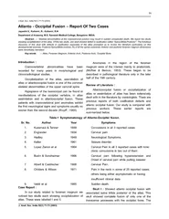
ve thoracic vertebrae (T 1 - T 5 ) of thirty columns. To overcome personal errors in judgment, the following procedures were adopted: Initially, all the measurements were taken by the author and criteria were established till the observations made individually coincided fully. Then the observations were repeated by a double blind method. Almost all the observations and RESULTS Measurements of the area of the inferior sur- face of the body, arch index and the ratio of arch index to inferior surface of the body are pre- sented in Table 1. The ratio showed a gradual decline from C 2 to T 5 level. Above C 7 level the size of the arch was greater than body area, but at T 1 the two were of almost equal size, below Int J Anat Res 2016, 4(2):2308-11. ISSN 2321-4287 2310 Komala B. ROLE OF RELA TIVE POSITION OF C OL UMN OF CER VICAL AND UPPER THORA CIC VER TEBRAE IN WEIGHT TRANSMISSION. which body area exceeded the arch size. In relation to the body area, neural arch size diminished considerably in the thoracic region (Figure 2). Table 1: The inferior body surface area, arch index and their ratio at each vertebral level (n=330). C 2 3.58±0.44 6.03±1.18 2.28±0.65 2.64 C 3 4.17±0.65 6.27±1.09 2.69±0.62 2.33 C 4 4.85±0.77 5.91±1.33 3.15±0.74 1.65 C 5 5.48±0.32 6.18±1.73 3.62±0.70 1.71 C 6 6.00±0.21 6.72±1.23 4.02±0.69 1.67 C 7 7.24±0.35 7.19±1.20 4.90±0.62 1.46 T 1 6.96±0.79 5.67±1.39 5.40±0.92 1.05 T 2 7.25±1.18 5.41±0.98 5.81±1.06 0.93 T 3 7.21±1.25 4.57±0.85 5.99±1.15 0.76 T 4 7.52±1.34 4.70±0.61 6.36±1.28 0.74 T 5 7.90±1.42 4.48±0.60 6.86±1.66 0.65 Arch index / Inferior body surface area Mean Inferior body surface area (cm 2 ) Total Inferior body surface area (body + 2 facets) (cm 2 ) Mean Arch index Vertebral levels Each value is Mean ± standard deviation. Fig. 1: Relative position of column of cervical and upper thoracic vertebrae in weight transmission. Neural arch index is obtained by the product of parameters a and b [A, B]; Diagrammatic representation of the positions of weight bearing pillars of the vertebral column [C, D]. a, cross section of anterior column formed by bodies; b and c, cross sections of posterior column formed by articular processes; d, cross section of posterior column formed by lamina. Fig. 2: Graph showing the arch index and the body surface area at vertebral levels. DISCUSSION Recently, the authors have developed a hypothesis according to which the vertebral column not only transmits weight through the bodies and intervertebral discs but also through the neural arch. In the cervical region, it is transmitted through three columns; an anterior column formed by the bodies and intervertebral discs and two posterior columns formed by the articular pillars [3]. This concept of a three column spine in the cervical region is similar to that of Louis [7]. Following the ideas of Davis [8] and on the basis of various mechanical principles, this hypothesis of weight transmission through the neural arch component of the cervical and upper thoracic vertebral column has been tested in the present investigation. By definition, stresss is equal to the load S divided by area a which resists the load or compressive force: s = S/a. In the vertebral column, the cross sectional area and load both increase from above downwards. Thus cross sectional area of the column at a particular level is correlated with its ability to resist longitudinal compression. If we consider that the load s is being transmitted th
rough three columns then the proportion of the load carried through each individual column will depend on its cross sectional area, according to the above principle. However, the position of these columns in relation to each other is also important. The resistance to overturning or bending or buckling increases in cube proportion as the columns are located away from the centre. This also helps to maintain stability. The columns of Figure 1(C) are more stable and efficient for weight bearing than the columns of Figure 1(D). The fact that the articular processes, through which it is postulated that weight transmission occurs, are placed at a wide distance from the body in the cervical region but are close to it in the thoracic region, is highly significant in relation to the geometrical property of columns discussed above. The arch index is maximal at C 7 (Table 1, Figure 2), indicating that the three columns are widest apart at this level. This is necessary because at this level a wide base must be formed against the fixed thoracic column for Int J Anat Res 2016, 4(2):2308-11. ISSN 2321-4287 2311 Komala B. ROLE OF RELA TIVE POSITION OF C OL UMN OF CER VICAL AND UPPER THORA CIC VER TEBRAE IN WEIGHT TRANSMISSION. support, stability and effective movements of the cervical column. The fact that in the upper thoracic region the posterior column becomes weaker and more closely placed to the anterior column (Tables 1) strongly supports the view that load from the posterior column has been transferred to the anterior column. The closer positions of the columns and the increased stress on the anterior column make the thoracic region more susceptible to bending or buckling deformity. CONCLUSION The arch index is maximal at C 7 , indicating that the three columns are widest apart at this level. This is necessary because at this level a wide base must be formed against the fixed thoracic column for support, stability and effective movements of the cervical column. The measurements obtained by the present study reveals the importance of neural arch in understanding the mechanics of spinal anatomy and its applications with respect to transmission of weight. Conflicts of Interests: None REFERENCES [4]. Cahill PJ, Iorio J, Samdani AF, Pahys JM, Betz RR. Anterior Growth Modulation Techniques: Vertebral Body Stapling. In The Growing Spine 2016 (pp. 731- 749). Springer Berlin Heidelberg. [5]. Richardson CA, Richardson DA, inventors; Gravity Fitness Australia Pty Ltd, assignee. Thoracic stabi- lizer. United States patent application US 14/ 422,067. 2013 Aug 19. [6]. Pal GP, Routal RV. A study of weight transmission through the cervical and upper thoracic regions of the vertebral column in man. J Anat 1986;148:245- 61. [7]. Louis R. Spinal stability as defined by three-column spine concept. Anatomia clinica 1985;7:33-42. [8]. Davis PR. Human lower lumbar vertebrae: Some mechanical and osteological considerations. J Anat. 1961;95(Pt 3):337-44. [1]. Cramer GD, Darby SA. Clinical anatomy of the spine, spinal cord, and ANS. Elsevier Health Sciences;2013. [2]. Ranson C, Harris PF. Anatomy for problem solving in sports medicine: The Back. M&K Update Ltd; 2015. [3]. Komala B. A study of relative magnitude of compressive forces passing through the cervical and upper thoracic vertebrae in man. Int J Anat Res 2016;4(1):1927-30. How to cite this article : Komala B. ROLE OF RELA TIVE POSITION OF C OL UMN OF CERVICAL AND UPPER THORACIC VERTEBRAE IN WEIGHT TRANSMISSION. Int J Anat Res 2016;4(2):2308-2311. DOI: 10.16965/ijar.2016.20