
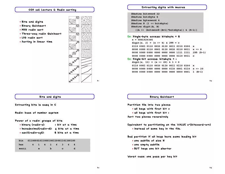
Golgi apparatus and vesicular transport Dr Mamoun Ahram Faculty of Medicine Second year Second semester 20142014 Principles of Genetics and Molecular Biology Functions of Golgi Further protein processing and modification ID: 438186
Download Presentation The PPT/PDF document "Lecture 3: Protein sorting" is the property of its rightful owner. Permission is granted to download and print the materials on this web site for personal, non-commercial use only, and to display it on your personal computer provided you do not modify the materials and that you retain all copyright notices contained in the materials. By downloading content from our website, you accept the terms of this agreement.


Slide1
Lecture 3: Protein sorting (Golgi apparatus and vesicular transport)
Dr. Mamoun AhramFaculty of MedicineSecond year, Second semester, 2014-2014
Principles of Genetics and Molecular BiologySlide2
Functions of Golgi
Further protein processing and modificationProtein sortingSynthesis of glycolipids and sphingomyelinSlide3
Structure of the Golgi
,
endosomesSlide4
Processing of N
-linked oligosaccharides in GolgiSlide5
O-linked glycosylation
Proteins can also be modified by the addition of carbohydrates to the side chains of acceptor serine and threonine residues.
The serine or
threonine
is usually linked directly to
N
-
acetylgalactosamine, to which other sugars can then be added. In some cases, these sugars are further modified by the addition of sulfate groups.Slide6
Lipid and Polysaccharide Metabolism in the Golgi
Transfer of phosphorylcholine group is from phosphatidylcholine
to
ceramide
.
Sphingomyelin is synthesized on the
lumenal
surface.Addition of sugar residues.Glucose is added to ceramide
on the cytosolic side and glucosylceramide then apparently flips and additional carbohydrates are added on the lumenal side of the membrane
Ceramide is synthesized in the ERSlide7
Protein Sorting and Export
In contrast to the ER, all of the proteins retained within the Golgi complex are associated with the Golgi membrane rather than being soluble proteins within the lumen
Continuous, unregulated secretion
Regulated secretion after
siganling
from specialized vesicles
Protein packaging mediated by cargo receptor
processing in Immature
secretory
vesiclesSlide8
Transport to the plasma membrane of polarized cells
This is accomplished by the selective packaging of proteins into transport vesicles from the trans Golgi or recycling
endosomes
.
Targeting is determined by special sequences (
basolatera
) or sugar modification (apical)Slide9
Processing of lumenal
lysosomal proteins
Addition of N-
acetylglucosamine
phosphates
Removal of N-
acetylglucosamine
The enzyme recognizes a signal patch (a three-dimensional structural determinant) not a sequence.Slide10
Transport of lysosomal proteins
Lumenal lysosomal
proteins marked by mannose-6-phosphates bind to a mannose-6-phospahte receptor.
The complexes are packaged into transport vesicles destined for late
endosome
, which mature into
lysosomes
.lysosomal membrane proteins are targeted by sequences in their
cytoplasmic tails, rather than by mannose-6-phosphates.Slide11
The mechanism of vesicular transport Slide12
How have we understood the mechanism?
Isolation of yeast mutants that are defective in protein transport and sorting (sec mutants) The role of Sec61 as translocation channel in the ER
Reconstitution of vesicular transport in cell-free systems
Biochemical analysis of synaptic vesicles
Tracing the path of GFP fusion proteins
Proteomics analysisSlide13
Formation and fusion of a transport vesicle
Coat disassembly
Vesicular docking & fusion
Vesicular transportSlide14
Coat proteinsSlide15
Formation of clathrin-coated vesiclesSlide16
Role of ARF1
Activation of Arf1 by GEF
Recruitment of AP1 (not shown) and
clathrin
Formation of Arf1-clathrin-receptor-cargo complex
Formation of vesicle
Budding and transport of vesicle
Inactivation and of Arf1 and disassembly of coat
Vesicle fusionSlide17
Players of vesicle fusion
The formation v-SNAREs-t-SNAREs complexes on the leads to membrane fusion.GTP-binding Rab proteins function in several steps of vesicle trafficking.
Different combinations of
Rab
proteins mark different organelles and transport vesicles
Effector proteins allow for specific interactionSlide18
The mechanism of fusion
Fusion
Closer vesicle-target
Disassembly of SNARE complex
Interaction of effector proteins
Tethering,
hydrolysis of GTP, SNARE interactionsSlide19
ExocytosisSlide20
Griscelli syndrome (GS)
A rare genetic conditionType GS: GS1, GS2, GS3Mutations in MYO5A, RAB27A and MLPH genes that encode the MyoVA-Rab27a-Mlph protein complex that function in
melanosome
transport and fusion.
Pigmentary
dilution of the skin, silver-grey hair, melanin clumps within hair shafts
Mature
melanosomes accumulatte in the centre of
melanocytesSlide21
LysosomesSlide22
Structure
Lysosomes are membrane-enclosed organelles that contain various enzymes that break down all types of biological macromolecules. Lysosomes degrade material taken up from outside and inside the cell.Slide23
Lysosomal enzymes
Lysosomes contain ~50 different acid hydrolases.
The enzymes are active at the acidic pH (about 5) that is maintained within
lysosomes
.
Levels of Protection:
Containment
Inactive if releasedA proton pump maintains lysosomal pH.Slide24
Lysosomal storage diseases
Glycolipidoses (sphingolipidoses)Oligosaccharidoses
Mucopolysaccharidoses
: deficiencies in
lysosomal
hydrolases
of glycosaminoglycans (heparan, keratan and dermatan sulfates, chondroitin
sulfates. They are chronic progressively debilitating disorders that lead to severe psychomotor retardation and premature death.Slide25
Glucocerebroside
Glucocerebroside is a glycosphingolipids (a monosaccharide attached directly to a ceramide
unit (a lipid)
It is a byproduct of the normal recycling of red blood cells during, which are
phagocytosed
by macrophages, degraded and their contents recycled to make new cells.Slide26
Types
Three types according to severity and nervous system involvementType I: (least severe, most common) the nervous system is not involved; spleen and liver enlargement, development of bone lesionsTypes II and III (more severe, much rarer): the only cells affected in
Gaucher's
disease are macrophages
Because macrophages function is to eliminate aged and damaged cells by
phagocytosis
by continually ingesting large amounts of lipids to be degraded in
lysosomesSlide27
Gaucher disease, type I (acid glucocerebrosidase deficiency)
Caused by mutation in the gene encoding acid-beta glucosidase
, or
glucocerebrosidase
, which catalyzes the hydrolysis of
glucocerebroside
to glucose and
ceramide.Slide28
Gaucher disease, type I (glucocerebrosidase deficiency-acid)
Gaucher's disease is the most common of the lysosomal storage diseases, which are caused by a failure of lysosomes to degrade substances that they normally break downThe resulting accumulation of nondegraded compounds leads to an increase in the size and number of lysosomes within the cellSlide29
Carbohydrate metabolismSlide30
No.
Type
Defective enzyme
Organ affected
Glycogen in the affected organ
Clinical features
0
glycogen synthase-2
Liver
hypoglycemia, early death, hyperketonia
I
Von Gierke disease
Glucose 6-phosphatase or transport system
Liver and kidney
Increased amount; normal structure.
Massive enlargement of the liver. Failure to thrive. Severe hypoglycemia, ketosis, hyperuricemia, hyperlipemia.
II
Pompe
disease
-1,4-Glucosidase (
lysosomal
)
All organs
Massive increase in amount; normal structure.
Cardiorespiratory
failure causes death, usually before age 2.
III
Cori disease
Amylo-1,6-glucosidase (debranching enzyme)
Muscle and liver
Increased amount; short outer branches.
Like type I, but milder course.
IV
Andersen disease
Branching enzyme
Liver and spleen
Normal amount; very long outer branches.
Progressive cirrhosis of the liver. Liver failure causes death, usually before age 2.
V
McArdle disease
Phosphorylase
Muscle
Moderately increased amount; normal structure.
Limited ability to perform strenuous exercise because of painful muscle cramps. Otherwise patient is normal and well developed.
VI
Hers disease
Phosphorylase
Liver
Increased amount.
Like type I, but milder course.
VII
Tarui Disease
Phosphofructokinase
Muscle
Increased amount; normal structure.
Like type V.
VIII
Phosphorylase kinase
Liver
Increased amount; normal structure.
Mild liver enlargement. Mild hypoglycemia.
IX
phosphorylase kinase, β-subunit
liver, leukocytes, muscle
like VI
Fanconi-Bickel, hepatorenal glycogenosis
glucose transporter-2 (GLUT-2)
liver
failure to thrive, hepatomegaly, rickets, proximal renal tubular dysfunction
OligosaccharidosesSlide31
Pompe disease (type II)
Lysosomes become engorged with glycogen because they lack α-1,4-glucosidase, a hydrolytic enzyme confined to these organellesGlycogen structure is normal, but its amount is excessiveSlide32
I-cell disease
Lack of targeting of lysosomal enzymes from GolgiA deficiency in tagging enzyme
Features: severe psychomotor retardation that rapidly progresses leading to death between 5 and 8 years of age.Slide33
TreatmentSlide34
Endocytosis
Molecules are taken up from outside the cell in endocytic vesicles, which fuse with early endosomes
.
Membrane receptors are recycled via
recyling
endosomes
.Early endosomes mature into late endosomes.
Transport vesicles carrying acid hydrolases from the Golgi fuse with late endosomes, which mature into lysosomes.The acid hydrolases dissociate from the mannose-6-phosphate receptor and the receptors are recycled to the Golgi.
Maturation
Recycling
endosomes
Maturationor fusionSlide35
Chloroquine
Anti-malarial agentIn the parasite’s vaculoe, hemoglobin is digested and
heme
is modified by
heme
polymerase.
If
heme is not modified, it is toxic to the parasite.Chloroquine inhibits the enzyme.It is a weak base that becomes charged at acidic pH
It crosses membranes into the malarial digestive vacuole.Slide36
Phagocytosis and autophagy