
Kim MJ Bancroft E Lehnkering E Donlan RM Mascola L Alcaligenes xylosoxidans Bloodstream Infections in Outpatient Oncology Office Emerg Infect Dis 200814710461052 httpsdoiorg103201eid1407070894 ID: 1042023
Download Presentation The PPT/PDF document "Figure 1 Figure 1. Scanning ele..." is the property of its rightful owner. Permission is granted to download and print the materials on this web site for personal, non-commercial use only, and to display it on your personal computer provided you do not modify the materials and that you retain all copyright notices contained in the materials. By downloading content from our website, you accept the terms of this agreement.


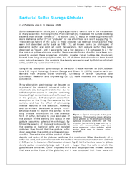


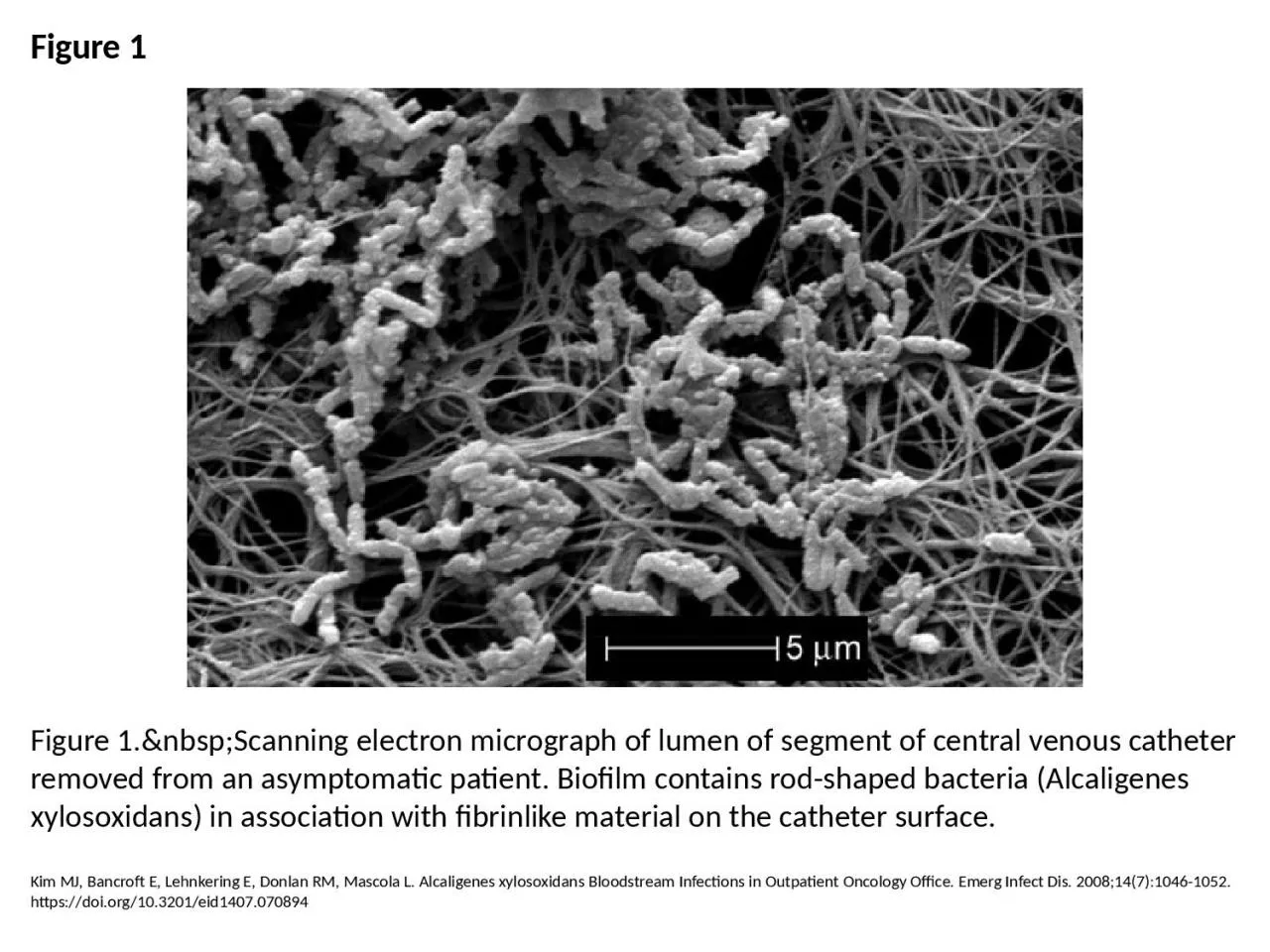
1. Figure 1Figure 1. Scanning electron micrograph of lumen of segment of central venous catheter removed from an asymptomatic patient. Biofilm contains rod-shaped bacteria (Alcaligenes xylosoxidans) in association with fibrinlike material on the catheter surface.Kim MJ, Bancroft E, Lehnkering E, Donlan RM, Mascola L. Alcaligenes xylosoxidans Bloodstream Infections in Outpatient Oncology Office. Emerg Infect Dis. 2008;14(7):1046-1052. https://doi.org/10.3201/eid1407.070894